Methods of clinical research of animals. General and special methods of clinical research
In healthy animals, vesicular or pulmonary respiration, physiological bronchial breathing in the tops of the lungs and laryngotracheal breathing in the larynx and trachea.
In pathological processes, a number of additional sounds may occur in the bronchi, alveoli of the lungs, pleura and pleural cavity, such as are not found in the norm.
Identification and differentiation of the main and additional noises that occur during pathology gives an idea of the physical state of the respiratory organs and often of their functional state. Changes in the work of the respiratory organs in pathology are sometimes so significant that they do not cause difficulties in their identification and diagnostic evaluation. However, it should be borne in mind that during auscultation of the respiratory organs, different animals have their own characteristics and difficulties.
auscultation technique. During direct auscultation, the animal's chest is covered with a towel. A towel is necessary not only for hygienic reasons, but also because it partly eliminates the friction noise of wool. To protect yourself from possible damage and reduce the movement of the animal, its head must be fixed, and in restless animals it is recommended to raise the thoracic limb.
Study of the left half chest performed with the right ear, and auscultation of the right half with the left. The specialist in this case becomes facing the head of the animal, and only with auscultation of the posterior parts of the lung can one become facing the rear of the animal. In order to create closer contact, as well as for the purpose of self-defense, the appropriate hand is placed on the withers or back of the animal and held during the entire examination. In small animals, they usually stand behind the animal; this allows auscultation to compare the auscultatory phenomena of both sides during auscultation. With unilateral damage to the lungs and bronchi, this is of great practical importance.
Auscultation is best done in a closed, small room with a soft floor. Auscultation on the street, in the common stables and cowsheds is greatly hampered by extraneous rustles, and in the summer, by insects that disturb the animals.
Despite the fact that the lungs are adjacent to the chest wall of the animal throughout, auscultation is possible only in places less covered by muscles. Parts of the lung, covered with a scapula and powerful muscles of the shoulder, give sounds either sharply weakened, or they are not heard at all. This is especially pronounced in large animals, in which, normally, breathing on the chest is much weaker than in small animals. This encourages auscultation of those parts of the chest where sound phenomena are more pronounced, and then move on to areas where they are less intense and hard to hear.
In the horse, auscultation of the lungs begins with the middle section of the chest, located directly behind the shoulder blade. After listening to this area, they proceed to the study of the mid-posterior chest, then auscultate the upper-middle and upper-posterior segments in turn. Next, the lower-posterior section of the lung is examined. The sections of the lung, hidden under massive muscles, are examined at the end and, often, after comparing the right and left sides. In cattle, the study is carried out in the same order, although it must be said that auscultation through the scapula is much easier than in the horse. In cattle, in addition, the prescapular region is available for auscultation. The study of this area gives an idea of the condition of the tops of the lungs. These departments often turn out to be the site of the development of tuberculous and peripneumonic processes. The area of the oscultation in the prescapular region can be expanded by abducting the leg back. In small animals, auscultation is available for all areas of the lung with the only difference being that in some places the breath sounds are heard better, while in others they are somewhat weaker.
2-3 phases of breathing are heard, and if no deviations from the norm are found, then they proceed to listening to another area. In case of detection of deviations from normal respiratory noise, it is necessary to establish the nature of these deviations. To do this, you can compare the breath noise with neighboring areas and with homologous points on the opposite side of the chest.
If breathing is not heard or heard very weakly, it is recommended to resort to increased breathing. In the horse, this can be achieved by leading, by lunging, or by closing the nostrils, turning off the breath for a while. In cattle, increased breathing is possible by closing the nostrils with a towel, hands, or using a Rautman clamp. This technique can cause an increase in respiratory noise and correctly assess the changes detected during auscultation.
During auscultation, it is necessary to establish the intensity of respiratory noises, their properties, and if there are pathological noises, to find out their nature and location.
All noises found on the chest from the side of the lungs are divided into main and adnexal. The main breath sounds include vesicular and physiological bronchial and laryngeal breathing; Airborne noises are subdivided into bronchopulmonary and extrapulmonary. Bronchopulmonary include dry and moist rales, crepitating rales, crepitus, bronchial abnormal breathing, amphoric breathing and the noise of a falling drop. Extrapulmonary, or pleural, in turn, are divided into pleural friction noise, splash noise and pulmonary fistula noise.
A number of methods are used to diagnose animal diseases. Among them are general, special (instrumental), laboratory and functional.General methods are divided into inspection, palpation, percussion, auscultation and thermometry. They are called general because they are used in the study of almost every patient, regardless of the nature of the disease.
Inspection [lat. inspectare - watch, observe] is carried out with the naked eye in good light or with the use of reflectors, endoscopic devices. Inspection can be group and individual, general and local, external and internal.
Group examination is carried out during the study a large number animals and with its help allocate sick or suspicious individuals for further comprehensive examination. Each sick animal admitted for treatment is subjected to an individual examination. General examination is carried out on the left and right, front and rear, and, if possible, also from above. At the same time, the habitus, the condition of the hairline, skin, the presence of superficial damage, the symmetry of various parts of the body are determined. Local examination allows you to explore the areas of localization of the disease process and can be external or internal (through lighting devices).
Palpation [lat. palpatio - groping]. The touch method is based on touch. Conduct a study first healthy parts of the body, and then the affected. At the same time, palpation should not cause pain to the animal or resemble tickling. Distinguish superficial and deep palpation.
Superficial examine the skin, subcutaneous tissue, muscles, joints, tendons and ligaments. By tightly applying the palm, for example, the temperature and humidity of the tissues are established, the state of the heart beat, the presence of tangible noises are assessed. The consistency and soreness of the tissues is determined by pressing the fingertips with increasing force until the animal responds. By stroking with the palm of the hand, the nature of the surface is established, and with the fingers, the shape and integrity of the bones and joints are determined. By gathering the skin into a fold, its elasticity is established and areas of increased pain sensitivity are identified.
Deep palpation examines the organs of the abdominal and pelvic cavity by determining their location, size, shape, consistency, pain. Deep palpation can be external and internal. The deep outer include:
Penetrating when pressed with fingers or a fist on abdominal wall and examine a specific organ, such as the liver, scar, etc.
Bimanual, i.e. with both hands, when it is possible to grasp the organ and determine its condition. The most informative in the study of small animals, foals and calves.
Jerking or balloting, when the pushes carried out on one side of the abdominal wall are caught by the palm on the other side. In this case, it is possible to detect the accumulation of fluid in the abdominal cavity, to establish the presence of a fetus in the uterus.
Deep internal palpation is carried out in large animals through the rectum (rectal examination) to obtain data on the state of the organs located in the pelvic and abdominal cavities.
Percussion [lat. percussio - tapping] - a research method for determining the condition and topography internal organs by the sound obtained when percussion of the surface of the body with a hammer or fingers. Striking the surface of the body causes oscillatory movements of superficial and deep-lying tissues, which are perceived by the researcher as sound. It is desirable to carry out percussion in a closed small room with silence. Distinguish between direct and mediocre, as well as digital and instrumental percussion.
Direct percussion is carried out with the tip of one or two (index and middle) fingers bent in the second phalanx. Impacts are applied directly to the surface being examined. The sound in this case is weak and fuzzy. Therefore, this type of percussion is used only when examining air cavities limited by bones (frontal, maxillary sinuses). Sometimes these cavities are tapped by applying light blows with the butt of the percussion mallet.
With mediocre percussion, blows are applied not to the surface under study, but to a finger or pessimeter pressed against the skin. In this case, the sound turns out to be louder and clearer, since it consists of a blow to a finger or a plessimeter, vibrations of the chest or abdominal wall and a column of air in the organ under study.
Small animals and young animals are examined with mediocre digital percussion. The index or middle finger of the left hand is firmly applied to the skin, and with the fingers right hand inflict staggering blows.
Mediocre instrumental percussion is carried out in large animals using a plessimeter and a hammer of various sizes and shapes. The plessimeter is tightly applied to the examined area of the body. The hammer is held with the index finger and thumb of the other hand without pinching the end of the handle. The blows are applied to the plessimeter perpendicularly, while they must be paired, short and jerky.
With topographic percussion, the blows should be of medium or weak strength, and the hammer is somewhat delayed on the plessimeter. The study is carried out, as a rule, along auxiliary lines.
When percussion to establish pathological changes in organs and tissues, percussion is carried out with strong, short and abrupt strokes. At the same time, the plessimeter is moved in the area of the organ projection onto the body surface from top to bottom and from front to back.
auscultation [lat. auscultatio - listening] - a method of examining internal organs by listening and evaluating the sounds generated during their work. Auscultation should be carried out whenever possible indoors and in complete silence. Listening is carried out directly by the ear or by means of special instruments.
With direct listening, the ear is applied to the body of the animal covered with a sheet, and safety precautions must be strictly observed. Thus, it is possible to auscultate large animals in a standing position. Listening to small and large recumbent animals is quite difficult.
Mediocre auscultation is carried out using stethoscopes, phonendoscopes or stethophonendoscopes. These tools create a closed acoustic system which makes sounds louder and more distinct. Listening begins from the center of the projection of the organ on the surface of the body (during auscultation of the lung - in the middle of the percussion triangle behind the scapula, the heart - in the place of the greatest severity of the cardiac impulse), and then sequentially assess the sounds in other areas.
thermometry [gr. thermos - warm + metreo - measure] - a method based on measuring the body temperature of an animal. Thermometry is mandatory when examining sick or suspicious animals. Carried out by means of thermometers of various designs (mercury, electric, registering infrared radiation of the body).
IN veterinary practice they mainly use the maximum veterinary mercury thermometer with a division scale from 34 to 44 ° C (medical one has a division scale up to 42 ° C). They measure the body temperature of animals in the rectum (in birds - in the cloaca) for 5-7 minutes. After each examination, the thermometer must be cleaned and disinfected.
Special (instrumental) methods require the use of various instruments. Among the methods of this group in veterinary practice, the following are most often used:
Endoscopy is a method of visual examination of abdominal and tubular organs devices with optics and electric lighting. The method finds more and more wide application for animal research, especially after the advent of fiber-optic flexible endoscopes.
Probing is a method of examining channels and cavities with special rubber or other (plastic, PVC) tubes called probes. They are administered to animals through the oral cavity or nasal passages. Probes also examine wound channels, fistulas, abscess cavities, etc. Probing allows you to establish the patency of the body, the presence foreign bodies and also get the contents, such as the stomach. In some cases, probes are used with therapeutic purpose– for extracting metal foreign objects from the mesh and scar in cattle, restoring the patency of the esophagus, gastric lavage.
Catheterization is carried out with special flexible or rigid tubes - catheters made of various materials. So, when examining the organs of urination by catheterization, patency is established urethra, get urine, wash bladder etc.
Graphical methods involve obtaining a document, it can be a graph, photograph, radiograph, etc. Rhinography (recording a stream of exhaled air) and pneumography (recording the respiratory movements of the chest) allow you to determine the frequency of respiratory movements in an animal, their strength, rhythm, which is important for recognizing shortness of breath. Gastrography and rumenography are used to assess the motor function of the stomach and scar, respectively. Sphygmography (recording of arterial pulse wave) is important for diagnosing arrhythmias. With the help of electrocardiography (recording of biopotentials of the heart), the functional state of the heart is assessed and almost all types of cardiac arrhythmias are recognized. With phonocardiography, sound phenomena are recorded in a beating heart. These and some other graphical methods, their diagnostic significance, are discussed in more detail in the sections devoted to the study. individual systems and organs.
X-ray methods are based on the use of electromagnetic oscillations of a certain wavelength - x-rays. Depending on the receiver used for these rays, a distinction is made between fluoroscopy (obtaining a shadow image of a body area on a fluoroscopic screen) and radiography (an x-ray image on a special photographic film, which, after development, is called a radiograph). There are also types of radiography - fluorography, electroroentgenography, X-ray photometry, etc.
Operative-surgical methods allow obtaining material for subsequent laboratory and other studies. Among them, the most commonly used biopsy and puncture. Biopsy is the excision of a piece of organ tissue in vivo. Puncture - a puncture of a body cavity or vessel in order to obtain biological fluid or tissue cells. Operative-surgical manipulations are carried out with special needles with mandrins or trocars of a certain design, with strict observance of the rules of surgical intervention.
It should be borne in mind that the list of special methods for studying animals is constantly expanding with the development of science and technology. In clinical practice, veterinary specialists successfully use echography (obtaining images of organs and tissues by means of ultrasonic waves), biotelemetry (recording information at a distance from the object of study), radioisotope studies (studying the function and structure of organs using radioactive substances introduced into the body) and many others. methods.
Laboratory methods involve the study of blood, urine, feces, expiration, punctures, secrets. General clinical analysis blood consists of determining the erythrocyte sedimentation rate, hemoglobin concentration, counting the number of erythrocytes, leukocytes and platelets, removing the leukogram and calculating color indicator. A biochemical study of blood includes the determination of indicators characterizing protein, carbohydrate, lipid, mineral, vitamin and other types of metabolism. Laboratory analysis urine, faeces, effusions, etc. carried out in the following areas: the study of the physical properties of the material (quantity, color, consistency, smell, impurities, relative density, etc.); chemical research to determine the presence of certain substances; microscopic examination.
Functional methods are used to assess the function of the body systems as a whole or its individual organs. They are usually used when the animal shows a decrease in productivity or performance, and clinical and laboratory studies fail to detect any major changes. In addition, knowledge of the functional capacity of the system is necessary to formulate the prognosis of the disease. In veterinary medicine, the methods of functional study of the cardiovascular, respiratory, digestive, nervous systems, urinary organs (kidneys), endocrine organs and hematopoietic organs.
To the main methods clinical trial the patient include: inspection, palpation, percussion, auscultation and thermometry.
Inspection best done in daylight. If necessary, you can use strong white diffused light (artificial). The study of the contours of the body and its individual parts is carried out with side lighting. In some cases, lighting devices (mirror, reflector, endoscope) can be used.
The examination is carried out in a certain sequence: first, the head, neck, then the chest, the iliac region, the abdomen, pelvis, limbs, etc. are examined. At the same time, they try to notice the existing abnormalities in the position of the animal, the shape, size, color and properties of the body surface. In the evaluation of examination data obtained by the method of examination, one often resorts to comparing one part of the body with the corresponding opposite.
Inspection can be a very valuable way of mass animal research. So, for example, in the background healthy livestock sick herds are easy to spot, which usually lag behind others, eat poorly, are depressed, and show other symptoms or signs of illness.
Palpation(palpation) of organs and tissues establish the nature of their surface, temperature, consistency, shape, size and sensitivity. This method provides objective data in a comparative study of the healthy side with the patient. It is better to start palpation from healthy areas and from the healthy side, and then move on to the diseased side and to the diseased area. Palpation can be done directly with the fingers and with instruments (eg, trial hoof forceps, probes). There are deep and superficial palpation.
superficial
palpation is performed by light touches and sliding on the skin of the palm. It can be used to determine the quality of the heart beat, chest vibration, skin surface temperature, pain reaction, muscle tension. It is used to study pulsating vessels.
Deep
palpation is the palpation of tissues and organs with the ends of the fingers by gradually increasing pressure. This type of palpation is used in the study of organs located in the abdominal cavity (stomach, intestines, liver, spleen and kidneys), especially in small and medium-sized animals. A variety of deep palpation includes penetrating and jerky palpation.
Penetrating palpation is performed with vertically placed fingers with constant but strong pressure in a limited place (according to the topographic location of the organ under study).
Jerky palpation consists of short and strong jolts applied with the fingers. It is used to determine the fluid in the cavities, as well as in the study of the liver and spleen. In the presence of fluid in the abdominal cavity, fluctuation is observed, and in case of disease said bodies their pain manifests itself.
Percussion(percussion) is the study of animals by the method of percussion. By the nature of the sounds resulting from percussion, it is possible to judge the state of the organs. Distinguish between direct and mediocre percussion.
Immediate
percussion consists in short blows to the examined part of the body, applied by one or more fingers folded together and slightly bent.
In this way, the frontal and maxillary sinuses are often examined. It is also used in cases where it is necessary to obtain not only sound, but also tactile impressions. Mediocre
percussion can be digital and instrumental.
Digital percussion is performed with a slightly bent middle finger of the right hand along the terminal phalanx of the index or middle finger of the left hand, tightly attached to the corresponding part of the body (it acts as a plessimeter). Blows should be short and jerky.
In veterinary practice, digital percussion is applicable to the study of small animals, calves and long-haired sheep and goats.
instrumental percussion - percussion with a percussion hammer and a plessimeter (Fig. 10). Used as a plessimeter various shapes and sizes of plates made of metal, wood, bone and plastic.
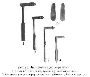
Percussion hammers have different sizes: for small animals they weigh from 60 to 75 g, and for large animals - from 100 to 160 g. The rubber pad in the hammer should be of medium elasticity and lie tightly in the screw head. During percussion, the plessimeter is held in the left hand and pressed tightly with the entire surface to the part of the body being examined. The percussion hammer is held large and index fingers right hand so that the handle can be slightly movable, and the strikes should be only due to the movement of the brush. In this case, the hammer bounces off the plessimeter more easily. Hammer blows should be short, jerky and applied perpendicular to the surface of the plessimeter. The strength of the blow is consistent with the thickness of the muscles. With a superficial location of small pathological foci in the lungs and determining the boundaries of organs, it is better to use weak or quiet percussion (through a plessimeter).
Percussion of well-fed animals, as well as suspected deep location of foci in the lungs, produces more strong blows. In this case, it is recommended to use metal pessimeters.
It is more convenient to perform percussion on a standing animal, in a small enclosed space. During percussion, the ear should be at the same height as the plessimeter.
Distinguish percussion staccato and legato. In the first case, they are tapped with jerky, short blows of a hammer bouncing off the surface of the plessimeter. This method is used to establish foci of inflammation in the lungs.
Legato percussion is performed with slow movements with the hammer held on the plessimeter. It is used to define the boundaries of organs. With strong (deep) percussion, the tissue vibrates to a depth of up to 7 cm, and on the surface by 4-0 cm; with a weak one - up to 4 cm in depth and 3 cm on the surface.
Percussion makes it possible to judge the state of organs by qualitatively different sounds. Organs containing air or gases give loud and prolonged sounds (tympanic, clear pulmonary).
Tympanic sound can be obtained by percussion of the caecum of a cow or horse, the upper part of the scar (in the hungry pits).
An atympanic, or clear lung, sound is produced by percussion of the chest in a healthy cow or horse.
Organs deprived of air, when percussion, give short and quiet sounds (dull, dull). Such sounds can be obtained by percussion of the muscles, croup and thigh.
Auscultation(listening) is a method of examining animals by listening. It allows you to capture the sounds that arise in the organs. According to the properties of the sounds that arise during the activity of certain organs, one can judge its functional and morphological state. There are direct and mediocre methods of auscultation.
Immediate auscultation is performed with the ear tightly attached to the body of the animal, and is widely used in veterinary practice. For this purpose, the animal is covered with a sheet or towel. Anterior part of the body of large animals right side listen with the left ear, and on the left side with the right.
To do this, you need to stand on the side of the animal, facing towards its head, put your hand on the withers or back and put your ear to the place of study. When examining organs located in the back of the body, they become facing the rear of the animal, placing their hand on its back. At the same time, listening should be done carefully, preventing the possibility of hitting the hind limb.
In restless horses, for this purpose, they raise the forelimb and hold the head of the animal well. Auscultation of sheep, goats, large dogs it is more convenient to produce on the table.
Mediocre auscultation is performed with stethoscopes or phonendoscopes.
Stethoscopes can be wood, metal or plastic. They conduct sound well and are especially valuable for auscultation of the heart. The disadvantage of hard stethoscopes is that they make it difficult to examine animals in any position, slide over the coat and force a person into uncomfortable positions. In contrast, flexible stethoscopes are more comfortable. They consist of a solid funnel-shaped part, from which two flexible rubber tubes extend, ending in ear cannulas. Flexible stethoscopes are suitable for auscultation of both small and large animals. However, they have not found wide use in veterinary practice, as they weaken and change the sound.
Phonendoscopes have received greater recognition in veterinary practice, allowing you to examine the animal in any position. They have a membrane tightly attached to the sound-receiving funnel-shaped part of the phonendoscope, which amplifies the sound, which is directed to the ear through rubber tubes. Phonendoscope systems are different. IN Lately the combined stethophonendoscope, which combines a stethoscope and a phonendoscope in its design, has become widespread (Fig. 11 and 12).
It should be noted that the comparative evaluation of auscultation methods is not without subjectivity. In the process of work, a subjective attitude is created not only to the methods of auscultation, but even to certain devices.
A general study begins after registration and anamnesis. It includes the definition of habitus, skin, lymph nodes, mucous membranes and measurement of body temperature.
Definition of habitus. Under the habitus understand the appearance of the animal at the time of the study: the position of the body in space, fatness, physique, constitution and temperament.
The position of the body in healthy animals can be naturally upright or naturally recumbent, in some diseases it is forcedly recumbent or upright. A forced position is characterized by the fact that animals cannot quickly change it in accordance with the changing situation. Thus, a forced lying position is noted in certain febrile diseases, when dogs and cats lie huddled in a corner and do not rise or rise when shouted.
Under the physique understand the degree of development of muscles and bones. When assessing it, the age and breed of the animal are taken into account. Distinguish between weak, medium and strong physique.
With a strong physique in animals, the chest is wide and deep, the legs are strong, strong, the ribs are steep with wide intercostal spaces.
With an average build, the muscles of the shoulder, thigh, limbs are well defined, the backbone is strong.
A weak physique is characterized by poor muscle development, a thin and long neck, a narrow chest, and long, thin limbs.
Fatness of dogs and cats determined by inspection and palpation. In short-haired animals, fatness is determined by examining the external forms of the body, and in long-haired animals, by palpation. Distinguish good, satisfactory and unsatisfactory fatness. With good fatness, the animals have rounded body contours, with unsatisfactory fatness - angular, with satisfactory - the muscles are moderately developed, the deposition of subcutaneous fat is palpable at the base of the tail, in the knee crease.
animal constitution- this is a set of anatomical and morphological features of the body, which are based on hereditary and acquired properties, which determine both its functional and reactive capabilities under the influence of environmental factors. There are four types of constitution: rough, tender, dense and loose. The definition of types is based on the development of the skeleton, muscles, skin and subcutaneous connective tissue.
When assessing temperament, attention is paid to the speed and degree of the animal's reaction to external stimuli, the behavior of dogs, the expression of the eyes, movements, ears and tail are observed. Depending on these factors, animals are distinguished with a lively and phlegmatic temperament. Dogs and cats with a lively temperament actively respond to external stimuli by playing with their ears, tail, changing the expression of their gaze, and head position. Their movements are fast and energetic. But with such animals, certain precautions should be observed, as they can be aggressive. Dogs and cats with a phlegmatic temperament are inactive and lazy.
Skin studies of dogs and cats
The skin is examined by inspection and palpation, determining the condition of the coat, skin moisture, its smell, temperature and elasticity. Inspection on non-pigmented areas establishes the color of the skin, its integrity, the nature of the lesions, as well as the condition of the coat (cleanliness, shine, tightness, density and uniformity).
Palpation determines the temperature of the skin, its moisture content and elasticity. To determine the temperature of the skin, the nose and the tip of the tail are palpated and compared with the temperature on the lateral surfaces of the chest. Skin moisture is determined by stroking with the palm of your hand on various parts of the animal's body. Shedding of scales of the epidermis at the same time, the absence of greasy deposits on the fingers indicates dry skin. Wetness of the fingers after palpation indicates sweating, and the presence of greasy plaque on the crumbs of the fingers indicates moderate skin moisture. To determine the elasticity of the skin on the back, it is gathered into a fold, pulled, and then released, trying to hold the hair between the fingers. In healthy animals, straightening of the fold occurs immediately. Loss or decrease in elasticity is accompanied by a delay in this process, and if there are no or less than 10 hairs left between the fingers, it is considered that the hair is well kept in the skin.
Examination of mucous membranes. In dogs and cats, the conjunctiva, the mucous membrane of the nose and mouth are examined. At the same time, attention is paid to their color, integrity, the presence of overlays, hemorrhages and secretion.
In carnivores, the conjunctiva is pale pink, but when animals are excited, it turns pink-red. The mucous membrane of the oral cavity is pale pink, very often has dark pigmentation. The study of the nasal mucosa is difficult due to the narrow nasal openings and the slight mobility of the wings of the nose, therefore, if necessary, they resort to the help of a rhinoscope.
For the study of the conjunctiva impose thumb one hand on the upper eyelid, and the other on the lower. Then press on the lower, while pulling up the upper eyelid. To study the mucous membrane of the lower eyelid, pressure is applied to the upper one, and the lower one is pulled down.
When analyzing the mucous membrane of the oral cavity, the lips, cheeks, gums, tongue and hard palate are examined. To do this, the dog's mouth is opened with the help of two ribbons applied to the upper and lower jaws behind the fangs, or the owner of the dog puts his hand under the lower jaw, covers it, pressing his fingers on the cheeks. The cheeks are pressed between the molars, the dog opens its mouth and cannot close it.
Measuring the body temperature of dogs and cats
Thermometry is an objective research method that facilitates the diagnosis of diseases.
Measure body temperature in dogs and cats with a maximum mercury thermometer. Before the introduction, it is shaken, holding the mercury reservoir with the index finger, after which it is lubricated with oil or petroleum jelly. Better place to measure body temperature - the rectum, where the thermometer is inserted with a slight rotational movement, then fixing it on the tail with the help of a tail pulp. Animals during this procedure are held by the head Measurement time - 8-10 minutes.
Normal body temperature in dogs fluctuates between 37.5-39.0 ° C, and in cats 38-39.5 ° C. It should be borne in mind that it depends on age, sex, breed, and external temperature. Puppies, kittens, bitches and cats have higher temperatures than adults and males. Minimum indicators it is observed in the second half of the night, and the maximum is recorded in the evening hours.
Examination of the circulatory organs of dogs and cats
The cardiovascular system is examined by inspection, palpation, percussion and auscultation.
Heart study. The heart of carnivores is located between the 3rd and 7th ribs, with 3/7 of the heart located on the right side of the chest. The anterior border runs along the anterior edge of the 3rd rib, the upper border is 2-3 cm below the horizontal line of the scapula shoulder joint, and the back reaches the 7th rib.
Animal heart study begin with examination and palpation of the cardiac region in order to determine the cardiac impulse, when examining the region of which, oscillatory movements of the chest are noticed. By palpation, the heart impulse is examined in the standing position of the animal. Determine its rhythm, strength, localization and distribution. The most intense cardiac impulse is felt in the region of the 5th intercostal space in the lower third of the chest. On the right, it is felt weaker and is fixed in the 4-5th intercostal space.
Percussion of the heart area is carried out in order to establish the boundaries of the organ. The upper border is determined by the rear vertical line of the anconeus. Percussion starts from the edge of the scapula and leads down to the transition of the pulmonary (atympanic) sound into a dull one. This line is the upper clinical border of the heart. Normally, it is 1-2 cm below the shoulder joint. Below the upper cardiac border is an area of absolute cardiac dullness. The posterior border is determined along the line connecting the ulnar tubercle and the maklok, while the plessimeter is moved to the next intercostal space stepwise up and back until the sound passes into the pulmonary one. Normally, the posterior border of the heart in dogs reaches the 7th rib. In addition, when the animal is in a sitting position, the part of the heart region covered with the sternum is also percussed.
In various diseases, there may be an increase, decrease and displacement of the boundaries of the heart. An increase in the boundaries is observed with hypertrophy of the heart, cardiac dropsy, pericarditis, expansion of the heart, and a decrease in alveolar emphysema, pneumothorax.
Auscultation of the heart determines the strength and clarity of tones, frequency and rhythm, as well as the presence or absence of noise. Auscultation is carried out in the region of the 4-6th intercostal space on the left and 4-6th on the right. Carnivores have loud, clear tones, it should be borne in mind that they normally have respiratory arrhythmia, and sometimes embryocardia, characterized by heart tones of equal strength and timbre with equal pauses. When diagnosing heart defects by auscultation, you should know the points of best audibility. Such a point for the bicuspid valve in dogs is the 5th intercostal space in the middle of the lower third of the chest, for the aortic semilunar valves - the 4th intercostal space under the horizontal line from the shoulder tubercle, and the pulmonary artery - on the left in the 3rd intercostal space along the upper edge of the sternum. best place audibility of the right AV valve is the 3rd-4th intercostal space in the lower half of the third of the chest.
In various diseases, changes in heart sounds can be noted in the form of their amplification, weakening, accentuation, rhythm of heart tones, and murmurs associated with cardiac activity.
Study of the arterial pulse of animals
The arterial pulse is examined by palpation with crumbs of 2-3 fingers of superficially located arteries, under which there is a solid base. Pay attention to the frequency, rhythm and quality of the pulse. To determine the pulse, examine the femoral artery in the groin, the brachial artery on the medial surface of the humerus above the elbow joint, or the artery of the saphenous immediately above the hock joint between the Achilles tendon and the deep flexor of the fingers. In newborn puppies pulse rate per minute is 180-200. In adult dogs- 70-120, at cats- 110-130. When determining the quality of the pulse, the filling of the arteries, the magnitude of the pulse wave, its shape, voltage are taken into account. vascular wall. Depending on the filling, a full pulse is distinguished (the diameter of the vessel during the filling period is twice the thickness of its two walls) and an empty pulse (the lumen of the artery is less than the thickness of its two walls).
According to the magnitude of the pulse wave, the blood filling of the artery and the tone of the vascular wall are judged. Depending on the size, a large pulse is distinguished, characterized by good content arteries, and a small pulse, in which the artery is poorly filled, its expansion is almost not expressed and is felt by the fingers in the form of weak shocks.
The rhythm of the pulse is judged by the periodicity in time and the correctness of the alternation of its phases in accordance with the rhythm of the heart. Based on this, there are rhythmic and arrhythmic pulses.
Respiratory studies of dogs and cats
The respiratory system is examined by methods of examination, palpation, auscultation, percussion. If necessary, they resort to special methods: radiography, fluoroscopy, fluorography, plegaphony, rhinography, etc. The upper respiratory tract and chest are examined.
The study of the upper respiratory tract begins with an examination of the nasal openings. Pay attention to the condition of the wings of the nose, the nature of the exhaled air, nasal discharge, examine accessory cavities nose. When examining exhaled air, attention is paid to its smell, which in some diseases can be putrid, sweetish, etc. In the presence of nasal discharges, their nature (mucous, serous, purulent, putrefactive, etc.), quantity (abundant, scarce,) frequency (constant or periodic), color, symmetry are determined.
Studies of the adnexal cavities are carried out by inspection, palpation and percussion. On examination, a change in the configuration of the sinuses is established. Palpation determines the sensitivity and softening of the bones of the maxillary and frontal sinuses. Percussion of the sinuses is carried out with the butt of a percussion hammer without a plessimeter. At the same time, they cover the eyes of the animal with the palm from the side from which the study is carried out. With the help of percussion, the nature of the sound is determined, by which the presence of exudate in the cavities is judged. In healthy animals, the adnexal cavities are filled with air and the sound during percussion is boxed, and when inflammatory processes(due to the presence of exudate), it becomes dull and blunt.
The larynx and trachea are examined by inspection, palpation and auscultation.
On examination, the presence of deformation and a change in the volume of these organs are revealed. In some diseases, edema is found in the larynx. An internal examination of the anterior parts of the larynx can be carried out through the oral cavity.
Palpation of the larynx begins from the lower part of the neck, moving the fingers forward to the intermaxillary space, feeling the larynx. At the same time, its pain, temperature, and the presence of swelling are determined.
Then, fingers are moved down from the larynx, feeling the trachea in order to establish changes in its integrity, sensitivity, and temperature.
Auscultation of the larynx and trachea is performed using a phonendoscope. In healthy dogs, inhalation and exhalation are heard, phonetically reproduced as the sound "x", called laryngeal breath sounds. In the region of the trachea, it is called tracheal breathing.
Chest examination of dogs and cats
The study of the chest begins with its examination, while establishing the shape and size, type, frequency, strength, symmetry and rhythm of respiratory movements. The shape of the chest in healthy dogs and cats is moderately round. With atelectasis of the lungs, it decreases in volume, becomes flat, and with emphysema - barrel-shaped.
The type of breathing is mixed-thoracic, although in some breeds it is predominantly chest. Disease respiratory system and related organs entails a change in the type of breathing. In animals with mixed type breathing chest type may be the result of a disease of the diaphragm, chest dropsy; with an injury or fracture of the ribs, the type of breathing becomes abdominal.
The respiratory rate is determined by the number of breaths per minute. In dogs, it ranges from 12 to 24, and in cats - 20-30. The number of respiratory movements is counted by the number of inhalations and exhalations according to the fluctuations of the chest or during auscultation of the trachea. The respiratory rate is influenced by age, breed, constitution and physiological state animal. Females and young dogs breathe faster than older and males.
Depending on the strength, breathing can be moderate, deep and shallow.
The determination of the symmetry of respiratory movements is carried out by comparing the excursions of the left and right chest walls. To do this, they stand in front of the animal so that both sides of the chest can be clearly seen. Uniform chest excursion on both sides indicates the symmetry of breathing.
Under the rhythm of breathing understand the sequential alternation of the phases of inhalation and exhalation. While inhaling like active phase, shorter than exhalation and the ratio between them is 1:1.6. Most frequent violations rhythm is shortness of breath. At the same time, if it is caused by a violation of inspiration, they speak of inspiratory dyspnea, exhalation - expiratory, and if difficulty in breathing occurs in both phases, then they speak of mixed dyspnea.
Palpation of the chest carried out in order to establish temperature, sensitivity, tangible vibration noise. Sensitivity is determined by pressing the knuckles along the intercostal spaces. In case of pain, the animals avoid palpation and show aggressiveness.
The temperature and tangible vibrations of the chest are determined by placing the palm on various parts of it. Local temperature increase is most often observed with pleurisy. At fibrinous pleurisy, pericarditis, when the surface of the pleura or pericardium becomes rough, a peculiar vibration of the chest is palpated.
Percussion of the chest carried out in order to establish topographic boundaries lungs, detection of pathological changes in them or the pleura. For percussion, it is better to put the dog on the table, using the digital method. To do this, the finger of one hand is pressed tightly against the chest wall in the intercostal space, and a medium-strength blow is applied with the finger of the other. Percussion determines the posterior border of the lungs along the lines of the maklok, ischial tuberosity and scapular-shoulder joint. Percussion from front to back. The posterior percussion border along the maklok line reaches the 12th rib, along the line of the ischial 11th tubercle - up to the 11th, and the scapular-shoulder joint - up to the 9th. Most often, an increase in the boundaries of the lung occurs with alveolar or interstitial emphysema, and a decrease occurs with intestinal flatulence, hypertrophic cirrhosis of the liver, and some other diseases.
: 1 - along the line of maklok; 2 - along the line of the ischial tuberosity; 3 - along the line of the scapular-shoulder joint.
Pathological changes in the lungs or pleura are detected by percussion from top to bottom along the intercostal spaces within the established boundaries of the lung. At the same time, in healthy animals, an atympanic or clear pulmonary sound is established. With pneumonia, pulmonary edema and other pathological conditions, accompanied by filling the lungs with fluid or the accumulation of the latter in the pleural cavity, the sound becomes dull or dull. With a significant expansion of the lungs due to an increase in residual air with alveolar emphysema, the percussion sound becomes boxy, and when formed in lung tissue air cavities, which is noted with interstitial emphysema, it acquires the character of a tympanic one.
Auscultation of the chest carried out in order to establish the nature of respiratory noise. For this, two methods are used: direct and instrumental. With the direct method, auscultation is carried out with the naked ear through a sheet or towel. Mediocre - carried out using a phonendoscope or stethoscope.
Auscultation should be carried out in a certain sequence: starting with listening to areas with the best audibility of breath sounds, followed by moving to places with poorer ones. To comply with this rule, it is recommended that the chest of the animal on each side be conditionally divided into three parts: upper, middle and lower. Then the upper and middle parts are divided into two halves by a vertical line. It turns out five zones of listening. Auscultation in them is carried out in the following order: anterior middle area, posterior middle, anterior superior, posterior superior and inferior.
On auscultation of the chest of healthy dogs, an intense and loud breath noise is heard during the inspiratory phase and partly at the beginning of the exit. This type of breathing is called vesicular. Immediately behind the scapular-shoulder girdle during the inhalation and exhalation phase, a loud respiratory noise is heard, phonetically resembling the letter "x" and called bronchial breathing.
In various diseases, the nature of physiological respiratory sounds may change and pathological ones may occur. This manifests itself in the form of strengthening or weakening vesicular breathing, appearance bronchial breathing in areas that are not characteristic of him, the occurrence of pathological noises (various wheezing, friction and pleural noises, etc.).
Examination of the digestive organs of dogs and cats
In the study of the digestive organs, methods of examination, examination, palpation, auscultation, percussion are used. If necessary, they resort to probing the esophagus and stomach, radiography and fluoroscopy, laboratory studies of gastric juice, feces, etc.
Studies of the digestive system are carried out according to the following scheme: the act of taking food and water, the oral cavity, pharynx, esophagus, abdomen, stomach and intestines, monitoring the act of defecation.
When examining the act of taking food and water, they turn Special attention on appetite and the act of swallowing.
Appetite is examined by observing the animal while eating. It is influenced by the physiological state of the animal, the environment, the quality and type of food, feeding time. There may be a lack, decrease, increase, perversion of appetite. It is reduced or absent various pathologies infectious, invasive and non-infectious origin. An increase in appetite accompanies some pathologies that occur with metabolic disorders (diabetes mellitus), and is also observed in the recovery stage after past illness. Perversion of appetite, characterized by eating inedible objects, is noted with a deficiency in the body of mineral salts, increased acidity in the stomach, rabies, etc.
In diseases of the central nervous system, lesions of the tongue, lips, teeth, chewing muscles, there is a disorder in the intake of food and water, which manifests itself in an unusual form of this process.
With lesions of the pharynx and esophagus, the act of swallowing is disturbed. This phenomenon is characterized by pain during swallowing food. Animals squeal, worry, sometimes there may be ejection of food masses through the nose (regurgitation). The complete impossibility of swallowing is noted with paralysis of the pharynx, rabies, botulism, encephalitis.
Vomiting may be the result of overfeeding. In this case, it is most often single, the vomit corresponds to normal content stomach. Frequent vomiting characteristic of lesions of the gastric mucosa, poisoning, diseases of the central nervous system, liver and other organs. In these cases, pay attention to the color and smell of vomit.
Examination of the oral cavity, pharynx and esophagus of dogs and cats
Oral cavity researched mainly by inspection. For internal examination, the upper jaw is captured between the thumb and forefinger, squeezing the lip between the teeth, and the lower jaw is somewhat pulled with the fingers of the other hand. For the same purpose, the Baicher mouth wedge or animal mouth fixator (FPZh-1) is used in dogs. Pay attention to the mucous membrane of the oral cavity, its color, moisture, integrity. Examine the tongue, teeth, determine the nature of salivation. When examining teeth, attention is paid to the correctness of their erasure, integrity, condition of the gums.
Throat examined by inspection and palpation. For examination, after setting the yawner, the base of the tongue is pressed against the lower palate with a spatula, after which the condition of the walls of the pharynx and tonsils is established. Palpation of the pharynx is carried out by squeezing the region of the upper edge of the jugular groove slightly above the larynx with the fingers of both hands, while paying attention to the soreness of the pharynx, the presence of tissue infiltration in its region and foreign bodies in its cavity.
Esophagus are examined by inspection, palpation, and also by setting the probe. By inspection, the patency of the food coma is established. Palpation - sensitivity of the esophagus, the presence of pathological infiltrates, tumors, foreign bodies. To detect narrowing and blockage of the esophagus, they resort to probing it. For this purpose, a set of Sharabrin rubber probes or medical probes of various numbers are used in dogs, depending on the size of the dog. For setting the probe or "x"-shaped yawn of the Sharabrin system. With the help of these yawns, the working end of the probe is directed along the hard palate, then its end bends down, then falling into the cavity of the pharynx and esophagus.
Examination of the abdomen, stomach, intestines and liver of dogs and cats
When researching belly methods of inspection, palpation, percussion and auscultation are used, and in necessary cases and trial puncture of the abdominal wall.
Inspection determines the volume and shape of the abdomen, the symmetry of its walls. An increase in the volume of the abdomen is observed with intestinal flatulence, gastric overflow, coprostasis, abdominal dropsy, enlarged liver, bladder. Local violations of the shape of the abdomen are observed with umbilical and mesenteric hernias, abscesses of the abdominal wall. A decrease in the volume of the abdomen occurs with exhaustion, prolonged diarrhea.
Palpation of the abdominal walls is carried out immediately on both sides with both hands. Soreness, tension of the abdominal wall, the state of some organs of the abdominal cavity are determined. Increased tension of the abdominal walls and severe soreness give rise to suspicion of peritonitis. In addition, palpation can establish an increase in the liver, the presence of intussusception and intestinal coprostasis.
Percussion examines the stomach, intestines, liver, and auscultation determines the nature of the peristalsis of the stomach and intestines.
The puncture of the abdominal wall is carried out in order to diagnose peritonitis and ascites. It is done in the lower abdomen in the area of the last two pairs of nipples, departing from the white line of the abdomen 1-1.5 cm.
Examination of the stomach carried out by inspection, palpation, auscultation, percussion, and, if necessary, radiography. The stomach is located in the left half of the abdominal cavity and in dogs reaches the abdominal wall near the 12th rib. With strong filling, it goes beyond the costal arch, lies on the abdominal wall and reaches umbilical region. Inspection determines the shape and volume of the abdomen. Palpation of the stomach is carried out in a standing position, pressing with the fingers of both hands, applied behind the costal arches on both sides, inward and forward. At the same time, the position of the stomach, its filling and soreness are determined.
When examining the intestines use auscultation, external palpation and examination in the abdomen. In this case, it should be borne in mind that the small intestine occupies mainly the right half of the abdominal cavity, and the thick one - the left.
On examination, pay attention to the abdominal wall in the region of the right and left hungry pits. A protrusion in the region of the left hungry fossa is usually characteristic of flatulence of the large intestine, and in the region of the right - of the small intestine.
The most important method for examining the intestines of animals is palpation. It is carried out in the standing position of the animal, evenly squeezing the lateral surfaces of the abdomen on both sides. At the same time, the degree of fullness and sensitivity of the intestine is established. Auscultation of the intestine makes it possible to judge the nature of peristalsis.
When examining the act of defecation, attention is paid to its frequency (in animals on a meat diet, once a day). The disorder of the act of defecation is manifested in the form of diarrhea, constipation, pain during defecation.
Liver examined by palpation and percussion. Palpation is carried out by placing the animal on its right side, as a result of which the liver is displaced to the abdominal wall. After that, they bring their hand to the right under the last rib and feel for the edge of the liver. Percussion of the organ is carried out with the animal in a standing position, immediately behind the posterior border of the lung. On the right side, the area of hepatic blunting in dogs is located within the 10-13th rib, and on the left - in the 11th intercostal space.
Study of the urinary system of animals
Includes the study of the process of urination, the study of the kidneys and bladder, if necessary, conduct a study of the urine of animals.
When examining the process of urination, attention is paid to the posture of the animal at this moment, duration, frequency, and also total urine and its appearance. The posture during urination depends on the sex: males raise the pelvic limb, females - the tail and squat. The number of urination depends on the conditions of detention. Usually dogs urinate 3-4 times a day, but in free keeping much more often.
kidneys are examined mainly by the method of external palpation through the abdominal wall. At the same time, attention is paid to the location of the kidneys, their size, shape, sensitivity, consistency, surface condition. For palpation, both thumbs are placed on lumbar region, the rest on the stomach on both sides behind the last rib. Then move the fingers up the abdominal wall to the last thoracic vertebra, evenly pressing them towards each other. The left kidney is found in the anterior left corner of the hungry fossa under the 2nd-4th lumbar vertebra. right kidney examine in the anterior corner of the hungry fossa under the first and third lumbar vertebrae. With various diseases, it is possible to establish an increase and decrease in the kidneys, a change in their surface, sensitivity. An increase can be observed with pyelonephritis, hydronephrosis, a decrease - with cirrhosis, pain - with inflammation and urolithiasis.
The main research method bladder in dogs and cats is palpation through the abdominal wall. The bladder is located in its lower region in front of the pubic fusion. The study is carried out with the animals in a sitting position, for which the fingers are placed on the abdominal wall in the area of the bladder and light pressure towards each other is probed. In decorative breeds of dogs and cats, the bladder is examined through the rectum. To do this, after appropriate treatment of the index finger, it is inserted into the rectum, and the opposite hand is pressed against the abdominal wall. The study of the bladder makes it possible to judge its filling, sensitivity. With inflammation, pain is noted during palpation, tumors are detected by the presence of dense bodies, and urinary stones- in the form of solid formations, shifting during palpation.
Study of the nervous system of dogs and cats
In the study of the nervous system, the behavior of the animal, the state of its skull and spinal column, sensory organs, skin sensitivity, motor sphere, and reflex activity are studied.
The behavior of an animal is judged by the results of observations of its reaction to external stimuli(calling, approaching stranger, giving food, etc.). Violation of behavior is manifested in excitation, depression, soporous or coma. The most characteristic increase in excitability in rabies, which turns into a riot. Dogs break loose from the chain, run away from home, cats attack people and animals. Oppression is accompanied by a delay in functions nervous activity. Animals are inactive, the reaction to stimuli is sharply reduced. When sporing, animals are in a state deep sleep, from which they can be withdrawn only when exposed to strong stimuli. A characteristic sign of a coma is the loss of reflexes and consciousness.
Research methods skull and spinal column are inspection, palpation and percussion.
During the examination, the shape and volume of the skull, its symmetry, as well as the presence of spinal deformities are determined.
On palpation, sensitivity, temperature of local tissues, hardness are established. bone formations, their deformation. The spinal column is palpated, starting from the cervical vertebrae and ending with the vertebrae of the tail root.
The skull is percussed with a finger, and in large dogs with the butt of a percussion hammer for small animals. At the same time, attention is paid to the nature of the sound and the reaction of the animal to percussion. In the presence of exudate in the sinuses, dullness of the sound is noted. The spinal column is percussed with a hammer without a plessimeter from the slope of the withers to the root of the tail, paying attention to the presence of pain.
Investigation of the sense organs of dogs and cats includes the study of vision, hearing, smell, taste.
The state of vision is judged by its organs (eyelids, eyeball), the reaction of the pupil to a light stimulus. The latter is determined by closing the examined eye for 2-3 minutes. In this case, in healthy animals, the pupil dilates and quickly returns to normal after the eye is opened. To test for a decrease or loss of vision in dogs, their eyes are alternately closed and led to an obstacle. With the loss of vision, the animal does not notice them.
Hearing is examined by closing the eyes of the animals and then reproducing the usual sound stimuli: whistling, shouting. With damage to the nerve hearing aid these sounds are perceived worse.
The sense of smell is also checked after elimination visual analyzers. Dogs and cats are brought objects or food, the smell of which they are well aware of. With a decrease in the sense of smell, animals do not react to these odors.
Taste is determined based on the reaction of animals to various feeds and unusual substances.
Study of skin sensitivity. In the study of skin sensitivity pay attention to the reaction skin when exposed to tactile, pain and temperature stimuli.
The study of tactile sensitivity is carried out after closing the eyes of the animal. Then, with a light touch, individual hairs are irritated in the region of the withers, abdomen, auricle or nostril. When tactile nerve endings are stimulated, a response of animals occurs in the form of contraction of the corresponding skin areas. Lack of reaction indicates the disappearance of tactile sensitivity.
Pain sensitivity is determined by tingling the skin with the tip of the needle. It starts from the distal parts of the limbs and goes up to the region of the croup or scapula, and then the spinal column and ends on the neck of the animal. healthy dogs and cats look around, tuck their ears in, fan their tails, bite, scratch.
Temperature sensitivity is determined by touching various sites skin of test tubes filled with hot or cold water which are applied alternately.
Study of the motor sphere of dogs and cats
Study motor sphere includes determining the activity of movements, muscle tone and coordination of movements.
The determination of the activity of movements is carried out by the method of inspection. In this case, partial (paresis) or complete loss of motor function (paralysis) may be noted.
Muscle tone is examined by palpation. Depending on muscle tension, it can be moderate, low or high. With reduced tone, the muscles are flabby, the range of motion of the limbs is wide, and the joints are often bent; with increased - there is a strong muscle tension, they become dense, and passive movements are made with difficulty.
In a clinical examination of the nervous system, superficial and deep reflexes are checked. Superficial reflexes include the skin and mucous membranes. From the skin, a tail reflex is determined, accompanied by pressing the tail to the body.
The abdominal reflex is also indicative, characterized by a strong contraction of the abdominal muscles in response to a light touch, as well as the anal reflex, which is manifested by contraction of the anal sphincter when touching the skin in the anus. Of the reflexes of the mucous membranes in dogs, the most indicative is sneezing. It is checked by irritating the nasal mucosa with a light object (feather, match).
The purpose of the lesson. To master the general methods of studying animals: examination, palpation, percussion, auscultation; master the technique of thermometry; familiarize yourself with special research methods.
Research objects and equipment. Horses, cows, dogs.
Percussion hammers, plessimeters, phonendoscopes, stethoscopes, sheets or towels for auscultation, thermometers.
Common methods of clinical examination include inspection, palpation, percussion, auscultation, and thermometry. Mastering these research methods is one of the main conditions that allow a specialist to identify pathology. In most cases, to clarify the diagnosis, along with common methods additionally have to apply special methods research: instrumental and laboratory methods.
Inspection (from lat. inspection- examination, inspection). This is the simplest and available method animal research. Inspection is preferably carried out in natural light (daylight) or in good artificial lighting. Inspection can be performed with the naked eye or with the use of special instruments. The animal is first subjected to a general and then a local examination. General inspection. Involves examination of the entire body of the animal, regardless of possible localization painful process. At the same time, it is possible to draw conclusions about the position of the body in space, fatness, physique, the condition of the skin and hair, superficial lesions, discharge from natural openings, the condition of the eyes, to identify excitation, depression or other signs characteristic of dysfunctions of organs and systems of the body.
Local inspection. This inspection discovered during general examination animal injuries or parts of the body where the disease process is mainly localized.
Local examination can be divided into external, when the outer integuments of the animal’s body are examined, and internal, when areas located not on the surface of the body, but in depth, such as the cavity of the larynx, pharynx, etc., are subject to examination.
In the study, various instruments can be used, including those equipped with light sources. With the use of instruments, the following can be examined: oral and nasal cavity, pharynx, larynx, vagina, rectum, bladder, etc.
Palpation (from lat. .palpatio- feeling). The method in which groping is applied, i.e. based on the sense of touch. The method of palpation allows you to determine the physical condition of the organs and tissues of the body.
Palpation is carried out with fingertips (pads), without causing pain to the animal. In some cases, palpation can be performed with a fist, hand, back side hands.
Palpation is divided into superficial and deep.
Superficial palpation is performed by placing the palm or fingertips on the study site, by gently pressing and sliding over the area under study. This method allows you to examine the skin, subcutaneous tissue, lymph nodes, superficial vessels, tendons, muscles, joints, etc. Palpation can determine local temperature, soreness, configuration and size of the formation, consistency, nature of the surface, etc.
Palpation should always begin with a healthy area of the body, gradually moving to the affected area. The movements of the fingers or palms should be smooth, soft, not causing additional pain to the animal.
Deep palpation is a method by which deeply located organs and foci are examined. Varieties of deep palpation include: penetrating; bimanual; jerky (balloting).
When conducting penetrating palpation fist or vertically placed fingers carry out a gradual, but strong pressure through the abdominal wall, reaching the organ under study and determining its physical condition. In this way, the scar, abomasum is examined.
Bimanual palpation(palpation with two hands) is performed on the stomach of small animals. The method consists in simultaneous, gradual pressure with both hands on both sides of the abdominal wall. Research is carried out while standing behind the animal. Bimanual palpation is also used in the study of the pharynx, especially in large animals.
If you suspect the presence of fluid, neoplasms or an enlarged organ in the abdominal cavity, apply jerky (balloting) palpation. With jerky palpation, studies are carried out with jerky movements of the fingers or fist. The palpation technique is as follows: they bring the fingers or fist to the wall of the abdominal cavity and perform a push, while at the end of the push the hand is not taken away from the abdominal wall (this is especially important if ascites is suspected), in the presence of a neoplasm or an enlarged organ, the hand immediately encounters this organ or neoplasm, and in the presence of fluid, a push through the abdominal wall is not felt immediately, but after some time (the return of the fluid that recoiled during the push).
Deep internal palpation allows diagnosing the state of organs located in cavities far from the surface of the body (pharynx, organs of the pelvic and abdominal cavities).
The study of the organs of the pelvic and abdominal cavities through the wall of the rectum is called rectal examination. In large animals, rectal examination is carried out with a hand inserted into the rectum, while in small animals it is possible to examine in best case organs of the pelvic cavity, as it is performed with a finger.
Percussion (from lat. percussio- tapping). The method is based on the ability of each tissue or organ to give a characteristic sound during percussion. Depending on the physical condition organ, this sound can change, and by the nature of the changed sound, one or another state of the organs and tissues of the body is judged.
Percussion is best done indoors, so as not to interfere with extraneous noise. Distinguish between direct and mediocre percussion.
Direct percussion consists in the fact that blows to the place of study are applied directly with a finger or hammer. The maxillary and frontal sinuses are subjected to direct percussion. In other areas of the body, direct percussion is ineffective, since the conditions for the appearance of sounds are negligible.
Direct percussion, carried out with a finger, is called digital, and performed with the help of a percussion hammer - instrumental.
With mediocre percussion, blows to the place of examination are not applied directly to the skin, but through a finger (digital) or a plessimeter (instrumental).
According to the method of striking, topographic and research percussion are distinguished. When, after a blow, a finger or a hammer is slightly delayed on a finger or a plessimeter, this method is called topographic and is used to determine the boundaries of an organ or pathological focus. Jerky percussion, without delay of the hammer or finger, is called staccato and is used to study an organ or focus.
Digital percussion is of particular value in the study of small animals (dogs, cats, small cattle, rabbits, birds, calves, foals, piglets, skinny adult pigs). In the study of large animals, digital percussion is not very informative, but can be used in the absence of instruments (plessimeter and hammer).
Digital percussion is carried out by pressing the middle finger of one hand to the place of study and applying short paired blows perpendicular to it with the middle finger of the other hand.
With instrumental percussion, instruments are used - a plessimeter and a percussion hammer. They can be different in shape, mass and performance (Fig. 1.12).
Rice. 1.12. Animal percussion instruments different types: A- percussion hammers; b- plessimeters
The hammer is taken with the thumb and forefinger, and the handle is pressed to the palm with the remaining fingers. The arm is bent at the wrist joint.
Plessimeters with a narrow working platform are most convenient to use, and the size of the malleus depends on the size of the animal. For the study of large animals, large hammers are preferable, for small animals - small ones.
When conducting instrumental percussion, the plessimeter is pressed tightly against the skin at the place of examination (for example, if it chest wall, then strictly in the intercostal spaces) and with a percussion hammer, pair blows are applied to it with a small gap and always of the same strength. The blows must be directed perpendicular to the plessimeter. The plessimeter during percussion should be moved one step. In the study of small animals or animals of unsatisfactory fatness, the blows should be weaker. Percussion of the same strength is used when determining the boundaries of an organ or a pathological focus. However, it must be remembered that the percussion method can be used to examine the animal's organs to a depth of up to 7 cm from the body surface, i.e. more deeply located organs and their parts, as well as lesions are inaccessible to percussion examination.
When conducting percussion, the following conditions must be observed: silence, the ear of the researcher (doctor) must be at the same level with the place of percussion, the force of impact and pressure of the pessimeter throughout the percussion must be the same, the animal is recommended to be placed at a distance of no closer than 1 m to avoid sound resonation from the wall.
Percussion determines the boundaries of the organ and focus, which makes it possible to establish their size, as well as to identify changes in the physical properties of organs.
Auscultation (from lat. auscultatio- listening). With the help of auscultation, you can listen to sounds that occur in the organs and cavities of the animal's body.
Distinguish between direct auscultation, when one or another organ is heard with the ear without instruments, and mediocre, when the organ is used with instruments (phonendoscope, stethoscope, stethophonendoscope) (Fig. 1.13).

Rice. 1.13.
- 1 - pelota; 2 - head of a phonendoscope; 3 - pilot holder;
- 4 - flexible sound duct; 5 - headband; 6 - membrane; 7 - horn of a stethoscope; # - phonendoscope head cover
Direct auscultation is performed as follows: the ear is applied through a sheet or towel (hygienic accessory) to the surface of the animal's body in accordance with the topography of the organs that need to be heard. The advantage of the method is that it can be performed under any conditions; the sounds emitted by the organs are not distorted; allows you to pick up sounds from a relatively large surface of the body (although this possibility is in some cases a disadvantage, since it makes it difficult to accurately determine the source of the sound).
Direct auscultation has found wide application in veterinary practice, especially in the study of large and calm animals.
Mediocre auscultation is carried out using stethoscopes, phonendoscopes, stethophonendoscopes. It allows you to listen to sounds from a more limited area than with direct auscultation. Stethoscopes are used to listen to individual components - heart sounds (for example, with defects), etc.
The narrow end of the cone-shaped extension is applied to the place of study, and the ear is applied to the wide end, the hand is taken away from the stethoscope.
If auscultation is carried out with a phonendoscope with a membrane, then it is necessary to press it tightly against the body of the animal in order to exclude friction of the membrane against the animal's hairline, which can introduce extraneous noise into the main ones coming from the organ under study. This is especially important to keep in mind when researching valve apparatus hearts.
Thermometry. This is a mandatory method of animal research, which is performed when an animal is received, regardless of the goals of the research.
Thermometry - very important method clinical research, since most diseases, especially infectious diseases, are initially manifested precisely by a change in body temperature.
To measure body temperature, you can use different thermometers (mercury, electronic). Each thermometer must be checked before use for correct readings. To do this, the tested thermometer is lowered into a vessel with water with a tested control thermometer, after 10 minutes they are removed and the readings of the tested and control thermometers are compared. A thermometer with incorrect readings is discarded.
The internal body temperature of the animal is measured in the rectal cavity. Before inserting the thermometer, the readings are checked, disinfected, lubricated with petroleum jelly or vaseline oil. The animal is preliminarily fixed, the tail is lifted and the thermometer is inserted into the rectum with rotational movements, giving it an inclined position so that the tip of the thermometer comes into contact with the mucous membrane, after which it is fixed to the tail with a tail bag, clamp or ribbons.
To measure body temperature in birds, a special “bird thermometer” is used, in which the temperature reading scale is designed for digital values greater than in animals.
Body temperature must be measured both at the initial reception of the animal, and at all subsequent examinations.
In cases where it is not possible to examine the body temperature in the rectum, the thermometer is inserted into the vagina, remembering that the temperature in the vagina is 0.3-0.5 ° C higher than the rectal temperature.
Special research methods. Special methods include: studies conducted in laboratories (laboratory) and performed using special tools and equipment (instrumental). Laboratory research exposed to body fluids and tissues. Among instrumental research most commonly performed electrocardiography (ECG), ultrasonography heart (EchoCG), x-ray diagnostics, ultrasound (ultrasound), endoscopy (many types), magnetic resonance imaging (MRI), computed tomography(CT), etc.
Special methods in any necessary combination are additional methods research and allow you to clarify the diagnosis.
Special research methods are described in the relevant chapters of this workshop.


