Laparoscopic cholecystectomy in dogs. Cholecystitis is a primary disease of the biliary system
It is no secret that our pets can suffer not only from their breed-specific diseases (read more about), but also suffer from completely human ailments. So, for example, your dog may be diagnosed with: cholecystitis. And here a lot of questions arise - how to treat cholecystitis in a dog, and how to prevent relapse of the disease...
Our publication will try to help you answer these questions...
Cholecystitis in dogs - description of the disease
A disease in which the bile ducts of an animal are affected, and such lesions are accompanied by inflammatory processes localized in gallbladder, called cholecystitis. This disease is difficult to detect in a timely manner, so when your pet is diagnosed this disease– it is, most often, already in a neglected state.
Causes of cholecystitis in dogs
Of course, after you hear such a diagnosis, you are interested in answers to questions about why your pet got sick, what caused the development of cholecystitis in a dog, whether you could somehow prevent the development of this disease... Well, similar Disease in animals can arise from several causes. And above all, The main causative agent of cholecystitis is microbes. Penetrating into the animal’s body from the intestines, by hepatic artery or through the biliary tract they enter the gallbladder. Also, microbes that cause cholecystitis can be transmitted by the lymphogenous route.
As you can see, there are many reasons that can lead to the development of the disease, however, it is not always possible to establish exactly what caused the development of cholecystitis in a dog in each specific case.
Symptoms of cholecystitis in dogs
As a rule, the initial and middle stage diseases occur in the animal’s body asymptomatically, only against the background of an exacerbation the dog’s appetite decreases, vomiting begins, indigestion begins, the mucous membranes of the mouth and nose may turn yellow, the dog itself looks lethargic and depressed, and when palpating the area of the liver and abdominal cavity the animal begins to whine , since there is pronounced pain in this place. Also, in sick animals there is a periodic increase in . And, as a result of difficulties with the outflow of bile, signs may appear obstructive jaundice.
Diagnosis of cholecystitis
Treatment of cholecystitis in dogs

- If the disease is advanced and the animal’s condition is serious, the dog is prescribed a series of therapeutic procedures, aimed at relieving the inflammatory process, normalizing the processes of bile secretion and digestion.
- For disinfection biliary tract and improving the outflow of bile itself, allohol, tincture is prescribed corn silk, cholagon, magnesium sulfate.
- To relieve spasms of the gallbladder and bile ducts Antispasmodics, atropine sulfate, and no-spa are prescribed.
- For pain relief, analgin, baralgin and other painkillers are used. However, a doctor should prescribe these medications, as well as determine their dosage, depending on the weight of the dog, its age, and its general condition.
- The final stage of treatment involves thermal physiotherapeutic procedures to improve the resorption of exudate, relieve pain and improve blood circulation.
One of the most difficult to diagnose and serious diseases of dogs is cholecystitis - an inflammatory process of the walls of the gallbladder and bile ducts.
The gallbladder in dogs is a small pear-shaped organ connected to the liver by bile ducts, through which bile flows into it and is stored there until the food mass enters the duodenum. Then the gallbladder contracts, due to which bile is discharged through the bile ducts into the intestines. Bile is necessary to break down fats, speed up the digestion of proteins and carbohydrates, and activate digestive enzymes.
With the development of inflammation motor function the gallbladder is disrupted, bile stops flowing into the intestines and corrodes the walls of the gallbladder, accumulated undissolved fats cause intoxication of the body. In advanced cases, ulcers appear in the walls of the gallbladder, bile penetrates into the abdominal cavity, which is dangerous due to the development of peritonitis and fatal animal.
Types of cholecystitis depending on the cause:
Obstructive. Develops as a result of compression of the bile ducts due to tumors of the liver and intestines, cholelithiasis, enlarged pancreas and mechanical injuries Oh.
The main reasons for the development of cholecystitis are:
Symptoms
Most often, cholecystitis in dogs is asymptomatic, and animals with advanced stage diseases. Owners are advised to show their pet to a specialist when the following symptoms diseases:
- Belching after every meal and frequent vomiting with undigested particles of food, mucus, and sometimes bile;
- Diarrhea, flatulence, bloating, constipation, light-colored feces;
- Increased body temperature;
- Deterioration in wool quality, hair loss, dry skin;
- Decreased appetite and weight loss;
- Weakness, apathy, drowsiness;
- Painful reaction to touch on the right side;
- The whites of the eyes, nasal mucosa and oral cavity yellow color;
- Urine is orange.
The nature of the course can be acute or chronic.
For acute course characterized by jaundice, intoxication, fever caused by blockage of the bile ducts and cessation of bile flow into the duodenum. The consequence of these processes may be rupture of the gallbladder with the development of peritonitis. To save the pet in these cases, urgent surgical intervention is necessary.
The chronic form is hidden; this form of the disease is characterized by: lethargy after feeding, vomiting, diarrhea or constipation, weight loss. With these symptoms, owners are advised to show the animal to a specialist.
Diagnostics
For staging accurate diagnosis the doctor examines the animal, notes signs of exhaustion, pain on palpation of the abdominal cavity, yellowness of the mucous membranes, dry skin, and a slight increase in body temperature.
To diagnose cholecystitis in a dog, laboratory tests must be carried out:
- general and biochemical blood tests, the disease is characterized by leukocytosis and changes in liver parameters;
- stool analysis - detects fatty acids and undigested fiber
- urine test - noted increased content bilirubin;
- Bile analysis - the pathogen is identified.
To clarify the diagnosis and determine the cause, radiography and ultrasound of the animal’s abdominal cavity are used, as well as a liver biopsy with cytological examination biopsy.
Treatment
Treatment methods depend on the nature of the course and severity of the disease. For conditions that are not life-threatening for the pet, use drug therapy If there is a suspicion of rupture of the gallbladder and the development of peritonitis, surgical treatment is indicated.
First of all, the dog is prescribed special diet. The animal is kept on a 12-hour fasting diet followed by therapeutic nutrition. The dog's diet should consist of carrots, pumpkin, lean beef, poultry, rice, buckwheat, low-fat cottage cheese, eggs and dairy products. The pet must be fed often, in small portions, food must be pureed.
Mandatory symptomatic therapy using anti-inflammatory, antispasmodic, analgesic, choleretic, hormonal, vitamin preparations and hepatoprotectors.
In case of severe intoxication and dehydration, the animal is prescribed intravenous infusions physiological solutions.
To relieve inflammatory edema and reduce pain, thermal procedures are used.
Prevention
Most often, you can prevent the development of cholecystitis by following simple preventive measures:
Cholecystitis in dogs can and should be treated. At the first symptoms of an intestinal disorder, a change in the color of feces or urine, it is advisable to show your pet to a doctor. Proper nutrition healthy food and active walks can prevent dangerous diseases and prolong the life of your four-legged friend.
A disease characterized by the occurrence of an inflammatory process in the gallbladder of a dog. Damage to the bile ducts occurs.
Cholecystitis is a rare disease in dogs, so it can be difficult to immediately determine its occurrence. Let's consider the symptoms, causes of the disease, how treatment is carried out, and other information.
Causes
There are several reasons. One of the most important is the bacteria cholecystitis, which penetrates from the intestines. Their path runs through the bile ducts and the hepatic artery.

In most cases, symptoms are difficult to notice in a dog with cholecystitis. However, by some signs you can still determine:
- Decreased appetite;
- The mucous membranes of the mouth and nose turn yellow;
- , stomach upset;
- depressed state;
- pain is expressed upon palpation (palpation) of the abdominal cavity, liver area;
- the increase is short-term;
- signs of jaundice, as the outflow of bile occurs with difficulty.
In any case, if these symptoms are detected, you must contact a veterinary clinic for examination and testing.
When the disease occurs, leukocytosis is detected in the blood - changes cellular composition blood. The level of bilirubin, a special bile pigment, increases in the blood and urine
Treatment

Treatment of cholecystitis involves prescribing dietary nutrition. If you feed your pet dry food, you will have to use special feed, dietary.
You may have to put your beloved pet on a fast for up to three days if unforeseen exacerbations occur during treatment. To relieve inflammation of cholecystitis use:
- ascorbic acid;
- salicylic acid;
- calcium.
If the case is severe and difficult to treat conventional treatment, use therapeutic procedures to relieve inflammation, improve digestion and bile secretion.

To disinfect the bile ducts so that bile is excreted normally, the following drugs are used:
- allohol;
- magnesium sulfate;
- hollagon;
- corn silk;
- hexamethylenetetramine.
To remove pain syndromes prescribe:
- analgin;
- baralgin;
- bellalgin;
- besalol.
To relieve spasm in the bile ducts, in the gallbladder itself veterinarian may prescribe atropine sulfate, noshpa. Subsequently, procedures are prescribed to resolve the exudate.
For proper blood circulation to occur, the pain would stop. Exudate is a special liquid that is released from small vessels blood vessels in tissue during inflammatory processes.
XPrevention
To avoid such unpleasant disease the dog should be fed correctly, given essential vitamins, minerals. Food must be served fresh.
Vaccinate against common diseases on time infectious diseases. And your pet will always delight you with its cheerful appearance and good mood.
Have you ever encountered this disease? What information do you have about him?
D.E. Mitrushkin. Vet clinic“Biocontrol”, Clinic of Experimental Therapy, State Scientific Research Center named after. N.N. Blokhin RAMS
Keywords: bile, gallstones, cholelithiasis, bile duct, cholelithiasis, gallbladder, cholecystolithiasis, liver, hepatic ducts
Abbreviations: ALT– alanine aminotransferase, CT – CT scan, RMJ- mammary cancer, Ultrasound- ultrasonography, ShchV- alkaline phosphatase, ECG– electrocardiogram
Introduction
Bile is a secretion that is constantly produced in the liver and enters the intrahepatic bile ducts, which, merging, form the right and left extrahepatic ducts, located near the porta hepatis. These ducts unite and form the common hepatic duct, which passes into the common bile duct, which flows into the duodenum. Bile enters the gallbladder (bile storage reservoir) from the common bile duct through the cystic duct and from it, as needed, is released back into the common bile duct.
Gallstone disease (cholelithiasis, from the Greek chole - bile and lithos - stone) is a metabolic disease of the hepatobiliary system, characterized by the formation of gallstones in the gallbladder (cholecystolithiasis), less often in the intrahepatic bile ducts (hepatic cholelithiasis) or the common bile duct (choledocholithiasis) .
Cholelithiasis is a rare disease in dogs and cats. Even its presence in animals is often asymptomatic and before the introduction of ultrasound into veterinary practice, it was often detected only during autopsy. The main cause of gallstone formation is a violation functional state liver (due to hepatitis, hepatosis or cirrhosis) and changes in connection with this physical and chemical properties bile (discholia). The formation of gallstones is associated with impaired metabolism of the main components of bile - cholesterol, phospholipids (lecithin, etc.), bile acids, bile pigments (bilirubin, biliverdin) and inorganic salts. Cholesterol in the bile of healthy animals is retained in a dissolved state due to cholesterol-retaining factors (bile acids and phospholipids). With the above liver pathologies, the amount of these two cholesterol-retaining factors falls below critical level and are created favorable conditions for the formation of colloidal solutions of cholesterol with the formation of thick heterogeneous bile (initial or prestone stage cholelithiasis) with further crystallization of cholesterol and the formation of stones. The formation of these stones may also be associated with increased secretion of cholesterol.
Predisposing factors for cholelithiasis include the presence of pathology (stenosis, tumor, adhesions, atrophy, dyskinesia, hypertrophy, etc.) of the biliary tract or gallbladder, leading to stagnation of bile (cholestasis) in both the liver and gallbladder. The entry of microorganisms or trematodes into stagnant bile creates the most favorable conditions for cholelithiasis, because in this case, mucus and dead bile are added to the stagnant bile epithelial cells. Risk factors for stone formation are also considered obesity, hemolytic anemia, poor feeding, insufficient exercise, hereditary factors and etc. .
Stones in the intrahepatic bile ducts in animals and humans are much less common than in the gallbladder or extrahepatic bile ducts. This is due to the fact that bile in the gallbladder is the most concentrated and the tendency to sediment appears in it first. In addition, bile in the intra- and extrahepatic bile ducts is constantly moving (flowing), and in the gallbladder it is at rest for a certain time.
Gallstones composition, appearance differ sharply from each other. In their chemical composition mainly includes three substances - cholesterol, calcium bilirubinate and calcium carbonate.
There are three main types of gallstones:
— cholesterol stones. They consist mainly of cholesterol. As a rule, solitary, yellowish-white in color, soft consistency. If the stones are in the bubble for a long time, they may become encrusted with calcium salts and become combined;
- pigment stones. They consist of calcium bilirubinate, cholesterol and bile acids. Most common in dogs. They are always multiple, black with a shiny surface, faceted in appearance. Most often of a loose consistency. Their appearance is associated with an excess of bile pigments, formed, in particular, in diseases accompanied by hemolysis;
- combined (cholesterol-pigment-calcareous) stones. They contain all three components in varying proportions, and the color and consistency of the stones depend on the predominance of one of them. Cholesterol gives yellowish tint, calcium bilirubinate is black-brown, calcium carbonate is white. Combination stones are always multiple. Their surface is usually smooth, irregular in shape, less often rounded. If there are few stones and they are large enough, between them a kind of articular surfaces– slightly concave on one stone and correspondingly convex on the next one.
In the presence of any stones, there is a likelihood of developing acute and chronic calculous cholecystitis, although with cholesterol and pigment stones inflammatory processes gallbladder are rare.
Small gallstones in chronic cholecystitis with dilation of the cystic duct can migrate from the bladder and, depending on their size, slip into the duodenum, get stuck in the cystic duct, common bile duct, or ascend into the hepatic ducts. The stone can act as a valve, obstructing the flow of bile into the duodenum or gallbladder. IN the latter case First, the bladder collapses, then the absorption of bile and swelling of the organ wall. If the outflow of bile from the gallbladder is disrupted, the bladder becomes overfilled with bile, blood circulation in it is disrupted as a result of compression of the feeding vessels, and destructive changes develop in the wall of the organ. If there are stones in the ducts, stones are constantly found in the bladder or liver. Isolated choledocholithiasis apparently does not exist. If stones are found in the ducts and there are no stones in the bladder or liver, it can be assumed that all the stones have passed into the ducts.
Streamlined bile duct stone may not cause clinical symptoms and morphological changes in the ducts, gall bladder and liver. But more often, the presence of a stone in the duct leads to serious consequences. First of all, the development of mechanical (cholestatic, obstructive, subhepatic) jaundice is possible. With incomplete obstruction, there may be intermittent jaundice, expansion of the overlying parts of the bile ducts and hypertrophy of their walls. Stagnation of bile also extends to the intrahepatic bile ducts; with prolonged obstruction, secondary bile ducts develop biliary cirrhosis liver, cholangitis. Complete obstruction of the bile ducts causes the development of the symptom complex of acute obstructive jaundice, which is characterized by cholemic syndrome and acholia syndrome.
Cholemic syndrome develops due to the entry of the main components of bile into the systemic circulation against the background of cholestasis (leading to increased pressure in the overlying bile ducts, stretching and increased permeability of bile capillaries or their rupture). Clinical manifestations of cholemia are jaundice (deposition of bilirubin gives the mucous membranes and sclera a characteristic icteric color), anorexia, vomiting, dehydration, pain on palpation of the right hypochondrium (due to spasm of the smooth muscles of the gallbladder and bile ducts), bradycardia and itchy skin(due to increased levels of bile acids in the blood). At biochemical analysis blood are determined high levels total bilirubin, ALT, alkaline phosphatase and cholesterol; when studying a coagulogram - a decrease in the rate of blood clotting; A clinical blood test may reveal moderate or severe leukocytosis (with a shift to the left) or anemia.
Stopping the flow of bile into the intestines (acholia syndrome) leads to discoloration feces, steatorrhea, dysbiosis and intestinal autointoxication.
Description clinical cases cholelithiasis
During the first half of 2009, three cases of cholelithiasis were reported among patients at the Biocontrol clinic. In three animals (a Cornish Rex cat, a miniature poodle and a Yorkshire terrier), the owners' complaints during initial treatment were associated with other pathologies (pyometra, convulsive syndrome, breast cancer and cough), and during examination and further treatment of the underlying disease concomitant disease cholelithiasis was diagnosed. In all three cases, the diagnosis was confirmed by pathological examination.
Clinical case 1. An 11-year-old Cornish Rex cat was admitted to the clinic with the owners' complaints of purulent discharge from the noose, periodic vomiting of bile and anorexia for 24 hours. Animal with established diagnosis– pyometra – supravaginal ovariohysterectomy was performed. 12 days after the operation, the animal was admitted in extremely serious condition. Body temperature 32.0 O C, pale mucous membranes, lethargy, anorexia, vomiting bile, convulsions, hard breathing sounds on auscultation.
Clinical blood test: leukocytes – 32.8 thousand/µl; red blood cells – 7.28 million/µl; hemoglobin – 101 g/l, hematocrit – 35.7%; platelets – 58 thousand/µl.
Biochemical blood test: glucose – 1.98 mmol/l; bilirubin - 9.9 µmol/l; ALT - 599 U/l; AST – 237 U/l; urea - 10.4 mmol/l; creatinine - 190 µmol/l; pancreatic amylase – 1734 U/l.
During an ultrasound, the animal was found to have many hyperechoic inclusions in the liver and gall bladder. On the same day, the cat underwent an exploratory laparotomy, during which the animal underwent cholecystotomy with removal of stones. During the operation, the animal suffered cardiac arrest.
A pathological examination revealed severe swelling, acute inflammation liver (Fig. 1); hepatic cholelithiasis (Fig. 2); interstitial nephroso-nephritis; severe fibrosis of the pancreas; myocardial edema; pulmonary atelectasis.
Rice. 1. Microphoto. Histological section of the liver. Severe swelling leukocyte infiltration. Hematoxylin and eosin staining, vol. ×40, approx. ×10

A

B

IN

G
Rice. 2. Macro photo. Hepatic cholelithiasis. Many combined stones of yellow and dark green color in the intrahepatic bile ducts. The stones are easily “squeezed out” by lightly squeezing the liver, which has a dense consistency (Fig. A, B, C). When cutting a stone, the layered structure and color change are clearly visible (shown by an arrow in Fig. D)
Clinical case 2. A dog, a miniature poodle breed, female, 17 years old, was admitted to the clinic with the owner’s complaints of seizures for 24 hours. Upon clinical examination, the general condition of the animal is serious. Body temperature 40 O C. Mucous membranes cyanotic pink. The ECG shows single extrasystoles. Pain on palpation of the abdominal wall. Ultrasound revealed parietal hyperechoic round formations with a diameter of up to 0.3 cm in the cavity of the gallbladder, diffuse changes liver and signs of chronic nephritis.
Clinical blood test: leukocytes – 23.5 thousand/µl; erythrocytes – 6.08 million/µl; hemoglobin – 128 g/l; hematocrit – 40.2%; platelets – 752 thousand/µl.
Biochemical blood test: glucose – 2.0 mmol/l; bilirubin – 0.9 µmol/l; ALT – 50 U/l; AST – 182 U/l; urea – 7.9 mmol/l; creatinine – 78 µmol/l; pancreatic amylase – 559 U/l.
The animal was admitted to the clinic's inpatient unit, where it received infusion therapy. The dog experienced epileptiform seizures lasting 15-30 seconds every 2 hours. On the 4th day of treatment due to extreme serious condition The animal was euthanized at the request of the owners.
A pathological examination revealed: massive intracerebral hemorrhage in the right frontal lobe of the brain, moderate internal hydrocephalus(Fig. 3); edema, plethora, fatty degeneration, perivascular sclerosis of the liver (Fig. 4); cholecystolithiasis (Fig. 5); macronodular cirrhosis of the body and head of the pancreas; bilateral large-focal nephroso-nephritis with cirrhosis and polycystic disease; myocarditis; a combination of emphysema, pneumosclerosis and congestive pulmonary congestion; hemosiderosis of the spleen.

Rice. 3. Macro photo. Frontal section of the brain. Massive intracerebral hemorrhage in the right parietal lobe of the brain (shown by arrow), moderate hydrocephalus

Rice. 4. Microphoto. Histological section of the liver. Edema, plethora, fatty degeneration, perivascular sclerosis of the liver. Hematoxylin and eosin staining, vol. ×40, approx. ×10


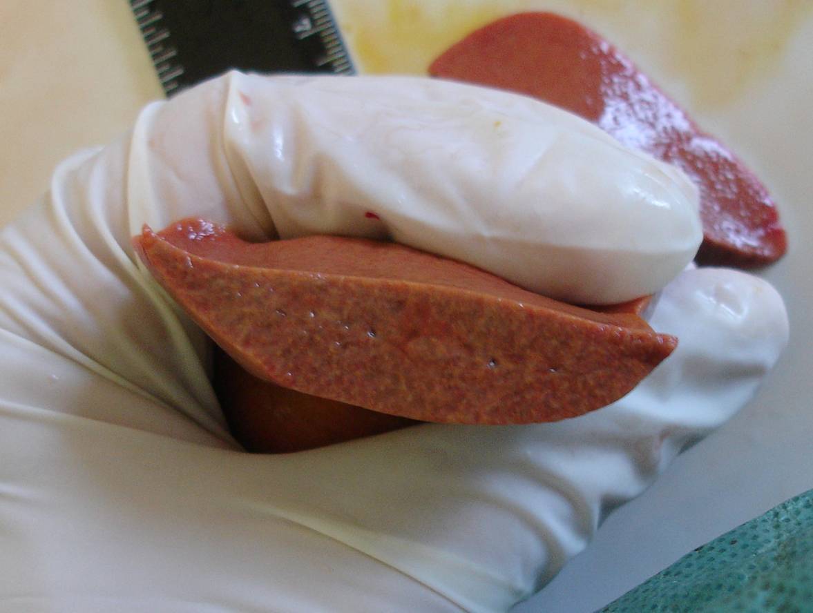
Rice. 5. Macro photo. Cholecystolithiasis. Multiple pigment stones with a diameter of up to 4 mm (shown by an arrow in Fig. A) in the unchanged gallbladder, loose consistency, crumbling under moderate compression (Fig. B).
Clinical case 3. A dog, Yorkshire terrier breed, female, age 5 years, was admitted to the clinic with complaints from the owners of a neoplasm of the mammary gland (noticed 6 months ago) and a cough for 3 months, worsening after physical activity. At clinical trial established: stage II breast cancer, cyanotic mucous membranes, the tracheal reflex is sharply positive, clear and vesicular breathing. Ultrasound reveals hyperechoic contents in the lumen of the gallbladder (Fig. 6), bilateral nephrolithiasis, diffuse changes in the liver. At x-ray examination: enlargement of the right heart, tracheal collapse.
A 
B 
Rice. 6. Ultrascanogram of the gallbladder in transverse (a) and longitudinal (b) sections. Hyperechoic contents in the lumen of the gallbladder (shown by an arrow)
The animal was treated in the clinic for 4 months: completion of the course radiation therapy, with further regional mastectomy and three courses chemotherapy. The condition worsened after the end of chemotherapy: persistent pancytopenia, epileptiform seizures, gastrointestinal bleeding.
Due to the extremely serious condition of the animal, at the request of the owners, it was euthanized.
Pathological and anatomical diagnosis: severe internal hydrocephalus (Fig. 7), fatty liver (Fig. 8, 9), cholecystolithiasis (Fig. 10), thrombosis of the right ventricular cavity, tracheal collapse III degree, bilateral nephrolithiasis, pinpoint hemorrhages in the small and large intestines.

Rice. 7. Macro photo. Segmental section of the brain. Dilatation of the ventricles of the brain
Rice. 8. Macro photo. Fatty liver degeneration. A yellowish-colored organ in section

Rice. 9. Microphoto. Fatty liver degeneration. Numerous fat droplets in the cytoplasm of hepatocytes, creating a fine mesh pattern. Hematoxylin and eosin staining, vol. ×40, approx. ×10
A 
B 
Rice. 10. Cholecystolithiasis. Pigment stones of the gallbladder in Fig. And are shown by arrows. The stones have a loose consistency and crumble under moderate pressure (Fig. B)
Discussion and conclusions
Gallstone disease is a rare disease in dogs and cats, often asymptomatic. In most cases, the pathology is concomitant with the development of the underlying disease. Only in one of the three clinical cases we described can we say that cholelithiasis was the main disease of the animal.
Main etiological factor pathology, both according to the veterinary literature and according to the above clinical cases, is liver pathology. Among the animals with cholelithiasis we studied, severe liver damage was confirmed (histologically) in all three cases. It represented like fatty degeneration, and hepatitis or perivascular cirrhosis.
Severe kidney pathologies (intermediate nephroso-nephritis, nephroso-nephritis with cirrhosis and polycystic disease, and nephrolithiasis, identified in each special case) and pancreas (fibrosis or cirrhosis of the organ, which we established in two out of three cases) may indicate a possible correlation of cholelithiasis with failure of these organs. It should be noted that in all three cases the disease was detected in females, and according to numerous medical literature data, the disease has a gender predisposition (in women, stones are 3-4 times more common).
Changes in hematological and biochemical parameters that appear during obstruction of the bile ducts with stones, leading to cholestasis, are more often manifested by leukocytosis and increased liver parameters.
The main instrumental method for studying the disease is ultrasound or CT, which makes it possible to identify the presence of stones, their size, quantity, location and, to a certain extent, structure.
In the presence of stones in the gallbladder, the main treatment method is cholecystotomy with removal of stones, and in case of serious pathology of the gallbladder, cholecystectomy. Becoming widespread in veterinary practice restoration of bile outflow by applying various anastomoses between the biliary system and the duodenum (cholecystoduodenostomy).
Bibliography
1. Kaliteevsky P.F. Macroscopic differential diagnosis pathological processes. Moscow, “Miklos”, 1993. p. 221-226.
2. Lyutinsky S.I. Pathological physiology animals. M.: KolosS, 2005. p. 351-352.
3. Paltsev M.A. Pathology: course of lectures. Volume 2. M., “Medicine”, 2007. p. 287-289.
4. Savoysky A.G., Baimatov V.N., Meshkov V.M. Pathological physiology. M.: KolosS, 2008, p. 409-411.
5. Buote N.J. The surgical treatment of cholelithiasis in cats: a study of nine cases. J Am Anim Hosp Assoc. 2002, 38(3): 290-6.
9. Fahie M.A., Martin R.A. Extrahepatic biliary tract obstruction: A retrospective study of 45 cases (1983–1993). J Am Anim Hosp Assoc. 1995, 31: 478–481.
10. Heidner G.L., Campbell K.L. Cholelithiasis in a cat. J Am Vet Med Assoc. 1985, 15; 186(2): 176-7.
11. Kirpensteijn J., Fingland R.B., Ulrich T., Sikkema D.A., Allen S.W. Cholelithiasis in dogs: 29 cases. J Am Vet Med Assoc. 1993, 202: 1137–1142.
12. Neer M.T. A review of disorders of the gallbladder and extrahepatic biliary tract in the dog and cat. J Vet Intern Med 1992; 6: 186–192.
13. Rege R.V., Prystowsky J.B. Inflammatory properties of bile from dogs with pigment gallstones. Am J Surg. 1996; 171(1):197–201.
14. Strombeck D.R., Guilford W.G. Small Animal Gastroenterology, 2nd ed. Davis, California: Stonegate Publ, 1990, p. 686–689.
15. Wolf A.M. Obstructive jaundice in a cat resulting from choledocholithiasis. J Am Vet Med Assoc. 1984, 1; 185(1): 85-7.
Summary
D.E. Mitrushkin. Cholelithiasis in dogs and cats. The frequency of cholelithiasis in dogs and cats is rare and often is subclinical, but can result in clinical signs such as icterus, anorexia, vomiting, dehydration, abdominal pain, bradycardia, skin itch and acholia. Values for bilirubin total, alanine aminotransferase, alkaline phosphatase, cholesterol and white blood cells are higher than normal at obstructive cholelithiasis. In this article three cases of cholelithiasis were presented. In all three cases we expressed found histopathological changes of liver, pancreas and kidneys. It is suggested that the pathology of these organs might have contributed to gallstones formation. The main method of treatment of disease is cholecystotomy, however, cholecystectomy is indicated if damage of gall bladder is severe.


