Acute inflammation of the pharynx. Acute inflammatory diseases of the pharynx: symptoms, treatment, signs, causes. Types of chronic ENT disease - laryngitis
Acute pharyngitis is an acute inflammation of the mucous membrane of all parts of the pharynx. This disease is often concomitant with respiratory infections of viral and microbial etiology (influenza, adenoviral, coccal).
The patient complains of a feeling of rawness or pain in the throat, soreness, dryness, hoarseness, and upon examination there is hyperemia of the mucous membrane of all parts of the pharynx, accumulation of viscous mucus on the back wall, sometimes of a hemorrhagic nature.
General symptoms - weakness, fever, discomfort - are caused by the underlying disease. For the treatment of acute pharyngitis, oil-balsamic nasal drops are recommended, a mixture of equal amounts of sea buckthorn, vaseline and menthol oils 3-5 times a day, warm alkaline inhalations, lubricating the pharyngeal mucosa with Lugol's solution on glycerin, analgesics and aspirin are prescribed orally.
Differential diagnosis of acute pharyngitis is carried out with diphtheria, scarlet fever, measles, rubella and other infectious diseases.
Sore throat is an acute inflammation of the tonsils and pharyngeal mucosa.
Sore throats, according to clinical data and pharyngoscopic picture, are divided into catarrhal, follicular, lacunar, ulcerative-membranous and necrotic.
Sore throat is a general nonspecific infectious-allergic disease of predominantly streptococcal etiology, in which local inflammatory changes are most pronounced in the lymphadenoid tissue of the pharynx, most often in the palatine tonsils and regional lymph nodes.
It manifests itself clinically in the form of catarrhal, follicular and lacunar tonsillitis.
Nonspecific sore throatNonspecific angina - catarrhal, when only the mucous membrane of the tonsils is affected, follicular - purulent damage to the follicles, lacunar - pus accumulates in the lacunae. Typically caused by group A streptococcus.
However, there are pneumococcal tonsillitis, staphylococcal tonsillitis and tonsillitis, the etiology of which is a mixed coccal flora. A type of this sore throat is alimentary sore throat, caused by epidemic streptococcus. The microbe is usually introduced when food preparation technology is violated by unscrupulous workers.
Catarrhal sore throat affects the mucous membrane of the tonsils and arches, and there is hyperemia in these areas of the pharynx, but there are no plaques.
The patient notes pain when swallowing, a burning sensation in the pharynx. Has a bacterial or viral etiology. The temperature is low-grade, fever is less common.
Regional lymph nodes may be moderately enlarged. The disease lasts 3–5 days. Treatment - rinsing with soda, sage, lubricating the tonsils with iodine-glycerin, taking aspirin orally.
Catarrhal sore throat must be distinguished from acute pharyngitis, which affects the entire mucous membrane of the pharynx, especially its posterior wall.
Follicular and lacunar tonsillitis are caused by the same pathogens and are similar both in clinical course and in the general reaction of the body and possible complications. The difference lies in the different forms of plaque on the tonsils.
With follicular angina, suppuration of the follicles occurs, and dead white leukocytes appear through the mucous membrane. With lacunar angina, inflammation begins from the lacunae, where pus accumulates, which then protrudes from the lacunae onto the surface of the tonsils.
After 1–2 days, plaque spreads over the entire surface of the tonsils, and it is no longer possible to distinguish between the two types of sore throat. Patients feel severe pain when swallowing, discomfort in the throat, and refuse food.
The cervical lymph nodes are sharply enlarged, the temperature rises to 39 and even 40 °C.
On days 2–3, a differential diagnosis with diphtheria is made. Already at the first examination, it is necessary to take a smear from the patient for diphtheria bacillus, and try to remove the plaque with a cotton brush.
If the plaque is removed, this speaks in favor of vulgar tonsillitis; if it is difficult to remove, and bleeding erosion remains in its place, this is most likely diphtheria.
In case of doubt, it is necessary to administer anti-diphtheria serum.
Treatment of follicular and lacunar tonsillitis consists of gargling, cervical semi-alcohol compress, prescribing analgesics, desensitizers (diphenhydramine, suprastin, tavegil), broad-spectrum antibiotics intramuscularly. A gentle diet is recommended for patients.
Sore throat caused by adenoviruses, occurs in the form of diffuse acute pharyngitis, although it may also be accompanied by plaque on the tonsils. Adenovirus infection is characterized by widespread damage to the lymph nodes and a very frequent combination with conjunctivitis.
This is especially true for adenovirus type 3, which causes pharyngoconjunctival fever. The influenza virus gives a similar picture, but in 10–12% of cases it can be combined with streptococcal sore throat.
Acute inflammation of the tonsils of another location. Sore throat of the lingual tonsil has characteristic symptoms - pain in the deep parts of the pharynx, which sharply intensifies when trying to stick out the tongue.
Diagnosis involves performing indirect laryngoscopy using a laryngeal speculum.
Sore throat of the nasopharyngeal tonsil. The pain is localized in the nasopharynx, thick mucous discharge is released from the nose, and an acute runny nose is noted. With posterior rhinoscopy, a swollen tonsil with a bluish color is visible, sometimes with plaque, and thick mucus flows down the back wall of the pharynx.
Sore throat as a syndrome of common infectious diseasesSore throat with scarlet fever may proceed in different ways. Most often it is catarrhal and lacunar tonsillitis.
In the classic course of scarlet fever, there is a characteristic redness of the soft palate in the circumference of the pharynx, which does not extend beyond the soft palate, swelling of the cervical lymph glands and a whitish thick coating on the tongue, followed by its clearing when the tongue takes on a bright color.
To make a diagnosis, it is necessary to take into account all the symptoms of the disease, primarily the scarlet fever rash in the area of the mastoid process and the flexor surfaces of the limbs.
There are severe forms of scarlet fever, occurring in the form of:
1) pseudomembranous tonsillitis with the formation of fibrinous exudate widespread on the mucous membrane of the tonsils, pharynx, nasopharynx and even cheeks in the form of a thick grayish film tightly fused to the underlying tissue. There is a bright hyperemia of the circumference of the pharynx, the rash appears already on the first day of the disease. The prognosis for this form of scarlet fever is unfavorable;
2) ulcerative necrotic tonsillitis, characterized by the appearance of grayish spots on the mucous membrane, quickly turning into ulcers. Deep ulceration may occur with the formation of permanent defects of the soft palate. The lateral cervical lymph nodes are affected by extensive inflammation;
3) gangrenous tonsillitis, which is rare. The process begins with the appearance of a dirty gray coating on the tonsils, followed by deep tissue destruction down to the carotid arteries.
Sore throat with diphtheria can occur in various clinical forms. With diphtheria, plaque extends beyond the arches. For tonsillitis, a strict boundary of the distribution of plaque within the tonsils is pathognomonic. If plaque spreads beyond the arches, the doctor must doubt the diagnosis of nonspecific tonsillitis. There is a simple diagnostic test. The plaque from the tonsil is removed with a spatula and dissolved in a glass of cold water.
If the water becomes cloudy and the plaque dissolves, it means a sore throat. If the water remains clear, but plaque particles float to the surface, then it is diphtheria.
Sore throat with measles occurs under the mask of catarrhal disease in the prodromal period and during the rash.
In the second case, the diagnosis of measles does not cause difficulties; in the prodromal period, it is necessary to monitor the appearance of measles enanthema in the form of red spots on the mucous membrane of the hard palate, as well as Filatov-Koplik spots on the inner surface of the cheeks at the opening of the Stenon's duct. The course of sore throat with rubella measles is similar to measles.
Sore throat with flu proceeds in the same way as catarrhal, but diffuse hyperemia affects the tonsils, arches, uvula, and back wall of the pharynx.
Erysipelas is a serious disease, often occurring together with facial erysipelas. It begins with a high fever and is accompanied by severe pain when swallowing. The mucous membrane is colored bright red with sharply defined borders of redness, it seems varnished due to swelling.
Sore throat with tularemia begins acutely - with chills, general weakness, redness of the face, enlarged spleen.
For differential diagnosis, it is important to establish contact with rodents (water rats, house mice and gray voles) or blood-sucking insects (mosquitoes, horseflies, ticks).
In most cases, tonsillitis with tularemia occurs when infected through the nutritional route - by consuming water or food after an incubation period of 6-8 days in an infected patient.
Another differential diagnostic sign is the formation of buboes - packets of lymph nodes in the neck, sometimes reaching the size of a chicken egg.
Lymph nodes may fester. The picture of the pharynx may resemble catarrhal or, more often, membranous sore throat, which is mistakenly diagnosed as diphtheria.
Sore throat with blood diseasesMonocytic tonsillitis(infectious mononucleosis or Filatov's disease) can have a varied clinical course - from catarrhal to ulcerative-necrotic. The etiology of this disease has not been fully elucidated. Clinically: enlarged liver and spleen (hepatolienal syndrome), the presence of compacted and painful to the touch lymph nodes (cervical, occipital, submandibular, axillary and inguinal, and even polylymphadenitis).
A pathognomonic symptom is the appearance of atypical mononuclear cells in the peripheral blood.
Agranulocytic tonsillitis associated with the complete or almost complete disappearance of granulocytes in the peripheral blood with the preservation of monocytes and lymphocytes against the background of severe leukopenia. The etiology of the disease is not clear; it is considered polyetiological. The disease is associated with excessive and uncontrolled use of drugs such as analgin, pyramidon, antipyrine, phenacytin, sulfonamides, antibiotics, chloramphenicol, Enap.
The clinical picture is usually severe and consists of symptoms of acute sepsis and necrotizing tonsillitis, since the microbes inhabiting the pharynx belong to the opportunistic flora and, when the leukocyte defense is turned off and other unfavorable circumstances, become pathogenic and penetrate the tissues and blood. The disease is severe, with high fever, stomatitis, gingivitis, and esophagitis. The liver is enlarged. The diagnosis is made on the basis of a blood test: severe leukopenia, below 1000 leukocytes in 1 mm 3 of blood, absence of granulocytes. The prognosis is serious due to the development of sepsis, laryngeal edema, necrosis of pharyngeal tissue with severe bleeding. Treatment consists of fighting secondary infection - prescribing antibiotics, vitamins, pharyngeal care (rinsing, lubricating, irrigation with antiseptic, astringent, balsamic solutions), intravenous transfusion of leukocyte mass. The prognosis for this disease is quite serious.
Alimentary-toxic aleukia characterized by the fact that, unlike agranulocytosis, when only granulocytes (neutrophils, eosinophils) disappear from the peripheral blood, the disappearance affects all forms of leukocytes. The disease is associated with the ingestion of a special fungus that multiplies in overwintered cereals left unharvested in the fields and contains a very toxic substance - poin, even a very small amount of which leads to contact lesions in the form of tissue necrosis, hemorrhagic ulcers affecting the entire gastrointestinal tract , and even contact with feces on the buttocks causes ulceration.
The poison is heat-stable, so heat treatment of flour (cooking baked goods, bread) does not reduce its toxicity.
From the side of the pharynx, necrotic sore throat is pronounced, when the tonsils look like gray dirty rags, and a sharp, nauseating odor is released from the mouth.
The number of leukocytes in the peripheral blood is up to 1000 or less, while granular leukocytes are completely absent. Characterized by high fever and the appearance of a hemorrhagic rash. Treatment in the early stage consists of gastric lavage, enemas, laxatives, a gentle diet, intravenous infusions of saline with vitamins, hormones, glucose, blood transfusions, and leukocyte mass.
At the stage of tonsillitis and necrosis, antibiotics are prescribed. With severe clinical manifestations of the disease, the prognosis is unfavorable.
Sore throats in acute leukemia occur with varying degrees of severity depending on the stage of leukemia. The onset of a sore throat (usually catarrhal) proceeds relatively favorably, begins against the background of apparent well-being, and only a blood test allows one to suspect acute leukemia at this early stage of the disease, which once again proves the mandatory blood test for sore throats.
Sore throats with developed leukemia, when the number of blood leukocytes reaches 20,000 or more, and the number of red blood cells drops to 1–2 million, sore throat is extremely severe in the form of an ulcerative-necrotic and gangrenous form with high fever and severe general condition. Nosebleeds, hemorrhages in organs and tissues, and enlargement of all lymph nodes occur. The prognosis is unfavorable, patients die within 1–2 years. Treatment of sore throat is symptomatic, local, antibiotics and vitamins are prescribed less often.
Sore throats with infectious granulomas and specific pathogensTuberculosis of the pharynx can occur in two forms - acute and chronic. The acute form is characterized by hyperemia with thickening of the mucous membrane of the arches, soft palate, and uvula, reminiscent of a sore throat; body temperature can reach 38 °C and higher. There are sharp pains when swallowing, the appearance of gray tubercles on the mucous membrane, then their ulceration. A characteristic medical history and the presence of other forms of tuberculosis help in the diagnosis.
Of the chronic forms of tuberculosis, the most common is ulcerative, developing from infiltration, often occurring without symptoms. The edges of the ulcer are raised above the surface, the bottom is covered with a gray coating, after its removal juicy granulations are found. Most often, ulcers are observed on the back wall of the pharynx. The course of processes in the pharynx depends on many reasons: the general condition of the patient, his diet, regimen, social conditions, timely and correct treatment.
In the acute miliary form of tuberculosis, the prognosis is unfavorable; the process develops very quickly with a fatal outcome in 2–3 months.
Treatment of pharyngeal tuberculosis, as well as its other forms, has become relatively successful after the advent of streptomycin, which is administered intramuscularly at 1 g per day for an average of 3 weeks. R-therapy sometimes gives good results.
Syphilis of the pharynx. Primary syphilis most often affects the tonsils. Chancroid is usually painless.
Usually, a hard infiltrate forms on the red limited background of the upper part of the tonsils, then erosion turns into an ulcer, its surface has a cartilaginous density. There are enlarged cervical lymph nodes on the affected side, painless on palpation.
Primary syphilis develops slowly, over weeks, usually on one tonsil.
The condition of patients with secondary angina worsens, fever and severe pain appear. If syphilis is suspected, the Wasserman reaction must be performed.
Secondary syphilis appears 2–6 months after infection in the form of erythema and papules. Erythema in the pharynx involves the soft palate, arches, tonsils, lips, surface of the cheeks, and tongue. The diagnosis of syphilis at this stage is difficult until papules appear from the lentil grain to the bean, their surface is covered with a coating with a hint of greasy sheen, the circumference is hyperemic.
Most often, papules are localized on the surface of the tonsils and on the arches.
The tertiary period of syphilis manifests itself in the form of gumma, which usually appears several years after the onset of the disease. Most often, gummas form on the back wall of the pharynx and soft palate. First, limited infiltration appears against the background of bright hyperemia of the pharyngeal mucosa. There may be no complaints during this period.
With further progression, paresis of the soft palate occurs, and food enters the nose. The course of tertiary syphilis is very variable, depending on the location and rate of development of the gumma, which can affect the bone walls of the facial skull, tongue, great vessels of the neck, causing heavy bleeding, and grows into the middle ear.
If syphilis is suspected, consultation with a venereologist is required to clarify the diagnosis and prescribe rational treatment.
Fusospirochetosis. The etiological factor is the symbiosis of a spindle-shaped rod and a spirochete in the oral cavity. A characteristic manifestation of the disease is the appearance of erosions on the surface of the palatine tonsils, covered with a grayish, easily removable coating.
In the initial stage of the disease, there are no subjective sensations, the ulcer progresses, and only after 2–3 weeks mild pain appears when swallowing, and regional lymph nodes on the affected side may enlarge.
Pharyngoscopy during this period reveals a deep ulcer of the tonsil, covered with a gray foul-smelling coating that is easily removed. General symptoms are usually not pronounced.
In differential diagnosis, it is necessary to exclude diphtheria, syphilis, tonsil cancer, blood diseases, for which a blood test, Wasserman reaction, and a smear for diphtheria bacillus are done.
Rarely, pharyngitis and stomatitis are associated with damage to the tonsils, and then the course of the disease becomes severe.
Treatment consists of rinsing with hydrogen peroxide, a 10% solution of Berthollet salt, and potassium permanganate. However, the best treatment is to generously lubricate the ulcer with a 10% solution of copper sulfate 2 times a day.
The beginning of ulcer healing is noted already on the third day, which, in turn, serves as a differential diagnosis with syphilis and blood diseases. The prognosis with timely treatment is favorable.
Candidomycosis pharynx is caused by yeast-like fungi, often in weakened patients or after uncontrolled use of large doses of antibiotics, causing dysbiosis in the pharynx and digestive tract.
There is a sore throat, fever, against the background of hyperemia of the mucous membrane of the pharynx, small white plaques appear with further extensive necrosis of the epithelium of the tonsils, arches, palate, and posterior wall of the pharynx in the form of grayish plaques, after removal of which erosion remains.
The disease must be differentiated from diphtheria, fusospirochetosis, and lesions due to blood diseases. The diagnosis is made on the basis of microscopy of smear materials with a coating of yeast fungi. Treatment involves the mandatory abolition of all antibiotics, irrigation of the pharynx with a weak soda solution, and lubrication of the lesions with Lugol's solution on glycerin.
This disease must be distinguished from pharyngomycosis, in which sharp and hard spines protruding to the surface are formed in the lacunae of the tonsils. Since there are no signs of inflammation of the surrounding tissues and subjective sensations, the disease may not be detected by the patient for a long time. Conservative treatment is ineffective. As a rule, the affected tonsils have to be removed.
Peritonsillar abscessBetween the tonsil capsule and the pharyngeal fascia there is paratonsillar fiber, and behind the pharyngeal fascia, laterally, there is fiber of the parapharyngeal space. These spaces are filled with fiber, the inflammation of which, and in the final stage – abscess formation, determine the clinical picture of the disease. An abscess is most often caused by nonspecific flora as a result of tonsillogenic spread of infection. The disease begins acutely, with the appearance of pain when swallowing, usually on one side.
Typically, a peritonsillar abscess occurs after a sore throat during the recovery period. When examining the pharynx, sharp swelling and hyperemia of the tissues around the tonsil (arches, soft palate, uvula), protrusion of the tonsil from the niche, and displacement to the midline are noted.
An abscess takes about 2 days to form on average. General symptoms are weakness, fever, enlarged cervical lymph nodes on the side of the abscess. The classic triad of peritonsillar abscess was noted: profuse salivation, trismus of masticatory muscles and open nasal sound (as a result of paralysis of the muscles of the velum).
Treatment of abscesses is prescribed in combination: intramuscular antibiotics, taking into account pain when swallowing and forced fasting, aspirin, analgesics, a semi-alcohol compress on the side of the neck (on the side of the abscess), antihistamines.
At the same time, surgical treatment is carried out. There are anterosuperior abscesses (pus accumulates behind the anterior arch and soft palate near the upper pole of the tonsil), posterior (with accumulation of pus in the area of the posterior arch), external (accumulation of pus between the tonsil capsule and the pharyngeal fascia). Anesthesia, as a rule, is local - lubricating the mucous membrane with a 5% solution of cocaine or a 2% solution of dicaine. A napkin is wrapped around the scalpel so that the tip protrudes no more than 2 mm, otherwise the main vessels of the carotid system can be injured.
The incision is made in the case of an anterior abscess strictly in the sagittal plane in the middle of the distance from the posterior molar to the uvula, then a blunt probe or a hemostatic clamp (Halsted) is inserted into the incision and the edges of the incision are spread apart for better emptying of the abscess.
When the pus is removed, the patient's condition usually improves significantly. A day later, the edges of the incision are again pulled apart with a clamp to remove accumulated pus. In the same way, the posterior abscess is opened through the posterior arch. It is more difficult and dangerous to open an external abscess, which lies deeper and requires greater caution due to the danger of injury to blood vessels. This can be helped by preliminary puncture with a syringe with a long needle, when, if pus is detected, the incision is made in the direction of the puncture. After any cut in the throat, rinse with furatsilin. A very rare occurrence is a retropharyngeal abscess - an accumulation of pus in the area of the back wall of the pharynx. In children, this is associated with the presence of lymph nodes in the retropharyngeal space, in adults – as a continuation of the external paratonsillar abscess.
Everyone in life has encountered various diseases of the ENT organs; the most common are viral or bacterial infections in the form of ARVI, influenza or sore throat. But there are a number of other pathologies, the symptoms of which need to be known in order to diagnose the disease in time.
Structure of the pharynx and larynx
To understand the essence of diseases, you should have a minimal understanding of the structure of the larynx and pharynx.
Regarding the pharynx, it consists of three sections:
- upper, nasopharynx;
- oropharynx, middle section;
- laryngopharynx, lower section.
The larynx is an organ that performs several functions. The larynx is the conductor of food to the digestive tube, and it is also responsible for the flow of air into the trachea and lungs. In addition, the vocal cords are located in the larynx, thanks to which a person is able to make sounds.
The larynx functions as a movement apparatus that has cartilage connected to ligaments and muscle joints. At the beginning of the organ is the epiglottis, the function of which is to create a valve between the trachea and the pharynx. At the moment of swallowing food, the epiglottis blocks the entrance to the trachea, so that food enters the esophagus and not into the respiratory organs.
What are the pathologies of the ENT organs?
According to their course, diseases are classified into: chronic and acute. In the case of an acute course of the disease, symptoms develop instantly and are pronounced. The pathology is more difficult to tolerate than in a chronic course, but recovery occurs faster, on average in 7-10 days.
Chronic pathologies arise against the background of a constant, untreated inflammatory process. In other words, the acute form becomes chronic without proper treatment. In this case, the symptoms do not arise so quickly, the process is sluggish, but complete recovery does not occur. With the slightest provoking factors, for example, hypothermia or a virus entering the body, a relapse of the chronic disease occurs. As a result of a constant focus of infection, a person’s immunity is weakened, because of this it is not difficult for a virus or bacteria to penetrate.
Diseases of the pharynx and larynx:
- epiglottitis;
- pharyngitis;
- tonsillitis;
- laryngitis;
- nasopharyngitis;
- adenoids;
- laryngeal cancer.
Epiglottitis
Diseases of the larynx include inflammation of the epiglottis (epiglottitis). The cause of the inflammatory process is the entry of bacteria into the epiglottis by airborne droplets. Most often, the epiglottis is affected by hemophilus influenza and becomes the cause of the inflammatory process. The bacterium can not only cause disease of the epiglottis, but is also the causative agent of meningitis, pneumonia, pyelonephritis and other pathologies. In addition to hemophilus influenza, the following can cause inflammation of the epiglottis:
- streptococci;
- pneumococci;
- candida fungus;
- burn or foreign body entering the epiglottis.
Symptoms of the disease develop rapidly, the main ones include: 
- difficult breathing with whistling. Swelling occurs in the epiglottis, which leads to partial closure of the larynx and trachea, which complicates the possibility of normal air flow;
- pain when swallowing, difficulty swallowing food with a feeling that there is something in the larynx;
- redness of the throat, pain in it;
- fever and increased body temperature;
- general weakness, malaise and anxiety.
Epiglottitis most often occurs in children aged 2 to 12 years, mostly boys. The main danger posed by inflammation of the epiglottis is the possibility of suffocation, therefore, at the first symptoms of the disease, you should immediately consult a doctor. There are acute and chronic inflammations of the epiglottis. If an acute form of pathology has developed, the child should be urgently taken to the hospital; transportation should be done in a sitting position.
Treatment consists of antibiotic therapy and maintaining patency of the upper respiratory tract. If life-threatening symptoms cannot be relieved, a tracheotomy is performed.
Rhinopharyngitis
Inflammation of the nasopharynx, which occurs when the throat and nose are infected by a virus, is called rhinopharyngitis. Symptoms of inflammation of the nasopharynx:
- nasal congestion, resulting in difficulty breathing;
- acute sore throat, burning sensation;
- difficulty swallowing;
- nasal voice;
- temperature increase.
 Children tolerate the inflammatory process in the nasopharynx more difficult than adults. Often, the inflammation from the nasopharynx spreads to the auricle, which leads to acute pain in the ear. Also, when the infection spreads to the lower respiratory tract, the symptoms are accompanied by cough and hoarseness.
On average, the course of the nasopharyngeal disease lasts up to seven days; with proper treatment, nasopharyngitis does not take a chronic form. Therapy is designed to eliminate painful symptoms. If the infection is caused by a bacteria, antibacterial drugs are prescribed, in case of a viral infection, anti-inflammatory medications are prescribed. It is also necessary to rinse the nose with special solutions and take antipyretics if necessary.
Diseases of the larynx include acute and chronic laryngitis. An acute form of the pathology, rarely develops in isolation; more often, laryngitis becomes a consequence of a respiratory disease. In addition, acute laryngitis can develop as a result of:
- hypothermia;
- when staying in a dusty room for a long time;
- as a result of an allergic reaction to chemical agents;
- the result of smoking and drinking alcoholic beverages;
- professional overload of the vocal cords (teachers, actors, singers).
Symptoms of such a disease of the larynx as laryngitis are characterized by:

Acute laryngitis with voice rest and the necessary treatment goes away within 7-10 days. If the doctor’s recommendations regarding treatment are not followed, the symptoms of the disease do not go away, and laryngitis itself becomes chronic. For laryngitis it is recommended:
- alkaline inhalations;
- voice rest;
- warm drink;
- antitussives;
- antiviral and immunomodulatory agents;
- antihistamines for severe swelling;
- gargling;
- hot foot baths, to drain blood from the larynx and reduce its swelling, etc.
Pharyngitis
Diseases of the pharynx most often manifest as pharyngitis. This infectious pathology often develops against the background of a viral or bacterial infection of the upper respiratory tract. Isolated pharyngitis occurs as a result of direct exposure to the pharyngeal mucosa of an irritant. For example, when talking for a long time in cold air, eating too cold or, conversely, hot food, as well as smoking and drinking alcohol.
Symptoms of pharyngitis are as follows:
- sore throat;
- pain when swallowing saliva;
- feeling of abrasion;
- pain in the ear when swallowing.
Visually, the pharyngeal mucosa is hyperemic, in places there may be an accumulation of purulent secretion, the tonsils are enlarged and covered with a whitish coating. It is important to differentiate acute pharyngitis from catarrhal tonsillitis. Treatment is mainly local: 
- gargling;
- inhalation;
- compresses on the neck area;
- dissolving lozenges for sore throat.
Chronic pharyngitis develops from acute, as well as against the background of chronic tonsillitis, sinusitis, dental caries, etc.
Diseases of the pharynx can be expressed as a sore throat. Inflammation of the lymphoid tissue of the tonsils is called tonsillitis or tonsillitis. Like other diseases of the pharynx, tonsillitis can be acute or chronic. The pathology is especially common and acute in children.
 The cause of tonsillitis is viruses and bacteria, mainly the following: staphylococcus, streptococcus, pneumococcus, fungi of the genus Candida, anaerobes, adenoviruses, influenza viruses.
Secondary tonsillitis develops against the background of other acute infectious processes, for example, measles, diphtheria or tuberculosis. The symptoms of sore throat begin acutely; they are similar to pharyngitis, but have certain differences. The tonsils greatly increase in volume, are painful to the touch, depending on the form of tonsillitis, are covered with purulent plaque, or their lacunae are filled with purulent contents. The cervical lymph nodes are enlarged and may be painful when pressed. Body temperature rises to 38-39 degrees. The throat feels pain when swallowing and soreness.
The classification of tonsillitis is quite extensive; the following forms are distinguished:
- catarrhal - superficial damage to the tonsils occurs. the temperature rises slightly, within 37-37.5 degrees. Intoxication is not severe;
- lacunar, the tonsils are covered with a yellowish-white coating, purulent secretion is observed in the lacunae. The inflammatory process does not spread beyond the lymphoid tissue;
- follicular, tonsils are bright scarlet, swollen, suppurating follicles are diagnosed in the form of whitish-yellowish formations;
- phlegmonous form, most often a complication of previous types of tonsillitis. Not only the tonsils are affected, but also the peritonsil tissue. The pathology occurs acutely, with sharp pain, most often the abscess occurs on one side. Regarding treatment, opening of the purulent sac and further antibacterial therapy is required.
Treatment is mainly medicinal, antibacterial and local action on the pharyngeal mucosa. In cases where the pathology becomes chronic, systematically recurrent tonsillitis or the presence of an abscess, these are indications for tonsil removal. Surgical excision of lymphoid tissue is resorted to in extreme cases, if drug therapy does not bring the desired results.
Adenoid vegetations
Adenoids are hypertrophy of the nasopharyngeal tonsil and occur in the nasopharynx. Most often diagnosed in children between 2 and 12 years of age. As a result of the growth of adenoid vegetation, nasal breathing is blocked and a nasal voice occurs; with the long-term presence of adenoids, hearing loss occurs. Hypertrophy of the nasopharyngeal tonsil has three stages, the second and third are not amenable to drug treatment and require surgical intervention - adenotomy.
Foreign bodies in the larynx or pharynx
The cause of a foreign body entering the throat is most often inattention or haste while eating. Children, left unattended by their parents, may try to swallow various small objects, such as toy parts.
Such situations can be extremely dangerous, it all depends on the shape and size of the foreign object. If an object gets into the larynx and partially blocks its lumen, there is a danger of suffocation. Symptoms of a person choking are:

This situation requires urgent medical care for the victim. Emergency assistance must be provided immediately, otherwise there is a high risk of suffocation.
Cancer of the pharynx or larynx
Diseases of the pharynx can be different, but the most terrible and certainly life-threatening is cancer. A malignant formation in the pharynx or larynx may not manifest itself in any way in the early stages, which leads to late diagnosis and, accordingly, untimely prescription of therapy. Symptoms of a tumor in the larynx are:
- persistent sensation of a foreign body in the larynx;
- desire to cough, disturbing object;
- hemoptysis;
- constant pain in the throat area;
- difficulty breathing when the tumor reaches a large size;
- dysphonia and even aphonia, when the formation is localized near the vocal cords;
- general weakness and loss of ability to work;
- lack of appetite;
- weight loss.
Oncological diseases are extremely life-threatening and have a disappointing prognosis. Treatment for laryngeal cancer is prescribed depending on the stage of the pathology. The main method is surgery and removal of the malignant tumor. Radiation and chemotherapy are also used. The prescription of one or another treatment method is purely individual.
Every disease, regardless of the complexity of its course, requires attention. You should not self-medicate, much less diagnose yourself. Pathology can be much more complex than you think. Timely diagnosis and compliance with all doctor’s instructions allows for a complete recovery and absence of complications.
![]()
Acute inflammatory diseases of the larynx and trachea often occur as a manifestation of acute inflammatory diseases of the upper respiratory tract. The cause may be a wide variety of flora - bacterial, fungal, viral, mixed.
4.4.1. Acute catarrhal laryngitis
Acute catarrhal laryngitis (laryngitis) - acute inflammationthinning the mucous membrane of the larynx.
As an independent disease, acute catarrhal laryngitis occurs as a result of activation of saprophytic flora in the larynx under the influence exogenous And endogenous factors. Among exogenous factors such as hypothermia, irritation of the mucous membrane by nicotine and alcohol, exposure to occupational hazards (dust, gases, etc.), prolonged loud conversation in the cold, consumption of very cold or very hot food play a role. Endogenous factors - reduced immune reactivity, gastrointestinal diseases, allergic reactions, age-related atrophy of the mucous membrane. Acute catarrhal laryngitis often occurs during puberty, when voice mutation occurs.
Etiology. Among the various etiological factors in the occurrence of acute laryngitis, bacterial flora plays a role - beta-hemolytic streptococcus, pneumococcus, viral infections; influenza A and B viruses, parainfluenza, coronavirus, rhinovirus, fungi. Mixed flora is often found.
Pathomorphology. Pathomorphological changes are reduced to circulatory disorders, hyperemia, small cell infiltration and serous impregnation of the laryngeal mucosa. When inflammation spreads to the vestibule of the larynx, the vocal folds may be covered by swollen, infiltrated vestibular folds. When the subglottic region is involved in the process, the clinical picture of false croup (subglottic laryngitis) occurs.
Clinic. It is characterized by the appearance of hoarseness, soreness, discomfort and a foreign body in the throat. Body temperature is often normal, less often it rises to low-grade levels. Violations of the voice-forming function are expressed in the form of varying degrees of dysphonia. Sometimes the patient is bothered by a dry cough, which is subsequently accompanied by expectoration of sputum.
Diagnostics. It does not present any particular difficulties, since it is based on pathognomonic signs: acute onset of hoarseness, often associated with a specific cause (cold food, ARVI, colds, speech stress, etc.); a characteristic laryngoscopic picture is more or less pronounced hyperemia of the mucous membrane of the entire larynx or only the vocal folds, thickening, swelling and incomplete closure of the vocal folds; absence of temperature reaction if there is no respiratory infection. Acute laryngitis should also include those cases when there is only marginal hyperemia of the vocal folds, since this is limited
the process, like a spilled one, tends to become chronic
In childhood, laryngitis must be differentiated from the common form of diphtheria. Pathological changes in this case will be characterized by the development of fibrinous inflammation with the formation of dirty gray films intimately associated with the underlying tissues.
Erysipelas of the mucous membrane of the larynx differs from the catarrhal process by clearly defined boundaries and simultaneous damage to the skin of the face.
Treatment. With timely and adequate treatment, the disease ends within 10-14 days; its continuation for more than 3 weeks most often indicates a transition to a chronic form. The most important and necessary therapeutic measure is compliance with the voice mode (silence mode) until the acute inflammatory phenomena subside. Failure to adhere to a gentle vocal regimen will not only delay recovery, but will also contribute to the process becoming chronic. It is not recommended to consume spicy, salty foods, alcoholic beverages, smoking, or alcohol. Drug therapy is mainly local in nature. Alkaline oil inhalations, irrigation of the mucous membrane with combined preparations containing anti-inflammatory components (Bioparox, IRS-19, etc.), infusion of medicinal mixtures of corticosteroids, antihistamines and antibiotics into the larynx for 7-10 days are effective. Effective mixtures for infusion into the larynx, consisting of 1% menthol oil, hydrocortisone emulsion with the addition of a few drops of 0.1% adrenaline hydrochloride solution. In the room where the patient is located, it is advisable to maintain high humidity.
For streptococcal and pneumococcal infections, accompanied by fever and intoxication, general antibiotic therapy is prescribed - penicillin drugs (phenoxymethylpenicillin 0.5 g 4-6 times a day, ampicillin 500 mg 4 times a day) or macrolides ( for example, erythromycin 500 mg 4 times a day).
The prognosis is favorable with appropriate treatment and compliance with the voice regime.
4.4.2. Infiltrative laryngitis
Infiltrative laryngitis (laryngitis inflltrativa) - acute inflammation of the larynx, in which the process is not limited tozygotic membrane, and spreads to deeper tissues. The process may involve the muscular system, ligaments, and upper ligament.
Etiology. The etiological factor is a bacterial infection that penetrates the tissue of the larynx during injury or after an infectious disease. A decrease in local and general resistance is a predisposing factor in the etiology of infiltrative laryngitis. The inflammatory process can occur in a limited or diffuse form.
Clinic. Depends on the extent and prevalence of the process. In the diffuse form, the entire mucous membrane of the larynx is involved in the inflammatory process; in the limited form, certain areas of the larynx are involved - the interarytenoid space, the vestibule, the epiglottis, and the subglottic cavity. The patient complains of pain that intensifies when swallowing, severe dysphonia, high body temperature, and poor health. There may be a cough with expectoration of thick mucopurulent sputum. Against the background of these symptoms, respiratory function is impaired. Regional lymph nodes are dense and painful on palpation.
With irrational therapy or a highly virulent infection, acute infiltrative laryngitis can turn into a purulent form - phlegmonous laryngitis { laryngitis phlegmonosa). In this case, pain symptoms sharply intensify, body temperature rises, general condition worsens, breathing becomes difficult, up to asphyxia. During indirect laryngoscopy, an infiltrate is detected, where a limited abscess can be seen through the thinned mucous membrane, which confirms the formation of an abscess. A laryngeal abscess can be the final stage of infiltrative laryngitis and occurs predominantly on the lingual surface of the epiglottis or in the area of one of the arytenoid cartilages.
Treatment. As a rule, it is carried out in a hospital setting. Antibiotic therapy is prescribed in the maximum dosage for a given age, antihistamines, mucolytics, and, if necessary, short-term corticosteroid therapy. Emergency surgery is indicated in cases where an abscess is diagnosed. After local anesthesia, the abscess (or infiltrate) is opened with a laryngeal knife. At the same time, massive antibiotic therapy, antihistamine therapy, corticosteroid drugs, detoxification and transfusion therapy are prescribed. It is also necessary to prescribe analgesics.
Usually the process stops quickly. Throughout the disease, you need to carefully monitor the condition of the lumen of the larynx and not wait for the moment of asphyxia.
In the presence of diffuse phlegmon spreading to the soft tissues of the neck, external incisions are made, always with wide drainage of purulent cavities.
It is important to constantly monitor respiratory function; whensigns of acute increasing stenosis require emergencytracheostomy.
4.4.3. Subglottic laryngitis (false croup)
Subglottic laryngitis -laryngitis subglottica(subchordal laryngitis- laryngitis subchordalis, false croup -false crop) - acute laryngitis with predominant localization of the process insubglottic cavity. It is usually observed in children under the age of 5-8 years, which is due to the structural features of the subvocal cavity: the loose tissue under the vocal folds in young children is highly developed and easily reacts to irritation with edema. The development of stenosis is also facilitated by the narrowness of the larynx in children and the lability of nervous and vascular reflexes. When the child is in a horizontal position, due to blood flow, the swelling increases, so the deterioration of the condition is more pronounced at night.
Clinic. The disease usually begins with inflammation of the upper respiratory tract, nasal congestion and discharge, low-grade fever, and cough. The general condition of the child during the day is quite satisfactory. At night, a sudden attack of suffocation, a barking cough, and cyanosis of the skin begin. Dyspnea is predominantly inspiratory, accompanied by retraction of the soft tissues of the jugular fossa, supra- and subclavian spaces, and epigastric region. This condition lasts from several minutes to half an hour, after which profuse sweating appears, breathing returns to normal, and the child falls asleep. Such conditions may recur after 2-3 days.
Laryngoscopy picture subglottic laryngitis appears in the form of a roll-shaped symmetrical swelling, hyperemia of the mucous membrane of the subglottic space. These ridges protrude from under the vocal folds, significantly narrowing the lumen of the larynx and thereby making breathing difficult.
Diagnostics. It is necessary to differentiate from true diphtheria croup. The term “false croup” indicates that the disease is opposed to true croup, i.e. diphtheria of the larynx, which has similar symptoms. However, with subglottic laryngitis, the disease is paroxysmal in nature - a satisfactory condition during the day is changed by difficulty breathing and an increase in body temperature at night. The voice with diphtheria is hoarse, with subglottic laryngitis it is not changed. With diphtheria there is no barking cough, which is characteristic of false croup. With subglottic laryngitis, there is no significant increase in
In the study of regional lymph nodes, there are no films characteristic of diphtheria in the pharynx and larynx. However, it is always necessary to conduct a bacteriological examination of smears from the pharynx, larynx and nose for diphtheria bacillus.
Treatment. Aimed at eliminating the inflammatory process and restoring breathing. Inhalation of a mixture of decongestant drugs is effective - 5% ephedrine solution, 0.1% adrenaline solution, 0.1% atropine solution, 1% diphenhydramine solution, hydrocortisone 25 mg and chymopsin. Antibiotic therapy is required, which is prescribed in the maximum dose for a given age, antihistamine therapy, and sedatives. The administration of hydrocortisone at a rate of 2-4 mg/kg of the child’s body weight is also indicated. Drinking plenty of water - tea, milk, alkaline mineral waters - has a beneficial effect; distracting procedures - foot baths, mustard plasters.
You can try to stop an attack of suffocation by quickly touching the back of the throat with a spatula, thereby causing a gag reflex.
In the case where the above measures are powerless, andsuffocation becomes dangerous, it is necessary to resort tonasotracheal intubation for 2-4 days, and if necessarytracheostomy is indicated.
4.4.4. Laryngeal sore throat
Laryngeal sore throat (angina laryngea), or submucosal laringit (laryngitis submucosa) - an acute infectious disease withdamage to the lymphadenoid tissue of the larynx, located in the ventricles of the larynx, in the thickness of the mucous membrane of the aryepigland -tan folds, at the bottom of the pyriform recess, as well as in the area of the lingual surface of the epiglottis. It is relatively rare and can occur under the guise of acute laryngitis.
Etiology. The etiological factors causing the inflammatory process are a variety of bacterial, fungal and viral flora. Penetration of the pathogen into the mucous membrane can occur through airborne droplets or alimentary routes. Hypothermia and trauma to the larynx also play a role in the etiology.
Clinic. In many ways, it is similar to the manifestations of tonsillitis of the palatine tonsils. I am worried about a sore throat, which gets worse when swallowing and when turning the neck. Dysphonia and difficulty breathing are possible. The body temperature with laryngeal sore throat is high, up to 39 ° C, the pulse is rapid. On palpation, regional lymph nodes are painful and enlarged.
Laryngoscopy reveals hyperemia and infiltration of the mucous membrane of the larynx, sometimes narrowing the lumen

respiratory tract, individual follicles with pinpoint purulent deposits. With a prolonged course, an abscess may form on the lingual surface of the epiglottis, aryepiglottic fold and other places where lymphadenoid tissue accumulates (Fig. 4.10).
Diagnostics. Indirect laryngoscopy with appropriate anamnestic and clinical data allows you to establish the correct diagnosis. Laryngeal sore throat should be differentiated from diphtheria, which may have a similar course.
Treatment. Includes broad-spectrum antibiotics (augmentin, amoxiclav, cefazolin, kefzol, etc.), antihistamines (tavegil, fenkarol, peritol, claritin, etc.), mucolytics, analgesics, antipyretics. If signs of respiratory distress occur, short-term corticosteroid therapy is added to treatment for 2-3 days. If the stenosis is significant, emergency tracheotomy is indicated.
4.4.5. Laryngeal edema
Laryngeal edema (oedema laryngea) - fast-growing va-zomotor-allergic process in the mucous membrane of the larynx,narrowing its lumen.
Etiology. The causes of acute laryngeal edema may be:
1) inflammatory processes of the larynx (subglottic laryngitis, acute laryngotracheobronchitis, chondroperichondritis and
acute infectious diseases (diphtheria, measles, scarlet fever, influenza, etc.);
laryngeal tumors (benign, malignant);
laryngeal injuries (mechanical, chemical);
allergic diseases;
pathological processes of organs adjacent to the larynx and trachea (tumors of the mediastinum, esophagus, thyroid gland, retropharyngeal abscess, phlegmon of the neck, etc.).
Clinic. Narrowing of the lumen of the larynx and trachea can develop immediately (foreign body, spasm), acutely (infectious
diseases, allergic processes, etc.) and chronically (against the background of a tumor). The clinical picture depends on the degree* of narrowing of the lumen of the larynx and the speed of its development. Whatever-| The faster the stenosis develops, the more dangerous it is. With inflammation! the etiology of edema is a sore throat that gets worse with! swallowing, foreign body sensation, change in voice. Ras-| extension of edema to the mucous membrane of the arytenoids! cartilage, aryepiglottic folds and subglottic cavity - [ ti causes acute laryngeal stenosis, causing severe! a picture of suffocation that threatens the patient’s life (see section! 4.6.1).
During laryngoscopic examination, swelling of the mucous membrane of the affected part of the larynx is determined in the form! watery or gelatinous swelling. Epiglottis at! this is sharply thickened, there may be elements of hyperemia, the process! extends to the area of the arytenoid cartilages. Voice-| When the mucous membrane swells, the gap sharply narrows, in! subglottic swelling looks like a bilateral pillow-| co-shaped protrusion.
It is characteristic that with the inflammatory etiology of edema on-| There are reactive phenomena of varying degrees of severity, hyperemia and injection of the vessels of the mucous membrane! spots, with non-inflammatory - hyperemia is usually absent-| howls.
Diagnostics. Usually no problem. Impaired breathing to varying degrees and a characteristic laryngoscopic picture allow one to correctly identify the disease.] It is more difficult to find out the cause of the edema. In some cases, the hyperemic, edematous mucous membrane covers a tumor, a foreign body, etc. in the larynx. Along with indirect laryngoscopy, it is necessary to do bronchoscopy, radiography of the larynx and chest, and other studies.
Treatment. It is carried out in a hospital setting and is aimed primarily at restoring external respiration. Depending on the severity of clinical manifestations, conservative and surgical treatment methods are used.
Conservative methods are indicated for compensated and subcompensated stages of narrowing of the airways and include the prescription of: 1) broad-spectrum antibiotics parenterally (cephalosporins, semi-synthetic penicillins, macrolides, etc.); 2) antihistamines (2 ml of pipolfen intramuscularly; tavegil, etc.); 3) corticosteroid therapy (prednisolone - up to 120 mg intramuscularly). It is recommended to administer intramuscularly 10 ml of a 10% solution of calcium gluconate, intravenously - 20 ml of a 40% solution of glucose simultaneously with 5 ml of ascorbic acid.
If the swelling is severe and there is no positive
dynamics, the dose of administered corticosteroid drugs can be increased. A faster effect is obtained by intravenous administration of 200 ml of isotonic sodium chloride solution with the addition of 90 mg of prednisolone, 2 ml of pipolfen, 10 ml of 10% calcium chloride solution, 2 ml of Lasix.
Lack of effect from conservative treatment, the appearance of decompensated stenosis requires immediate tracheo-stpomy. In case of asphyxia, an emergency conicotomy is performed,
and then, after restoration of external respiration,- tracheo-ostomy
4.4.6. Acute tracheitis
Acute tracheitis (tracheitis acuta) - acute inflammation of the mucous membrane of the lower respiratory tract (trachea and bronchi). It is rare in isolated form; in most cases, acute tracheitis is combined with inflammatory changes in the upper respiratory tract - the nose, pharynx and larynx.
Etiology. The cause of acute tracheitis is infections, the pathogens of which saprophyte in the respiratory tract and are activated under the influence of various exogenous factors; viral infections, exposure to unfavorable climatic conditions, hypothermia, occupational hazards, etc.
Most often, when examining tracheal discharge, bacterial flora is detected - Staphylococcus aureus, H. in- fluenzae, Streptococcus pneumoniae, Moraxella catarrhalis and etc.
Pathomorphology. Morphological changes in the trachea are characterized by hyperemia of the mucous membrane, edema, focal or diffuse infiltration of the mucous membrane, blood filling and dilation of the blood vessels of the mucous membrane.
Clinic. A typical clinical sign of tracheitis is a paroxysmal cough, especially at night. At the beginning of the disease, the cough is dry, then sputum of a mucopurulent nature, sometimes streaked with blood, appears. After a coughing attack, pain of varying severity is observed behind the sternum and in the larynx. The voice sometimes loses sonority and becomes hoarse. In some cases, subfebrile body temperature, weakness, and malaise are observed.
Diagnostics. The diagnosis is established based on the results of laryngotracheoscopy, anamnesis, patient complaints, microscopic
robotic examination of sputum, lung radiography.
Treatment. The patient must be provided with warm, moist air in the room. Prescribe expectorants (licorice root, mucaltin, glycyram, etc.) and antitussives (libeksin, tusuprex, sinupret, broncholitin, etc.), mucolytic drugs (acetylcysteine, fluimucil, bromhexin), antihistamines (suprastin, pipolfen, claritin, etc.), paracetamol. The simultaneous administration of expectorants and antitussives should be avoided. The use of mustard plasters on the chest and foot baths has a good effect.
When body temperature rises, antibacterial therapy (oxacillin, augmentin, amoxiclav, cefazolin, etc.) is recommended to prevent descending infection.
Forecast. With rational and timely therapy, the prognosis is favorable. Recovery occurs within 2-3 weeks, but sometimes a protracted course is observed and the disease can become chronic. Sometimes tracheitis is complicated by a descending infection - bronchopneumonia, pneumonia.
4.5. Chronic inflammatory diseases of the larynx
Chronic inflammatory disease of the mucous membrane and submucosa of the larynx and trachea occurs under the influence of the same reasons as acute: exposure to unfavorable household, professional, climatic, constitutional and anatomical factors. Sometimes an inflammatory disease becomes chronic from the very beginning, for example, in diseases of the cardiovascular and pulmonary systems.
There are the following forms of chronic inflammation of the larynx: catarrhal, atrophic, hyperplastic; diffuseny or limited, subglottic laryngitis and pachydermalarynx.
4.5.1. Chronic catarrhal laryngitis
Chronic catarrhal laryngitis (laryngitis chronica catar- rhalis) - chronic inflammation of the mucous membrane of the larynx. This is the most common and mildest form of chronic inflammation. The main etiological role in this pathology is played by long-term stress on the vocal apparatus (singers, lecturers, teachers, etc.). The impact is also important
unfavorable exogenous factors - climatic, professional, etc.
Clinic. The most common symptom is hoarseness, a disorder of the voice-forming function of the larynx, fatigue, and a change in voice timbre. Depending on the severity of the disease, you may also experience a feeling of soreness, dryness, a sensation of a foreign body in the larynx, and a cough. There is a smoker's cough, which occurs against the background of prolonged smoking and is characterized by a constant, rare, mild cough.
At laryngoscopy moderate hyperemia and swelling of the mucous membrane of the larynx are determined, more pronounced in the area of the vocal folds; against this background, there is pronounced injection of the vessels of the mucous membrane.
Diagnostics. It does not present any difficulties and is based on the characteristic clinical picture, medical history and data from indirect laryngoscopy.
Treatment. It is necessary to eliminate the influence of the etiological factor; it is recommended to maintain a gentle vocal regime (exclude loud and prolonged speech). Treatment is mainly local. During an exacerbation, infusion of an antibiotic solution with a suspension of hydrocortisone into the larynx is effective: 4 ml of isotonic sodium chloride solution with the addition of 150,000 units of penicillin, 250,000 units of streptomycin, 30 mg of hydrocortisone. This composition is poured into the larynx 1 - 1.5 ml 2 times a day. The same composition can be used for inhalation. The course of treatment is carried out for 10 days.
When using drugs locally, antibiotics can be changed after culture of the flora and detection of sensitivity to antibiotics. You can also exclude hydrocortisone from the composition, and add chymopsin or flu-imupil, which has a secretolytic and mucolytic effect.
The administration of aerosols for irrigation of the laryngeal mucosa with combined preparations, which include an antibiotic, an analgesic, and an antiseptic (Bioparox, IRS-19), has a beneficial effect. The use of oil and alkali-oil inhalations must be limited, since these drugs have a negative effect on the ciliated epithelium, inhibiting and completely stopping its function.
A major role in the treatment of chronic catarrhal laryngitis belongs to climatotherapy in conditions of a dry sea coast.
The prognosis is relatively favorable with proper therapy, which is repeated periodically. Otherwise, a transition to a hyperplastic or atrophic form is possible.
4.5.2. Chronic hyperplastic laryngitis
Chronic hyperplastic (hypertrophic) laryngitis
(laryngitis chronica hyperplastica) is characterized by limitedor diffuse hyperplasia of the laryngeal mucosa. There are the following types of hyperplasia of the laryngeal mucosa:
singers' nodules (singers' nodules);
pachyderma of the larynx;
chronic subglottic laryngitis;
prolapse, or prolapse, of the laryngeal ventricle.
Clinic. The patient's main complaint is varying degrees of severe persistent hoarseness, voice fatigue, and sometimes aphonia. During exacerbations, the patient is bothered by soreness, a sensation of a foreign body when swallowing, and a rare cough with mucous discharge.
Diagnostics. Indirect laryngoscopy and stroboscopy can detect limited or diffuse hyperplasia of the mucous membrane, the presence of thick mucus both in the interarytenoid and in other parts of the larynx.
In the diffuse form of the hyperplastic process, the mucous membrane is thickened, pasty, and hyperemic; the edges of the vocal folds are thickened and deformed throughout, which prevents their complete closure.
In a limited form (singing nodules), the mucous membrane of the larynx is pink without any special changes; on the border between the anterior and middle thirds of the vocal folds there are symmetrical formations in the form of connective tissue outgrowths (nodules) on a wide base with a diameter of 1-2 mm. These nodules prevent complete closure of the glottis, resulting in a hoarse voice (Fig. 4.11).
With pachyderma of the larynx, in the interarytenoid space the mucous membrane is thickened, on its surface there are limited epidermal outgrowths that externally resemble small tuberosities, granulations are localized in the posterior third of the vocal folds and the interarytenoid space. There is scanty viscous discharge in the lumen of the larynx, and crusts may form in places.
Prolapse (prolapse) of the laryngeal ventricle occurs as a result of prolonged voice strain and inflammation of the ventricular mucosa. During forced exhalation, phonation, and coughing, the hypertrophied mucous membrane protrudes from the ventricle of the larynx and partially covers the vocal folds, preventing complete closure of the glottis, causing the hoarse sound of the voice.
Chronic subglottic laryngitis with indirect

Rice. 4.11. Limited form of hyperplastic laryngitis (singing nodules).
My laryngoscopy resembles a picture of false croup. In this case, there is hypertrophy of the mucous membrane of the subglottic cavity, narrowing the glottis. Anamnesis and endoscopic microlaryngoscopy help clarify the diagnosis.
Differential diagnosis. Limited forms of hyperplastic laryngitis must be differentiated from specific infectious granulomas, as well as from neoplasms. Appropriate serological reactions and biopsy followed by histological examination help in establishing the diagnosis. Clinical experience shows that specific infiltrates do not have a symmetrical localization, as in hyperplastic processes.
Treatment. It is necessary to eliminate the impact of harmful exogenous factors and mandatory adherence to a gentle vocal regime. During periods of exacerbation, treatment is carried out as for acute catarrhal laryngitis.
In case of hyperplasia of the mucous membrane, the affected areas of the larynx are shaded every other day with a 5-10% solution of silver nitrate for 2 weeks. Significant limited hyperplasia of the mucous membrane is an indication for endolaryngeal removal followed by histological examination of the biopsy specimen. The operation is performed using local topical anesthesia with 10% lidocaine solution, 2% cocaine solution, 2% solution di- Cain. Currently, such interventions are carried out With using endoscopic endolaryngeal methods.
4.5.3. Chronic atrophic laryngitis
Chronic atrophic laryngitis (laryngitis chronica atro phied) characterized by degeneration of the mucous membrane of the larynx with its pallor, thinning, formation of viscous secretion and dry crusts.
The disease in isolated form is rare. The cause of the development of atrophic laryngitis is most often atrophic rhinopharyngitis. Environmental conditions, occupational hazards, gastrointestinal diseases
tract, the lack of normal nasal breathing also contributes to the development of atrophy of the laryngeal mucosa.
Clinic and diagnostics. The leading complaint with atrophic laryngitis is a feeling of dryness, soreness, a foreign body in the larynx, and varying degrees of dysphonia. When coughing up, there may be streaks of blood in the sputum due to a violation of the integrity of the epithelium of the mucous membrane at the time of the cough impulse.
During laryngoscopy, the mucous membrane is thinned, smooth, shiny, and in places covered with viscous mucus and crusts. The vocal folds are somewhat thinned. During phonation, they do not close completely, leaving an oval-shaped gap, in the lumen of which there may also be crusts.
Treatment. Rational therapy includes eliminating the cause of the disease. It is necessary to avoid smoking, eating irritating foods, and maintain a gentle voice mode. Medications are prescribed to help thin mucus and facilitate its easy expectoration: pharyngeal irrigation and inhalation of an isotonic sodium chloride solution (200 ml) with the addition of 5 drops of a 5% alcohol solution of iodine. The procedures are carried out 2 times a day, using 30-50 ml of solution per session, in long courses for 5-6 weeks. Inhalations of 1-2% menthol oil are periodically prescribed. This solution can be poured into the larynx daily for 10 days. To enhance the activity of the glandular apparatus of the mucous membrane, a 30% solution of potassium iodide is prescribed, 8 drops 3 times a day orally for 2 weeks (before prescribing, it is necessary to determine iodine tolerance).
In case of an atrophic process simultaneously in the larynx and nasopharynx, a good effect is obtained by submucosal infiltration into the lateral sections of the posterior wall of the pharynx of a solution of novocaine and aloe (1 ml of a 1% solution of novocaine with the addition of 1 ml of aloe). The composition is injected under the mucous membrane of the pharynx, 2 ml in each direction at a time. Injections are repeated at intervals of 5-7 days, for a total of 7-8 procedures.
4.6. Acute and chronic stenosis of the larynx and trachea
Laryngeal stenosis Andtrachea is expressed in the narrowing of their lumen,which prevents the passage of air into the underlyingrespiratory tract, leading to severe external disordersbreathing up to asphyxia.
The general phenomena for stenosis of the larynx and trachea are almost the same, and the treatment measures are also similar. Therefore, it is advisable to consider laryngeal and tracheal stenoses together. Acute or chronic stenosis of the larynx - not
a separate nosological unit, but a symptom complex of any disease of the upper respiratory tract and adjacent areas. This symptom complex develops rapidly and is accompanied by severe disturbances in the vital functions of the respiratory and cardiovascular systems, requiring emergency assistance. Delay in providing it can lead to the death of the patient.
4.6.1. Acute laryngeal stenosis and tracheitis
Acute laryngeal stenoses are more common than tracheal stenoses. This is explained by the more complex anatomical and functional structure of the larynx, a more developed vascular network and under the mucous tissue. Acute narrowing of the airways in the area of the larynx and trachea immediately causes severe disruption of all basic life support functions, up to their complete shutdown and death of the patient. Acute stenosis occurs suddenly or in a relatively short period of time, which, unlike chronic stenosis, does not allow the body to develop adaptive mechanisms.
The main clinical factors that are subject to immediate medical assessment in acute laryngeal stenosis are:
degree of external respiration insufficiency;
the body's reaction to oxygen starvation.
With stenosis of the larynx and trachea, adapternew(compensatory and protective) and pathological mechanismWe. Both are based on hypoxia and hypercapnia, which disrupt tissue trophism, including the brain. And nervous, which excites the chemoreceptors of the blood vessels of the upper respiratory tract and lungs. This irritation is concentrated in the corresponding parts of the central nervous system and, as a response, the body’s reserves are mobilized.
Adaptive mechanisms have less opportunity to form during the acute development of stenosis, which can lead to depression up to complete paralysis of one or another vital function.
Adaptive reactions include:
respiratory;
hemodynamic (vascular);
blood;
fabric.
Respiratory manifested by shortness of breath, which leads to increased pulmonary ventilation; in particular, is happening deepening
slowing down or increasing breathing, attracting additional muscles to perform the respiratory act - the back, shoulder girdle, neck.
TO hemodynamic compensatory reactions include tachycardia, increased vascular tone, which increases the minute volume of blood by 4-5 times, accelerates blood flow, increases blood pressure, and removes blood from the depot. All this enhances the nutrition of the brain and vital organs, thereby reducing oxygen deficiency and improving the removal of toxins caused by laryngeal stenosis.
Bloody And tissue adaptive reactions are the mobilization of red blood cells from the spleen, increased vascular permeability and the ability of hemoglobin to be completely saturated with oxygen, and increased erythropoiesis. The ability of the tissue to absorb oxygen from the blood increases, and a partial transition to the anaerobic type of metabolism in cells is noted.
All these mechanisms can, to a certain extent, reduce hypoxemia (lack of oxygen in the blood), hypoxia (in tissues), as well as hypercapnia (increased CO 2 content in the blood). Insufficiency of pulmonary ventilation can be compensated provided that a minimum volume of air enters the lung, which is individual for each patient. An increase in stenosis, and therefore hypoxia, under these conditions leads to the progression of pathological reactions, the mechanical function of the left ventricle of the heart is disrupted, hypertension appears in the pulmonary circle, the respiratory center is depleted, and gas exchange is sharply disrupted. Metabolic acidosis occurs, the partial pressure of oxygen drops, oxidative processes decrease, hypoxia and hypercapnia are not compensated.
Etiology. Etiological factors of acute stenosis of the larynx and trachea can be endogenous and exogenous. Among the first local inflammatory diseases - swelling of the larynx and trachea, subglottic laryngitis, acute laryngotracheobronitis, chondroperichondritis of the larynx, laryngeal tonsillitis. Non-inflammatory processes - tumors, allergic reactions, etc. General diseases of the body - acute infectious diseases (measles, diphtheria, scarlet fever), heart disease, vascular disease, kidney disease, endocrine diseases. Among the latter, the most common are foreign bodies, injuries of the larynx and trachea, the condition after bronchoscopy, intubation.
Clinic. The main symptom of acute stenosis of the larynx and trachea is shortness of breath, noisy, intense breathing. Depending on the degree of narrowing of the respiratory tract, upon examination, retraction of the supraclavicular fossa, retraction of the intercostal spaces, and disturbance of the breathing rhythm are observed. These signs are associated with an increase in negative pressure in the mediastinum during inspiration. It should be noted that with stenosis on
At the level of the larynx, shortness of breath is inspiratory in nature, the voice is usually changed, and with narrowing of the trachea, expiratory shortness of breath is observed, the voice is not changed. A patient with severe stenosis develops a feeling of fear, motor agitation (he rushes about, tries to run), facial hyperemia, sweating occur, cardiac activity, secretory and motor function of the gastrointestinal tract, and urinary function of the kidneys are impaired. If the stenosis continues, increased heart rate and cyanosis of the lips, nose and nails occur. This is due to the accumulation of CO 2 in the body. There are 4 stages of airway stenosis:
I - stage of compensation; II - stage of subcompensation;
Stage of decompensation;
Asphyxia stage (terminal stage).
In the compensation stage, due to a decrease in oxygen tension in the blood, the activity of the respiratory center increases, and at the same time, an increase in CO 2 content in the blood can directly irritate the cells of the respiratory center, which is manifested by a decrease and deepening of respiratory excursions, shortening or loss of pauses between inhalation and exhalation, and a decrease number of pulse beats. The width of the glottis is 6-7 mm. At rest there is no lack of breathing; shortness of breath appears when walking and physical activity.
In the stage of subcompensation, the phenomena of hypoxia deepen, and the performance of the respiratory center weakens. Already at rest, inspiratory shortness of breath appears (inhalation is difficult) with the inclusion of auxiliary muscles in the act of breathing. In this case, there is retraction of the intercostal spaces, soft tissues of the jugular, supraclavicular and subclavian fossae, swelling (fluttering) of the wings of the nose, stridor (breathing noise), pallor of the skin, and the restless state of the patient. The width of the glottis is 4-5 mm.
In the stage of decompensation, stridor is even more pronounced, and the tension of the respiratory muscles becomes maximum. Breathing is rapid and shallow, the patient takes a forced semi-sitting position, and tries to hold on to the headboard or other object with his hands. The larynx makes maximum excursions. The face acquires a pale bluish color, a feeling of fear, cold sticky sweat, cyanosis of the lips, tip of the nose, distal (nail) phalanges appear, the pulse becomes frequent. The width of the glottis is 2-3 mm.
In the stage of asphyxia with acute stenosis of the larynx, breathing is intermittent, according to the Cheyne-Stokes type, gradually the pauses between respiratory cycles increase and stop completely. The width of the glottis is 1 mm. There is a sharp drop in cardiac activity, the pulse is frequent, thread-like,
blood pressure is not determined, the skin is pale gray due to spasm of small arteries, the pupils dilate. In severe cases, loss of consciousness, exophthalmos, involuntary urination, defecation are observed. And death comes quickly.
Diagnostics. Based on the described symptoms, data from indirect laryngoscopy, tracheobronchoscopy. It is necessary to find out the causes and location of the narrowing. There are a number of clinical signs to distinguish between laryngeal and tracheal stenosis. With laryngeal stenosis, it is mainly inhalation that is difficult, i.e. shortness of breath is inspiratory in nature, and with tracheal - exhalation (expiratory type of shortness of breath). The presence of an obstruction in the larynx causes hoarseness, while a narrowing in the trachea causes the voice to remain clear. Acute stenosis should be differentiated from laryngospasm, bronchial asthma, and uremia.
Treatment. It is carried out depending on the cause and stage of acute stenosis. In the compensated and subcompensated stages, it is possible to use drug treatment in a hospital setting. For laryngeal edema, dehydration therapy, antihistamines, and corticosteroid drugs are used. For inflammatory processes in the larynx, massive antibiotic therapy and anti-inflammatory drugs are prescribed. For diphtheria, for example, it is necessary to administer specific anti-diphtheria serum.
The most effective way to carry out drug destenosis, the scheme of which is outlined in the relevant sections on the treatment of laryngeal edema.
In the decompensated stage of stenosis urgently needed tracheostomy, and in the stage of asphyxia, a conicotomy is urgently performed, and then a tracheostomy.
It should be noted that with appropriate indicationsthe doctor is obliged to perform these operations in almost anyconditions and without delay.
In relation to the isthmus of the thyroid gland, depending on the level of the incision, there are upper tracheostomy -above the isthmus of the thyroid gland (Fig. 4.12), lower below itand middle through the isthmus, with its preliminary dissection anddressing. It should be noted that this division is conditional due tovarious options for the location of the isthmus of the thyroid gland in relation to the trachea. More acceptable is the division depending on the level of the tracheal rings incision. At toptracheostomy cut 2-3 rings, with an average of 3-4 rings andat lower 4-5 rings.
The technique of upper tracheostomy is as follows. The patient's position is usually supine; a cushion must be placed under the shoulders to protrude the larynx and facilitate orientation.

Rice. 4.12. Tracheostomy.
a - midline skin incision and separation of the wound edges; b - exposure of rings
trachea; c - dissection of the tracheal rings.
Sometimes, with rapidly developing asphyxia, the operation is performed in a semi-sitting or sitting position. Local anesthesia - 1% novocaine solution mixed with 0.1% adrenaline solution (1 drop per 5 ml). The hyoid bone, the inferior notch of the thyroid and the arch of the cricoid cartilage are palpated. For orientation, you can use brilliant green

Rice. 4.12. Continuation.
d - formation of a tracheostomy.
mark the midline and level of the cricoid cartilage. A layer-by-layer incision of the skin and subcutaneous tissue is made from the lower edge of the thyroid cartilage at 4-6 cm, vertically downwards strictly along the midline. The superficial plate of the cervical fascia is dissected, under which the white line is found - the junction of the sternohyoid muscles. The latter is incised and the muscles are gently removed using a blunt method. After this, part of the cricoid cartilage and the isthmus of the thyroid gland are observed, which is dark red in color and soft to the touch. Then an incision is made into the capsule of the gland that fixes the isthmus, the latter is shifted downwards and held with a blunt hook. After this, the tracheal rings covered with fascia become visible. Careful hemostasis is necessary to open the trachea. To fix the larynx, the excursions of which are significantly pronounced during asphyxia, a sharp hook is inserted into the thyrohyoid membrane. To avoid severe coughing, a few drops of a 2-3% dicaine solution are injected into the trachea. Using a pointed scalpel, 2-3 rings of the trachea are opened. The scalpel must not be inserted too deeply so as not to injure the posterior wall of the trachea, which is devoid of cartilage, and the adjacent anterior wall of the esophagus. The size of the incision should correspond to the size of the tracheotomy tube. To form a tracheostomy, the skin around the wound on the neck is separated from the underlying tissue and sutured with four silk threads to the perichondrium of the dissected tracheal rings. The edges of the tracheostomy are spread apart using a Trousseau dilator and a tracheostomy tube is inserted. The latter is fixed with a gauze bandage on the neck.
In some cases - in pediatric practice, with stenosis caused by diphtheria of the larynx and trachea, naso(oro) is used
tracheal intubation with a flexible tube made of synthetic material. Intubation is carried out under the control of direct laryngoscopy, its duration should not exceed 3 days. If a longer period of intubation is necessary, a tracheostomy is performed, since a long stay of the endotracheal tube in the larynx causes ischemia of the mucous membrane of the wall, followed by ulceration, scarring and persistent stenosis of the organ.
4.6.2. Chronic stenosis of the larynx and trachea
Chronic stenosis of the larynx and trachea- long-term and irreversible narrowing of the airway lumen, causing a number of severe complications from other organs and systems. Persistent morphological changes in the larynx and trachea or in areas adjacent to them usually develop slowly over a long time.
The causes of chronic stenosis of the larynx and trachea are varied. The most common are:
surgical interventions and injuries during laryngotracheal operations, long-term tracheal intubation (over 5 days);
benign and malignant tumors of the larynx and trachea;
traumatic laryngitis, chondroperichondritis;
thermal and chemical burns of the larynx;
prolonged presence of a foreign body in the larynx and trachea;
dysfunction of the lower laryngeal nerves as a result of toxic neuritis, after strumectomy, with compression by a tumor, etc.;
congenital defects, scar membranes of the larynx;
specific diseases of the upper respiratory tract (tuberculosis, scleroma, syphilis, etc.).
Often in practice, the development of chronic laryngeal stenosis is associated with the fact that tracheostomy is performed with a gross violation of the surgical technique: instead of the second or third ring of the trachea, the first one is cut. In this case, the tracheotomy tube touches the lower edge of the cricoid cartilage, which always quickly causes chondroperichondritis with subsequent severe stenosis of the larynx.
Prolonged wearing of a tracheotomy tube and its incorrect selection can also cause chronic stenosis.
Clinic. Depends on the degree of narrowing of the airways and the cause of the stenosis. However, the slow and gradual increase in stenosis gives time for the development of the body’s adaptive mechanisms, which allows even in conditions
insufficiency of external respiration to maintain life support functions. Chronic stenosis of the larynx and trachea has a negative effect on the entire body, especially children, which is associated with oxygen deficiency and changes in reflex influences emanating from receptors located in the upper respiratory tract. Impaired external respiration leads to sputum retention and frequent recurrent bronchitis and pneumonia, which ultimately leads to the development of chronic pneumonia with bronchiectasis. With a long course of chronic stenosis, these complications are accompanied by changes in the cardiovascular system.
Diagnostics. Based on characteristic complaints and anamnesis. Examination of the larynx to determine the nature and localization of stenosis is carried out using indirect and direct laryngoscopy. Diagnostic capabilities have expanded significantly in recent years thanks to the use of bronchoscopy and endoscopic methods, which make it possible to determine the level of the lesion, its extent, the thickness of the scars, the appearance of the pathological process, and the width of the glottis.
Treatment. Minor scar changes that do not interfere with breathing do not require special treatment. Scar changes that cause persistent stenosis require appropriate treatment.
For certain indications, expansion (bougienage) of the larynx with bougies increasing in diameter and special dilators is sometimes used for 5-7 months. If there is a tendency to narrowing and long-term dilatation is ineffective, the lumen of the airways is restored surgically. Surgical plastic interventions on the upper respiratory tract are usually performed in an open manner and represent various options for laryngopharyngotracheofissure. These surgical interventions are complex and multi-stage in nature.
4.7. Diseases of the nervous system of the larynx
Among the diseases of the nervous system of the larynx there are:
sensitive;
movement disorders.
Depending on the location of the main process, disorders of the innervation of the larynx can be of central or peripheral origin, and in nature - functional or organic.
4.7.1. Sensitivity disorders
Laryngeal sensitivity disorders can be caused by central (cortical) and peripheral causes. Central disturbances, caused, as a rule, by a violation of the ratio of excitation and inhibition processes in the cerebral cortex, are bilateral in nature. At the heart of the naru-; Neuropsychic diseases (hysteria, neurasthenia, functional neuroses, etc.) are responsible for the sensitive innervation of the larynx. Hysteria, according to I.P. Pavlov, is the result of a breakdown of higher nervous activity in people with insufficient coherence of the signaling systems, expressed in the predominance of the activity of the first signaling system and the subcortex over the activity of the second signaling system. In easily suggestible persons, dysfunction of the larynx, which arose under the influence of nervous shock or fear, can be fixed, and these disorders take on a long-term character. Sensory impairment manifests itself hypoesthesia(decreased sensitivity) of varying severity, up to anesthesia, or hyperesthesia(increased sensitivity) and paresthesia(perverted sensitivity).
Hypesthesia or anesthesia larynx is more often observed with traumatic injuries of the larynx or superior laryngeal nerve, during surgical interventions on the organs of the neck, with diphtheria, with anaerobic infection. Decreased sensitivity of the larynx usually causes minor subjective sensations in the form of sore throat, awkwardness in the throat, and dysphonia. However, against the background of a decrease in the sensitivity of the reflexogenic zones of the larynx, there is a danger of pieces of food and liquid getting into the respiratory tract and, as a result, the development of aspiration pneumonia, impaired external respiration, even asphyxia.
Hyperesthesia can be of varying severity and is accompanied by a painful sensation when breathing and talking, often there is a need to cough up mucus. With hyperesthesia, examination of the oropharynx and larynx becomes difficult due to a pronounced gag reflex.
Paresthesia is expressed by a wide variety of sensations in the form of tingling, burning, sensation of a foreign body in the larynx, spasm, etc.
Diagnostics. Based on medical history, patient complaints and laryngoscopic picture. In diagnosis, a method for assessing the sensitivity of the larynx during probing can be used: touching the mucous membrane of the laryngopharynx wall with a probe with cotton wool causes an appropriate response. Along with this, consultation with a neurologist or psychotherapist is necessary.
Treatment. It is carried out jointly with a neurologist. By-
Since sensitivity disorders are based on disorders of the central nervous system, therapeutic measures are aimed at their elimination. Sedative therapy, pine baths, vitamin therapy, and sanatorium treatment are prescribed. In some cases, novocaine blockades are effective both in the area of nerve nodes and along the conduction pathways. Physiotherapeutic agents for peripheral lesions include intra- and extralaryngeal galvanization, acupuncture, and homeopathic remedies.
4.7.2. Movement disorders
Motor disorders of the larynx manifest themselves in the form of partial (paresis) or complete (paralysis) loss of its functions. Such disorders can arise as a result of the inflammatory and regenerative process in both the laryngeal muscles and the laryngeal nerves. They can be central And peripheral origin. Distinguish myogenic And neuro-gene paresis And paralysis.
♦ Central laryngeal paralysis
Paralysis of central (cortical) origin develops with traumatic brain injury, intracranial hemorrhage, multiple sclerosis, syphilis, etc.; can be unilateral or bilateral. Paralysis of central origin is often associated with damage to the medulla oblongata and is combined with paralysis of the soft palate.
Clinic. Characterized by speech disorders, sometimes breathing problems and convulsions. Movement disorders of central origin often develop in the last stage of severe brain disorders, the cure of which is difficult to hope for.
Diagnostics. Based on the characteristic symptoms of the underlying disease. With indirect laryngoscopy, a violation of the mobility of one or both halves of the larynx is observed.
Treatment. Aimed at eliminating the underlying disease. Local disorders in the form of difficulty breathing sometimes require surgical intervention (a tracheostomy is performed). In some cases, it is possible to use physiotherapy in the form of electrophoresis of drugs and electrical stimulation of the laryngeal muscles. Climatic and phonopedic treatment has a beneficial effect.
♦ Peripheral laryngeal paralysis
Peripheral paralysis of the larynx, as a rule, is unilateral and is caused by a violation of the innervation of muscles by the laryngeal, mainly recurrent, nerves, which is explained by
the topography of these nerves, the proximity to many organs of the neck and chest cavity, diseases of which can cause dysfunction of the nerve.
Paralysis of the muscles innervated by the recurrent laryngeal nerves is most often caused by tumors of the esophagus or mediastinum, enlarged peribronchial and mediastinal lymph nodes, syphilis, and cicatricial changes in the area of the apex of the lung. The causes of damage to the recurrent nerve can also be an aortic arch aneurysm for the left nerve and an aneurysm of the right subclavian artery for the right recurrent laryngeal nerve, as well as surgical interventions. The left recurrent laryngeal nerve is most often affected. With diphtheria neuritis, paralysis of the larynx is accompanied by paralysis of the soft palate.
Clinic. Hoarseness and weakness of the voice of varying severity are characteristic functional symptoms of laryngeal paralysis. With bilateral damage to the recurrent laryngeal nerves, breathing disturbance is observed, while the voice remains sonorous. In childhood, choking occurs after eating, associated with the loss of the protective reflex of the larynx.
During laryngoscopy, characteristic disturbances in the mobility of the arytenoid cartilages and vocal folds are determined, depending on the degree of motor disorders. In the initial stage of unilateral paresis of the muscles innervated by the recurrent laryngeal nerve, the vocal fold is somewhat shortened, but retains limited mobility, moving away from the midline during inspiration. In the next stage, the vocal fold on the affected side becomes motionless and is fixed in the middle position, occupying the so-called cadaveric position. Subsequently, compensation appears on the side of the opposite vocal fold, which extends beyond the midline and approaches the vocal fold of the opposite side, which maintains a sonorous voice with slight hoarseness.
Diagnostics. If the innervation of the larynx is impaired, it is necessary to identify the cause of the disease. An X-ray examination and computed tomography of the chest organs are performed. To exclude syphilitic neuritis, it is necessary to examine the blood according to Wasserman. Vocal fold paralysis, accompanied by spontaneous rotatory nystagmus on one side, indicates damage to the nuclei of the medulla oblongata.
Treatment. In case of motor paralysis of the larynx, the underlying disease is treated first. For paralysis of inflammatory etiology, anti-inflammatory therapy and physiotherapeutic procedures are carried out. For toxic neuritis, for example syphilis, special
physical therapy. Persistent disorders of laryngeal mobility caused by tumors or cicatricial processes are treated surgically. Plastic surgeries are effective - removal of one vocal fold, excision of vocal folds, etc.
♦ Myopathic paralysis
Myopathic paralysis is caused by damage to the muscles of the larynx. In this case, the constrictors of the larynx are predominantly affected. Paralysis of the vocal muscle is most common. With bilateral paralysis of these muscles during phonation, an oval-shaped gap is formed between the folds (Fig. 4.13, a). Paralysis of the transverse arytenoid muscle is laryngoscopically characterized by the formation of a triangular-shaped space in the posterior third of the glottis due to the fact that when this muscle is paralyzed, the bodies of the arytenoid cartilages do not come together completely along the midline (Fig. 4.13, b). Damage to the lateral cricoarytenoid muscles causes the glottis to take on a diamond shape.
Diagnostics. Based on medical history and laryngoscopic picture.
Treatment. Aimed at eliminating the cause of paralysis of the laryngeal muscles. Physiotherapeutic procedures (electrotherapy), acupuncture, food and voice therapy are used locally. To increase the tone of the muscles of the larynx, faradization and vibration massage have an effect. Phonopedic treatment has a good effect, in which, with the help of special sound and breathing exercises, the speech and respiratory functions of the larynx are restored or improved.

Rice. 4.13. Motor disorders of the larynx.
Laryngospasm
Convulsive narrowing of the glottis, which involves almost all the muscles of the larynx - laryngospasm, occurs more often in childhood. The cause of laryngospasm is hypo-calcemia, a lack of vitamin D, while the calcium level in the blood decreases to 1.4-1.7 mmol/l instead of the normal level of 2.4-2.8 mmol/l. Laryngospasm can be of a hysterical nature.
Clinic. Laryngospasm usually occurs suddenly after a strong cough or fright. Initially, there is a noisy, uneven long breath, followed by intermittent shallow breathing. The child's head is thrown back, the eyes are wide open, the neck muscles are tense, and the skin is cyanotic. Cramps of the limbs and facial muscles may appear. After 10-20 seconds, the respiratory reflex is restored. In rare cases, the attack ends in death due to cardiac arrest. Due to increased muscle excitability, the performance of surgical interventions - adenotomy, opening of a retropharyngeal abscess, etc., in such children is associated with dangerous complications.
Diagnostics. Glottic spasm is recognized based on the clinical picture of the attack and the absence of any changes in the larynx in the interictal period. At the time of an attack, with direct laryngoscopy, one can see a curled epiglottis, the aryepiglottic folds converge along the midline, the arytenoid cartilages are brought together and inverted.
Treatment. Laryngospasm can be eliminated by any strong irritant of the trigeminal nerve - an injection, a pinch, pressing on the root of the tongue with a spatula, spraying the face with cold water, etc. For prolonged spasm, intravenous administration of a 0.5% novocaine solution is beneficial.
In threatening cases, tracheotomy or conicotomy should be resorted to.
In the post-attack period, restorative therapy, calcium supplements, vitamin D, and exposure to fresh air are prescribed. With age (usually by 5 years) these phenomena disappear.
4.8. Injuries of the larynx and trachea
Injuries to the larynx and trachea, depending on the damaging factor, can be mechanical, thermal, radiation And chemical. There are also open and closed injuries.
In peacetime, injuries to the larynx and trachea are relatively rare.
♦ Open injuries
Open injuries, or wounds, to the larynx And tracheas, as a rule, are of a combined nature; they damage not only the larynx itself, but also the organs of the neck, face, and chest. There are cut, stab and gunshot wounds. Incised wounds occur as a result of damage caused by various cutting instruments. Most often they are inflicted with a knife or razor for the purpose of murder or suicide (suicide). Based on the level of the incision, the following are distinguished: 1) wounds located under the hyoid bone, when the thyrohyoid membrane is cut; 2) injuries to the subglottic area. In the first case, due to the contraction of the cut neck muscles, the wound, as a rule, gapes wide, thanks to which the larynx and part of the pharynx can be examined through it. With such wounds, the epiglottis always moves upward, breathing and voice are preserved, but speech with a gaping wound is absent, since the larynx becomes disconnected from the articulatory apparatus. If in this case you move the edges of the wound, thereby closing its lumen, then speech is restored. When food is swallowed, it comes out through the wound.
Clinic. The general condition of the patient is significantly impaired. Blood pressure drops, pulse quickens, and body temperature rises. When the thyroid gland is injured, significant bleeding occurs. Consciousness, depending on the degree and nature of the injury, can be preserved or confused. When the carotid arteries are injured, death occurs immediately. However, the carotid arteries are rarely crossed in suicidal wounds; suicides throw their heads back strongly, protruding their necks, and the arteries are displaced posteriorly.
Diagnostics presents no difficulties. It is necessary to determine the level of the wound. Examination through the wound And Probing allows us to determine the condition of the cartilaginous skeleton of the larynx, the presence of edema, and hemorrhages.
Treatment surgical, includes stopping bleeding, ensuring adequate breathing, replenishing blood loss and primary wound treatment. Particular attention should be paid to respiratory function. As a rule, a tracheostomy is performed, preferably a lower one.
If the wound is located in the area of the thyrohyoid membrane, the wound should be sutured in layers with the obligatory suturing of the larynx to the hyoid bone with chrome-plated catgut. Before suturing the wound, it is necessary to stop the bleeding most carefully by ligating or suturing the vessels. To reduce tension and ensure
bringing the edges of the wound closer together, the patient’s head is tilted anteriorly while suturing. If necessary, the wound must be incised widely for complete revision. If the mucous membrane of the larynx is damaged, it may be sutured, a laryngostomy is formed, and a T-shaped tube is inserted. In order to protect against infection, the patient's nutrition is provided using a gastric tube inserted through the nose or mouth. At the same time, anti-inflammatory and restorative treatment is prescribed, including the administration of massive doses of antibiotics, antihistamines, detoxification drugs, hemostatics, and anti-shock therapy.
■ Gunshot injuries to the larynx and trachea. These injuries are rarely isolated. More often they are combined with damage to the pharynx, esophagus, thyroid gland, blood vessels and nerves of the neck, spine, spinal cord and brain.
Gunshot wounds of the larynx and trachea are divided into end-to-end,blindAndtangents (tangential).
With a through wound, as a rule, there are two openings - an entrance and an exit. It must be taken into account that the inlet rarely coincides with the course of the wound canal, the site of damage to the larynx and the outlet, since the skin And the tissues on the neck are easily displaced.
In blind wounds, a fragment or bullet gets stuck in the larynx or in the soft tissues of the neck. Once in the hollow organs - larynx, trachea, esophagus, they can be swallowed, spat out, or aspirated into the bronchus.
With tangential (tangential) injuries, the soft tissues of the neck are affected without compromising the integrity of the mucous membrane of the larynx, trachea, and esophagus.
Clinic. Depends on the depth, degree, type and forward force of the wounding projectile. The severity of the wound may not correspond to the size and force of the wounding projectile, since concomitant organ contusion, disruption of the integrity of the skeleton, hematoma and swelling of the internal lining aggravate the patient’s condition.
The wounded person is often unconscious, shock is often observed, as the vagus nerve is injured And sympathetic trunk and, in addition, when large vessels are injured, large blood loss occurs. An almost constant symptom is difficulty breathing due to injury And compression of the airways by edema and hematoma. Emphysema occurs when the wound opening is small and quickly sticks together. Swallowing is always impaired and accompanied by severe pain; food entering the respiratory tract contributes to coughing and the development of inflammatory complications in the lung.
,...■,.■■■. ■ . ■■■ ■ . ■ 309
Diagnostics. Based on medical history and examination. The cervical wound is mostly wide, with torn edges, with significant loss of tissue and the presence of foreign bodies - metal fragments, pieces of tissue, particles of gunpowder in the wound, etc. When wounded at close range, the edges of the wound are burned, and there is hemorrhage around it. In some wounded people, soft tissue emphysema is detected, which indicates penetration of the wound into the cavity of the larynx or trachea. Hemoptysis may also indicate this.
Laryngoscopy (direct and indirect) in a wounded person is often practically impossible due to severe pain, inability to open the mouth, fractures of the jaw, hyoid bone, etc. In the following days, during laryngoscopy, it is necessary to determine the condition of the area of the vestibule of the larynx, glottis and subglobal cavity. Hematomas, ruptures of the mucous membrane, damage to the cartilage of the larynx, and the width of the glottis are detected.
The X-ray method of examination and computed tomography data are informative in diagnosis, with the help of which you can determine the condition of the skeleton of the larynx, trachea, the presence and localization of foreign bodies.
Treatment. For gunshot wounds, it includes two groups of measures: 1) restoring breathing, stopping bleeding, primary treatment of the wound, combating shock; 2) anti-inflammatory, desensitizing, restorative therapy, anti-tetanus (possibly other) vaccination.
To restore breathing and prevent further impairment of respiratory function, as a rule, a tracheotomy is performed to form a tracheostomy.
Bleeding is stopped by applying ligatures to the vessels in the wound, and if large vessels are damaged, the external carotid artery is ligated.
The fight against painful shock includes the administration of narcotic analgesics, transfusion therapy, blood transfusions of the same group, and cardiac drugs.
Primary surgical treatment of the wound, in addition to stopping bleeding, includes gentle excision of crushed soft tissues and removal of foreign bodies. In case of extensive damage to the larynx, a laryngostomy should be formed with the introduction of a T-shaped tube. After emergency measures, it is necessary to administer anti-tetanus serum according to the regimen (if serum was not administered before the operation).
The second group of measures includes the prescription of broad-spectrum antibiotics, antihistamines, dehydration and corticosteroid therapy. Patients are fed through a nasoesophageal tube. When inserting a probe, you should be careful not to get it into the respiratory tract, which is determined by coughing and difficulty breathing. " ■ >
♦ Closed injuries
Closed injuries of the larynx and trachea occur when various foreign bodies, metal objects, etc. enter the laryngeal cavity and subglottic cavity or when a blunt blow from the outside falls on the larynx. Often the mucous membrane of the larynx is injured by a laryngoscope or endotracheal tube during anesthesia. At the site of injury, an abrasion, hemorrhage, and disruption of the integrity of the mucous membrane are found. Sometimes swelling appears at the site of the injury and around it, which can spread, and then it poses a threat to life. If an infection occurs, a purulent infiltrate may appear at the wound site; the possibility of developing phlegmon and chondroperichondritis of the larynx cannot be ruled out.
With prolonged or rough exposure of the endotracheal tube to the mucous membrane, in some cases a so-called endotracheal granuloma is formed. The most common location is the free edge of the vocal fold, since in this place the tube is in most close contact with the mucous membrane.
Clinic. With a closed injury to the mucous membrane of the larynx and trachea by a foreign body, sharp pain occurs, which intensifies when swallowing. Swelling and tissue infiltration develop around the wound, which can lead to difficulty breathing. Due to severe pain, the patient cannot swallow saliva or eat food. The addition of a secondary infection is characterized by the appearance of pain on palpation of the neck, increased pain when swallowing, and increased body temperature.
With external blunt trauma, swelling of the soft tissues of the larynx on the outside and swelling of the mucous membrane, often in its vestibular region, are noted.
Diagnostics. Based on medical history and objective research methods. During laryngoscopic examination, you can see swelling, hematoma, infiltrate or abscess at the site of injury. In the pyriform pocket or in the fossae of the epiglottis on the affected side, saliva may accumulate in the form of a lake. Radiography in frontal and lateral projections, as well as using contrast agents, allows in some cases to detect a foreign body and determine the level of a possible fracture of the laryngeal cartilage.
Treatment. The tactics of patient management depend on the examination data of the patient, the nature and area of damage to the mucous membrane, the state of the lumen of the respiratory tract, the width of the glottis, etc. If there is an abscess, it is necessary to open it with a laryngeal (hidden) scalpel after preliminary application of anesthesia. When expressed
significant breathing disorders (stenosis II- III degree) emergency tracheostomy is required.
In case of edematous forms, drug destenosis (corticosteroids, antihistamines, dehydration drugs) is prescribed to eliminate stenosis.
In all cases of closed laryngeal injuries occurring against the background of a secondary infection, antibacterial therapy, antihistamines and detoxification agents are necessary.
The term “throat diseases” in everyday life most often means ENT diseases of the pharynx (the part of the digestive and respiratory systems that communicates with the nasal cavity, oral cavity and larynx).
As in the case of other organs, throat diseases can be the result of infection (viral, bacterial or fungal) - both acute and chronic, various injuries, harmful external influences (caustic and toxic substances, dust, tobacco smoke).
Classification
ENT diseases of the throat can be divided into acute inflammatory, chronic inflammatory and their complications. Diseases of the larynx and throat also include hypertrophy of the palatine and pharyngeal tonsils, foreign bodies, wounds and burns of the pharynx. Let's look at them in more detail separately.
Symptoms
Acute inflammatory diseases of the pharynx
This group includes acute pharyngitis and various sore throats, almost the most common throat diseases in children.

Acute pharyngitis is an acute inflammation of the pharyngeal mucosa, developing due to exposure to microorganisms or harmful environmental factors, such as smoking, alcohol, etc.
With this disease, the patient most often complains of a burning sensation, dryness, rawness in the throat, suffocation, sensations described as a “lump in the throat.” Temperature is usually either pain.
Sore throat is a common acute infectious-allergic disease that develops when the lymphoid tissue of the pharyngeal ring is damaged. The most common cause is group A beta-hemolytic streptococcus.
There are common forms of tonsillitis (catarrhal, follicular and lacunar), atypical forms, as well as specific tonsillitis in some infectious diseases and blood diseases.

– the mildest form, characterized by pain and sore throat, a feeling of “coma,” slight pain when swallowing and a slight increase in temperature.
Follicular tonsillitis– occurs more severely with severe pain radiating to the ear, headache, weakness, sometimes vomiting, suffocation. The temperature can rise to 39°C.
Lacunar is the most severe of the banal forms. All tonsils are covered with plaque, the lacunae are filled with a yellowish-white coating, pain when swallowing, fever and symptoms of intoxication, including the feeling of a “lump in the throat,” are also observed.
With various infectious diseases, tonsillitis can also develop as one of the components of the main process.
Symptoms of sore throat include:
- diphtheria (then the tonsils are covered with a dense white-gray coating, the development of croup - suffocation) is possible;
- scarlet fever;
- measles;
- agranulocytosis;
- leukemia;
- herpetic sore throat (with small blisters on the tonsils and unilateral conjunctivitis).
A fungal infection may be involved.
A separate form of sore throat is Simanovsky-Plaut-Vincent angina. It is caused by a symbiosis of a spindle-shaped bacterium and an oral spirochete, leading to the development of a greenish coating, a feeling of “lump” in the throat, putrid breath and high fever.
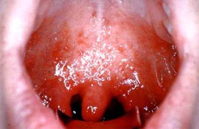
Sore throats can occur with complications, such as paratonsillitis, para- and retropharyngeal abscesses.
Peritonsillitis is an inflammation of the peri-tonsillary tissue, manifested in a strong increase in temperature to 39-40 ° C, the inability to eat and swallow saliva due to very severe pain, “coma in the throat,” suffocation; trismus is also characteristic - a symptom in which a person cannot fully open his mouth due to a tonic spasm of the masticatory muscles. In the oral cavity, in the projection of the tonsil, a large bulge is detected.
Parapharyngeal abscess is suppuration of parapharyngeal tissue, and repharyngeal abscess is retropharyngeal. Their symptoms are in many ways similar to paratonsillitis (except for the characteristic bulging), the differential diagnosis should be carried out by an ENT doctor.
Tonsil hypertrophy
This term refers to the proliferation of lymphadenoid tissue. Most often, hypertrophic processes occur in the palatine and pharyngeal tonsils.
Enlarged tissues can make breathing difficult, cause suffocation, interfere with diction and food intake, and cause a feeling of a “lump” in the throat.
Children with this disease sleep poorly, cough at night, and some may develop neuropsychiatric disorders because of this.
Chronic inflammatory diseases of the pharynx
These include chronic forms of pharyngitis and tonsillitis.
Chronic pharyngitis– inflammation of the pharyngeal mucosa – occurs due to insufficiently effective treatment of acute forms. There are catarrhal, hypertrophic (lateral and granulosa) and atrophic forms.

Patients complain of rawness, soreness, tickling, a “lump” in the throat, suffocation, foreign body sensation, and blocked ears.
The temperature may not rise. They often need a sip of water to swallow something.
Chronic tonsillitis– persistent infectious-allergic disease with local manifestations in the form of inflammation of the tonsils. Most often it occurs as a complication of other infectious processes (such as tonsillitis and caries).
The simple form is characterized by frequent (1-2 times a year) sore throats with corresponding complaints: pain, “lump in the throat,” coughing, fever.
In the toxic-allergic form, symptoms of intoxication and allergization are added to sore throats; associated diseases such as rheumatism, glomerulonephritis, polyarthritis, endocarditis and others are often found.
Foreign bodies, wounds and burns of the throat
Foreign bodies most often enter the throat when talking or laughing while eating, as well as in children while playing. Sometimes foreign bodies in older people are dentures. Patients complain of a lump in the throat, pain and difficulty breathing and swallowing.

Throat wounds can be external and internal, penetrating and non-penetrating, isolated and combined, blind and through.
Symptoms most often include bleeding, breathing problems, speech problems, difficulty swallowing due to a “coma,” suffocation, and severe pain.
Burns can develop due to thermal and chemical damage to the throat wall. Thermal burns are most often caused by exposure to temperature - exposure to hot food and drink, less often - hot air or steam.
Chemical burns occur when exposed to hydrochloric, acetic, nitric acids, caustic soda or potassium.
Burns can be of three degrees - from the first, the mildest, accompanied by redness of the mucous membrane, to the third, with necrosis of the deep layers of tissue.
Burns are most often accompanied by pain, drooling, and general intoxication. Due to numerous complications, throat burns are a life-threatening condition.
Treatment
Treatment of acute pharyngitis is usually carried out on an outpatient basis, it is prescribed by a therapist or ENT doctor. It includes rinses with antiseptics (chlorophyllipt, chamomile infusion), aerosols (Polydex), desensitizing and immunostimulating drugs. Antibiotics are rarely prescribed.

Banal sore throats are usually treated on an outpatient basis by an ENT doctor, in severe cases - in a hospital.
Antibiotics from the penicillin group, antihistamines (Tavegit, Telfast), Bioparox inhalations, rinses and non-steroidal anti-inflammatory drugs are prescribed.
Treatment of infectious diseases and blood diseases accompanied by tonsillitis should be carried out not by an ENT specialist, but by an infectious disease specialist or hematologist in the appropriate hospitals.
Important to remember! Any suspicion of diphtheria is an undeniable indication for examination and, possibly, hospitalization, since diphtheria is a very dangerous disease.
For Simanovsky-Plaut-Vincent angina, antibiotic therapy with penicillin preparations, restorative and vitamin therapy are carried out; sanitize the oral cavity and cleanse the tonsils from necrotic lesions.

The tactics for managing paratonsillitis and other abscesses include antibiotic therapy and mandatory surgical intervention to sanitize purulent foci.
Chronic pharyngitis is treated on an outpatient basis by excluding exposure to harmful factors (alcohol, smoking), inhalations, lubricating the throat with Collargol (done by an ENT doctor), and sucking caramels with antiseptics (Hexalize, Faringosept). In the treatment of chronic pharyngitis, both conservative and surgical methods are used. The first involves washing the lacunae of the tonsils (10-15 procedures), lubricating their surface with iodinol or collargol, rinsing and physiotherapeutic procedures (UHF or microwave therapy).
Surgical methods include tonsillectomy. A similar, but less radical method - tonsillitis - or adenotomy, respectively, treat hypertrophy of the palatine and lingual tonsils.

Foreign bodies are removed by an ENT doctor using special forceps or loops. You should not remove the foreign body yourself using tweezers, as you can aggravate the process and cause asphyxia.
Surgical treatment of wounds is also carried out by an ENT specialist if the necessary tools and equipment are available, most often in a hospital setting.
Treatment of throat burns is a difficult and multi-stage process, involving both ENT specialists and other specialists. At first, all measures are usually aimed at preserving the patient’s life, then at preventing the formation of adhesions.
In the acute period, anti-shock and detoxification measures are carried out, the fight against respiratory disorders, hemostasis and antibiotic therapy are carried out.
In the long-term period, the most common procedure is bougienage - expanding the lumen of the throat to restore its patency.
Prevention
Throat diseases are varied, so their prevention is also different. You should avoid traumatic situations, monitor the food and drinks you consume, and do not talk while eating.
You should also treat all acute diseases in a timely manner, and in no case leave the process untreated.
Activation of natural immunity, for example, with the help of the drug Immunity, will also have a positive effect.
It helps cope with viral and bacterial infections in just two days, promotes immune activation and removes toxins from the body, reducing recovery time.
Chronic nonspecific inflammation of the pharynx is a common disease. Various unfavorable professional and everyday factors that cause the occurrence of acute inflammation of the pharynx and upper respiratory tract, with repeated exposure, lead to the development of chronic inflammation. In some cases, the cause of the disease may be metabolic diseases, diseases of the gastrointestinal tract, liver, diseases of the hematopoietic organs, etc.
3.6.1. Chronic pharyngitis
Chronic pharyngitis(pharyngitis chronica)- chronic inflammation of the pharyngeal mucosa, developing as a consequence of acute inflammation with inadequate treatment and unresolved etiological factors. There are chronic catarrhal, hypertrophic (lateral and granulosa) and atrophic pharyngitis.
Etiology. The occurrence of chronic pharyngitis in most cases is caused by local long-term irritation of the pharyngeal mucosa. Contributing to the occurrence of chronic pharyngitis are repeated acute inflammations of the pharynx, inflammation of the tonsils, nose and paranasal sinuses, prolonged disturbance of nasal breathing, unfavorable
pleasant climatic and environmental factors, smoking, etc. In some cases, the cause of the disease may be diseases of the gastrointestinal tract, endocrine and hormonal disorders, dental caries, alcohol consumption, acute irritants and excessively hot or cold foods. Finally, chronic pharyngitis can also occur with a number of chronic infectious diseases, such as tuberculosis.
Pathomorphology. The hypertrophic form of pharyngitis is characterized by thickening of all layers of the mucous membrane and an increase in the number of rows of epithelium. The mucous membrane becomes thicker and denser, blood and lymphatic vessels are dilated, and lymphocytes are detected in the perivascular space. Lymphoid formations, normally scattered throughout the mucous membrane in the form of barely noticeable granules, significantly thicken and expand, often due to the fusion of adjacent granules; hypersecretion is noted, the mucous membrane is hyperemic. The hypertrophic process may primarily relate to the mucous membrane of the posterior pharyngeal wall - granulosa pharyngitis, or to its lateral parts - lateral hypertrophic pharyngitis.
Atrophic chronic pharyngitis is characterized by sharp thinning and dryness of the pharyngeal mucosa; in pronounced cases it is shiny, “varnished”. The size of the mucous glands and their number are reduced. Desquamation of the epithelial cover is observed.
With catarrhal pharyngitis, persistent diffuse venous hyperemia is detected, the mucous membrane is pasty due to the expansion and stasis of small-caliber veins, and perivascular cellular infiltration is observed.
Clinic. Catarrhal and hypertrophic forms of inflammation are characterized by a feeling of rawness, soreness, tickling, awkwardness in the throat when swallowing, the sensation of a foreign body that does not interfere with food intake, but forces frequent swallowing movements. With hypertrophic pharyngitis, all these phenomena are more pronounced than with the catarrhal form of the disease. Sometimes there are complaints of stuffy ears, which disappears after several swallowing movements.
The main complaints with atrophic pharyngitis are a feeling of dryness in the throat, often difficulty swallowing, especially with the so-called empty throat, and often bad breath. Patients often have a desire to take a sip of water, especially during prolonged exercise.
It should be noted that the patient’s complaints do not always correspond to the severity of the process: in some cases, with minor pa-ogic changes and even in the apparent absence of them
A number of unpleasant side effects arise that force the patient to undergo long-term and persistent treatment, while for others, on the contrary, severe changes go almost unnoticed.
Pharyngoscopically The catarrhal process is characterized by hyperemia, some swelling and thickening of the mucous membrane of the pharynx, in places the surface of the posterior wall is covered with transparent or cloudy mucus.
Granular pharyngitis is characterized by the presence of granules on the back wall of the pharynx - semicircular elevations the size of millet grains of a dark red color, located against the background of a hyperemic mucous membrane, superficial branching veins. Lateral pharyngitis appears in the form of cords of varying thickness located behind the palatine arches.
The atrophic process is characterized by thinning, dryness of the mucous membrane, which has a pale pink color with a dull tint, covered in places with crusts and viscous mucus.
Outpatient treatment is aimed primarily at eliminating local and general causes of the disease, such as chronic purulent process in the nasal cavity and paranasal sinuses, tonsils, etc. It is necessary to exclude exposure to possible irritating factors - smoking, dust and air pollution, irritating food, etc.; carry out appropriate treatment of common chronic diseases that contribute to the development of pharyngitis. Sanitation of the oral cavity is of no small importance.
The most effective local therapeutic effect is on the mucous membrane of the pharynx in order to cleanse it of mucus and crusts.
For hypertrophic forms, rinse with warm isotonic or 1% sodium chloride solution. The same solution can be used for inhalation and pharyngeal spraying. Reduces swelling of the mucous membrane by lubricating the back wall of the pharynx with a 3-5% solution of silver nitrate, 3-5% solution of protargol or collargol; rinsing with an infusion of sage, celandine, bicarmint, hexorol, miramistin, octenisept can be recommended. A positive effect is achieved by the use of antiseptics in the form of caramels for resorption in the mouth, which have a bacteriostatic effect - faringo-sept, hexalize. Large granules can be effectively removed using cryotherapy, cauterization with a concentrated 30-40% solution of silver nitrate, or vagotil.
Treatment of atrophic rhinitis includes daily removal of mucopurulent discharge and crusts from the nasal cavity. It is better to do this with an isotonic or 1% sodium chloride solution with the addition of 4-5 drops of a 5% alcohol solution of iodine per 200 ml of liquid, a Rotocan solution. Systematic and long-term irrigation of the pharynx with these solutions relieves irritation of the mucous membrane and reduces the severity of symptoms of pharyngitis. Courses are held periodically


