What forms the bronchi of the lung. Anatomy of the human lungs. General patterns of the structure of the bronchial tree
Breathing is one of the main functions that ensure human life. Without water, life will last several days, without food - up to several weeks. If there is no breathing for more than 5 minutes, brain damage from oxygen starvation are irreversible, and with further lack of air access, death occurs. That is why it is necessary to know the structure of the respiratory organs, the functions of the human bronchi, take care of their health and promptly seek help for any ailments.
What do the bronchi look like?
The respiratory system consists of several sections and organs. The mouth, nose, and nasopharynx are involved in saturating the body with oxygen—this is called the upper respiratory tract. Next are the lower respiratory tract, which includes the larynx, trachea, bronchial tree and the lungs themselves.
The bronchi and the bronchial tree are one and the same. This organ received this name due to its appearance and structure. Smaller and smaller “branches” extend from the central trunks; the ends of the branches approach the alveoli. With the help of bronchoscopy, you can view the bronchi from the inside. The picture of the mucous membrane shows that they gray, cartilaginous rings are also clearly visible.
The division of the bronchi, left and right, is explained by the fact that their structure clearly corresponds to the size of the lung. The right one is wider, in accordance with the lung, it has about 7 cartilaginous rings. It is located almost vertically, continuing the trachea. The left bronchus is narrower. It contains 9-12 rings of cartilage tissue.
Where are the bronchi located?
The bronchial tree cannot be seen with the naked eye. It is hidden in the chest. The left and right bronchi begin at the point where the trachea branches into two trunks. This is the 5-6 thoracic vertebra, if we talk about the approximate level. Next "branches" bronchial tree penetrate and branch, forming a whole tree.
The bronchi themselves conduct air to the alveoli, each to its own lung. Human anatomy suggests asymmetry; accordingly, the left and right bronchi are also of different sizes.
 The bronchial tree has a branched structure. It consists of several departments:
- Bronchi of the first order. This is the largest part of the organ and has the most rigid structure. The length of the right one is 2-3 cm, the left one is about 5 cm.
- Zonal extrapulmonary - depart from the bronchi of the first order. There are 11 of them on the right, 10 on the left.
- Intrapulmonary subsegmental areas. They are noticeably narrower than the bronchi of the first order, their diameter is 2-5 mm.
- The lobar bronchi are thin tubes, about 1 mm in diameter.
- Respiratory broncheoles are the endings of the “branches” of the bronchial tree.
The branching ends at the broncheoli, because they are connected directly to the alveoli, the final components of the pulmonary parenchyma. Through them, the blood in the capillaries is saturated with oxygen and begins to move throughout the body.
The tissue itself that makes up the bronchial tree consists of several layers. Structural features - the closer to the alveoli, the softer the walls of the bronchial tree.
- Mucous membrane - lines the bronchial tree from the inside. On the surface there is ciliated epithelium. Its structure is not uniform; the mucosa contains different cells: goblet cells secrete mucus, neuroendocrine cells secrete serotonin, and basal and intermediate cells restore mucous membranes.
- Fibromuscular - acts as a kind of framework for the lungs. It is formed by cartilaginous rings connected by fibrous tissue.
- Adventitia - the outer membrane of the bronchi, consists of loose connective tissue.
From thoracic aorta The bronchial arteries are separated, and it is they that provide nutrition to the bronchial tree. In addition, the structure of the human bronchi includes a network of lymph nodes and nerves.
Functions of the bronchi
The importance of the bronchi cannot be overestimated. At first glance, the only thing they do is carry oxygen to the alveoli from the trachea. But the functions of the bronchi are much broader:
- The air passing through the bronchial tree is automatically cleansed of bacteria and tiny dust particles. The cilia of the mucous membrane trap everything unnecessary.
- The bronchi are able to cleanse the air of some toxic impurities.
- When dust enters the bronchial system or mucus forms, the cartilaginous framework begins to contract, and the cilia remove harmful substances from the lungs by coughing.
- The lymph nodes of the bronchial tree are of no small importance in immune system person.
- Thanks to the bronchi, warm air that has reached the required level of humidity enters the alveoli.
Thanks to all these functions, the body receives pure oxygen, vital for the functioning of all systems and organs.
Diseases that affect the bronchi
Diseases of the bronchi are necessarily accompanied by a narrowing of the lumen, increased secretion mucus and difficulty breathing.
 Asthma is a disease that involves difficulty breathing caused by a reduction in the lumen of the bronchial tube. Usually attacks are provoked by some kind of irritant.
Most common reasons occurrence of asthma:
- Congenital high risk allergies.
- Bad ecology.
- Constant inhalation of dust.
- Viral diseases.
- Disturbances in the endocrine apparatus of the body.
- Eating chemical fertilizers along with fruits and vegetables.
Sometimes a predisposition to asthmatic reactions is inherited. Sick man suffering frequent attacks suffocation, accompanied by a painful cough, clear slime, actively released during an attack. Some people note that repeated sneezing sometimes occurs before asthma attacks.
First aid for a patient is the use of an aerosol prescribed by a doctor. This measure will help restore normal breathing or at least relieve it until the ambulance arrives.
Asthma is a serious disease that requires mandatory consultation with a doctor who will conduct an examination, prescribe tests and, based on their results, prescribe treatment. Seizures that are not stopped can lead to complete closure of the lumen of the bronchi and suffocation.
Bronchitis
Bronchitis affects the bronchial mucosa. It becomes inflamed, the lumen of the bronchiole narrows, and a lot of mucus is released. The patient is tormented by a suffocating cough, which is dry at first, then becomes moist, less harsh, and sputum comes out. There are 2 stages:
- Acute – bronchitis is accompanied by high fever, most often caused by viruses and bacteria. An increase in temperature is observed. This condition lasts for several days. With proper treatment acute form passes with virtually no consequences.
- Chronic – caused not only by viruses, but also by smoking, an allergic reaction, and working in harmful conditions. Usually high temperature is not observed, but this type of bronchitis causes irreversible consequences. Other organs suffer.
It is very important to treat promptly acute stage Chronic bronchitis is difficult to treat, relapses occur quite often, putting a strain on a person’s heart.
Measures to prevent bronchial diseases
 People of any age, especially children, are susceptible to bronchial diseases. Therefore, it is necessary to take care of their health in advance so that you do not have to purchase and take medications, risking suffering from side effects:
- Immunoprophylaxis is the most important component of bronchitis prevention. Organism with strong immunity is able to cope with bacteria that have entered the bronchi and remove them with mucus, while a weakened person will not be able to fight the infection. Among these measures correct mode day, timely rest, absence of constant overload.
- Decline harmful effects for the lungs – people with harmful conditions workers should wear appropriate respirators and masks, smokers should reduce or eliminate tobacco consumption.
- During the epidemic season, you should not visit entertainment events and shopping centers, as well as other places with big amount of people. If necessary, you need to wear protective medical masks, constantly changing them to fresh ones.
The health of the bronchial tree is the key to proper breathing. Oxygen is vital to the body, so it is important to take care of the respiratory system. If you suspect a disease or worsening breathing, you should immediately consult a doctor.
The word "bronchi" comes from the Greek "bronchos", which literally translates as "breathing tubes". This organ occupies one of the leading roles in the human respiratory system, because through it oxygen-rich air enters the lungs and waste air exits. Without the bronchi, full metabolism would be impossible.
Where are they located?
The human bronchi are paired organ, which is a continuation of the trachea. At approximately the level of the 4th (male) and 5th (female) vertebrae, the trachea divides, forming two tubes directed into the lungs. After entering the lung, they are divided again: the right one into three branches, and the left into two, which corresponds to the lobes of the lung.
The shape and size of the main branches are not the same: the right one is shorter and wider, and the left one is slightly longer, but narrower, which is clearly visible in the photo.

After this, the bronchi divide even more, forming a bronchial tree, in which strictly. It consists of:
- Zonal, or lobar bronchi, which are the largest in size;
- Segmental and subsegmental (medium size);
- Small shares;
- Bronchioles - (the smallest microscopic tubes that pass into the alveoli).
The area of the entire human bronchial tree is colossal, and its cross section is about 11800 cm3.
What are the bronchi needed for?
The bronchi resemble a kind of tubular ventilation system that ensures full human life. This body is responsible for the following functions:
- The movement of air through the respiratory system during inhalation and exhalation. The neuromuscular bronchial apparatus, which regulates the lumen in the bronchus, is responsible for the uniformity of air supply;
- Protective and drainage functions, thanks to which respiratory system small ones are removed foreign bodies that got inside with air, or pathogenic microorganisms. The bronchial epithelium contains goblet cells that secrete mucus, to which foreign bodies stick, and the cilia of the epithelium set this mucus in motion and bring the object out. This process causes in a person;
- Heating of incoming air, which is regulated with the help of muscle fibers of the bronchus, making the lumen narrower or wider. For example, at sub-zero temperatures, the lumen narrows, the air moves through it more slowly, which ensures its normal heating;
- Humidification of the air occurs due to the secretion secreted by the mucous membrane. This prevents drying out of the lungs.

How to keep your bronchi healthy?
The healthy state of the bronchi ensures the full functioning of a person. If there are problems with them, breathing problems occur, which is incompatible with vigorous activity.
To prevent this, you need to follow some rules:
- Refuse bad habits, especially from smoking, which provokes asthma and cancer.
- Eat healthy food. In order for the bronchi to be in excellent condition, it is advisable to consume foods rich in the following vitamins and minerals:
- C (strengthens the walls of blood vessels and is destructive to microbes);
- A (increases the body's resistance);
- E (improves metabolism in the respiratory system);
- Calcium (inhibits inflammatory processes);
- Magnesium (tones the respiratory organs);
- Potassium (improves the functionality of the respiratory system).

You should not overuse coffee, tea, spices, or meat broths, as they provoke the synthesis of histamine, which significantly increases the amount of mucus secreted. Excessive salt consumption worsens bronchial passage.
- Regularly perform simple breathing exercises, which will help:
- While walking, inhale for two steps and exhale for three steps;
- Place your hands with your palms parallel to each other at chest level. Inhale, and as you exhale, you need to close your palms and press firmly;
- Inhale and exhale slowly by raising your arms up, placing them behind your head and spreading them out to the sides;
- Inhale - raise the dumbbells to your shoulders, exhale - lower them;
- Breathe regularly using your abdominal muscles.
- More often found on fresh air and visit the sea coast once a year.
- IN for preventive purposes drink infusion of coltsfoot, which strengthens the body.

What can happen to the bronchi?
- , which are caused by the presence of inflammation of the walls of the bronchial tubes. It can occur due to exposure to viruses, bacteria, allergens, and smoking. Bronchitis can occur in acute or chronic form;
- Bronchial, characterized by attacks of suffocation that occur with a certain frequency. Provoke occurrences bronchial asthma Maybe allergic reaction, polluted air, consumption of products grown using large quantity chemicals, various infections;
- Bronchial tuberculosis, which is accompanied by severe cough with the discharge of large quantities of sputum and difficulty breathing. This disease develops after infections enter the body;
- Bronchial candidiasis, developing with weakened protective functions body. When inflamed, candida produces purulent cavities that provoke severe pain in the chest, bronchospasm, the appearance of a small amount of blood in the sputum;
- , the cause of which in most cases is smoking. Accompanied by illness persistent cough, in which the sputum is colored light pink, hyperthermia, weakness, loss of body weight, and the appearance of swelling.

Prevent the appearance of these serious illnesses, you can take care of your health. This includes a balanced diet, giving up bad habits, timely treatment correctly selected drugs.
The right main bronchus is a continuation of the trachea. Its length is from 28 to 32 mm, the lumen diameter is 12-16 mm. The left main bronchus is 40-50 mm long and has a width from 10 to 13 mm.
Towards the periphery, the main bronchi are dichotomously divided into lobar, segmental, subsegmental and further down to the terminal and respiratory bronchioles. However, there is also a division into 3 branches (trifurcation) or more.
The right main bronchus is divided into the upper lobe and intermediate, and the intermediate - into the middle lobe and lower lobe. The left main bronchus is divided into the upper lobe and lower lobe. Total airway generations are variable. Starting from the main bronchus and ending with the alveolar sacs, the maximum number of generations reaches 23 - 26.

The main bronchi are the bronchi of the first order, the lobar bronchi are the second, the segmental bronchi are the third order, etc.
Bronchi from the 4th to the 13th generation have a diameter of about 2 mm, total number There are 400 such bronchi. In the terminal bronchioles, the diameter ranges from 0.5 to 0.6 mm. The length of the airways from the larynx to the acini is 23-38 cm.

The right and left main bronchi (bronchi principles dexter et sinister) begin from the bifurcation of the trachea at the level of the upper edge of the fifth thoracic vertebra and go to the gates of the right and left lungs, respectively. In the area of the hilum of the lungs, each main bronchus is divided into lobar (second-order bronchi). Above the left main bronchus is the aortic arch, above the right is the azygos vein. The right main bronchus has more vertical position and shorter in length (about 3 cm) than the left main bronchus (4-5 cm in length). The right main bronchus is wider (diameter 1.6 cm) than the left (1.3 cm). The walls of the main bronchi have the same structure as the walls of the trachea. The inside of the walls of the main bronchi are lined with mucous membrane, and the outside is covered with adventitia. The basis of the walls are cartilages that are not closed at the back. The right main bronchus has 6-8 cartilaginous half-rings, the left one has 9-12 cartilages.
Innervation of the trachea and main bronchi: branches of the right and left recurrent laryngeal nerves and sympathetic trunks.
Blood supply: branches of the inferior thyroid, internal thoracic artery, thoracic aorta. Venous drainage occurs in the brachiocephalic veins.


Lymphatic drainage: into the deep cervical lateral (internal jugular) The lymph nodes, pre- and paratracheal, upper and lower tracheobronchial lymph nodes.
Histological structure of the bronchi
Outside, the trachea and large bronchi are covered with a loose connective tissue sheath - adventitia. The outer shell (adventitia) consists of loose connective tissue containing in large bronchi fat cells. Blood vessels pass through it lymphatic vessels and nerves. The adventitia is not clearly demarcated from the peribronchial connective tissue and, together with the latter, provides the possibility of some displacement of the bronchi in relation to the surrounding parts of the lungs.
Further inward are the fibrocartilaginous and partially muscular layers, the submucosal layer and the mucous membrane. In addition to cartilaginous half-rings, the fibrous layer contains a network of elastic fibers. The fibrocartilaginous membrane of the trachea is connected to neighboring organs using loose connective tissue.
The anterior and lateral walls of the trachea and large bronchi are formed by cartilage and annular ligaments located between them. The cartilaginous skeleton of the main bronchi consists of half-rings of hyaline cartilage, which, as the diameter of the bronchi decreases, decreases in size and acquires the character of elastic cartilage. Thus, only large and medium bronchi consist of hyaline cartilage. Cartilages occupy 2/3 of the circumference, the membranous part - 1/3. They form a fibrocartilaginous skeleton, which ensures the preservation of the lumen of the trachea and bronchi.
Muscle bundles are concentrated in the membranous part of the trachea and main bronchi. There is a superficial, or outer, layer, consisting of rare longitudinal fibers, and a deep, or internal, layer, which is a continuous thin shell formed by transverse fibers. Muscle fibers are located not only between the ends of the cartilage, but also enter the interannular spaces of the cartilaginous part of the trachea and, to a greater extent, the main bronchi. Thus, in the trachea, bundles of smooth muscles with a transverse and oblique arrangement are found only in the membranous part, i.e., there is no muscle layer as such. In the main bronchi, sparse groups of smooth muscles are present throughout the entire circumference.
With a decrease in the diameter of the bronchi, the muscle layer becomes more developed, and its fibers run in a somewhat oblique direction. Muscle contraction causes not only a narrowing of the lumen of the bronchi, but also some shortening of them, due to which the bronchi participate in exhalation by reducing the capacity of the airways. Muscle contraction allows you to narrow the lumen of the bronchi by 1/4. When you inhale, the bronchus lengthens and expands. The muscles reach the 2nd order respiratory bronchioles.
Inward from the muscle layer is the submucosal layer, consisting of loose connective tissue. It contains vascular and nervous formations, the submucosal lymphatic network, lymphoid tissue and a significant part of the bronchial glands, which belong to the tubular-acinous type with mixed mucous-serous secretion. They consist of terminal sections and excretory ducts, which open as flask-shaped extensions on the surface of the mucous membrane. The relatively large length of the ducts contributes to the long course of bronchitis during inflammatory processes in the glands. Atrophy of the glands can lead to drying of the mucous membrane and inflammatory changes.
The largest number of large glands is present above the bifurcation of the trachea and in the area of division of the main bronchi into lobar bronchi. U healthy person up to 100 ml of secretion is released per day. It consists of 95% water and 5% equal amount proteins, salts, lipids and inorganic substances. The secretion is dominated by mucins (high molecular weight glycoproteins). To date, there are 14 types of glycoproteins, 8 of which are found in the respiratory system.
Bronchial mucosa
The mucous membrane consists of cover epithelium, basement membrane, lamina propria and lamina muscularis mucosa.
The bronchial epithelium contains high and low basal cells, each of which is attached to the basement membrane. The thickness of the basement membrane ranges from 3.7 to 10.6 µm. The epithelium of the trachea and large bronchi is multirow, cylindrical, ciliated. The thickness of the epithelium at the level of segmental bronchi ranges from 37 to 47 microns. In its composition, there are 4 main types of cells: ciliated, goblet, intermediate and basal. In addition, serous, brush, Clara and Kulchitsky cells are found.
Ciliated cells predominate on the free surface of the epithelial layer (Romanova L.K., 1984). They have an irregular prismatic shape and an oval vesicular nucleus located in the middle part of the cell. The electron optical density of the cytoplasm is low. There are few mitochondria, the endoplasmic granular reticulum is poorly developed. Each cell bears on its surface short microvilli and about 200 ciliated cilia 0.3 µm thick and about 6 µm long. In humans, the density of cilia is 6 µm2.
Spaces are formed between neighboring cells; The cells are connected to each other by finger-shaped outgrowths of the cytoplasm and desmosomes.
The population of ciliated cells is divided into the following groups according to the degree of differentiation of their apical surface:
- Cells in the phase of formation of basal bodies and axonemes. At this time, there are no cilia on the apical surface. During this period, the accumulation of centrioles occurs, which move to the apical surface of the cells, and the formation of basal bodies, from which cilia axonemes begin to form.
- Cells in the phase of moderate ciliogenesis and cilia growth. On the apical surface of such cells appears a small amount of cilia, the length of which is 1/2-2/3 of the length of the cilia of differentiated cells. In this phase, microvilli predominate on the apical surface.
- Cells in the phase of active ciliogenesis and cilia growth. The apical surface of such cells is already almost entirely covered with cilia, the sizes of which correspond to the size of the cilia of cells in the previous phase of ciliogenesis.
- Cells in the phase of completed ciliogenesis and cilia growth. The apical surface of such cells is entirely covered with densely arranged long cilia. Electron diffraction patterns show that the cilia of adjacent cells are oriented in the same direction and curved. This is an expression of mucociliary transport.
All these groups of cells are clearly visible in photographs obtained using light electron microscopy (SEM).
Cilia are attached to basal bodies located in the apical part of the cell. The cilium axoneme is formed by microtubules, of which 9 pairs (doublets) are located along the periphery, and 2 singles (singlets) are located in the center. Doublets and singlets are connected by nexin fibrils. On each of the doublets, on one side there are 2 short “handles” that contain ATPase, which is involved in the release of ATP energy. Thanks to this structure, the cilia rhythmically oscillate with a frequency of 16-17 in the direction of the nasopharynx.
They move the mucous film covering the epithelium at a speed of about 6 mm/min, thereby ensuring continuous drainage function of the bronchus.
Ciliated epithelial cells, according to most researchers, are at the stage of final differentiation and are not capable of dividing by mitosis. According to the modern concept, basal cells are the precursors of intermediate cells that can differentiate into ciliated cells.
Goblet cells, like ciliated cells, reach the free surface of the epithelial layer. In the membranous part of the trachea and large bronchi, the share of ciliated cells accounts for up to 70-80%, and the share of goblet cells - no more than 20-30%. In those places where there are cartilaginous semirings along the perimeter of the trachea and bronchi, zones with different ratios of ciliated and goblet cells are found:
- with a predominance of ciliated cells;
- with an almost equal ratio of ciliated and secretory cells;
- with a predominance of secretory cells;
- with full or almost complete absence ciliated cells (“unciliated”).
Goblet cells are single-celled glands of the merocrine type that secrete a mucous secretion. The shape of the cell and the location of the nucleus depend on the phase of secretion and the filling of the supranuclear part with mucus granules, which merge into larger granules and are characterized by low electron density. Goblet cells have an elongated shape, which, during the accumulation of secretions, takes the form of a glass with a base located on the basement membrane and intimately connected with it. The wide end of the cell protrudes dome-shaped on the free surface and is equipped with microvilli. The cytoplasm is electron-dense, the nucleus is round, the endoplasmic reticulum is of a rough type, well developed.
Goblet cells are unevenly distributed. Scanning electron microscopy revealed that different zones of the epithelial layer contain heterogeneous areas consisting either only of ciliated epithelial cells or only of secretory cells. However, continuous accumulations of goblet cells are relatively few in number. Along the perimeter of a section of the segmental bronchus of a healthy person there are areas where the ratio of ciliated epithelial cells to goblet cells is 4:1-7:1, and in other areas this ratio is 1:1.
The number of goblet cells decreases distally in the bronchi. In the bronchioles, goblet cells are replaced by Clara cells, which are involved in the production of serous components of mucus and alveolar hypophase.
In small bronchi and bronchioles, goblet cells are normally absent, but may appear in pathology.
In 1986, Czech scientists studied the reaction of the epithelium of the airways of rabbits to the oral administration of various mucolytic substances. It turned out that the target cells of mucolytics are goblet cells. After mucus is cleared, goblet cells typically degenerate and are gradually removed from the epithelium. The degree of damage to goblet cells depends on the substance administered: lasolvan has the greatest irritant effect. After administration of broncholysin and bromhexine, massive differentiation of new goblet cells occurs in the epithelium of the airways, resulting in goblet cell hyperplasia.
Basal and intermediate cells are located deep in the epithelial layer and do not reach the free surface. These are the least differentiated cellular forms, due to which the physiological regeneration. The shape of the intermediate cells is elongated, the basal cells are irregularly cubic. Both have a round, DNA-rich nucleus and a small amount of cytoplasm, which has a high density in the basal cells.
Basal cells are capable of giving rise to both ciliated and goblet cells.
Secretory and ciliated cells are combined under the name “mucociliary apparatus”.
The process of mucus moving through the airways of the lungs is called mucociliary clearance. The functional effectiveness of the MCC depends on the frequency and synchronism of the movement of the cilia of the ciliated epithelium, and also, which is very important, on the characteristics and rheological properties mucus, i.e. from the normal secretory ability of goblet cells.
Serous cells are few in number, reach the free surface of the epithelium and are distinguished by small electron-dense granules of protein secretion. The cytoplasm is also electron dense. Mitochondria and rough reticulum are well developed. The nucleus is round, usually located in the middle part of the cell.
Secretory cells, or Clara cells, are most numerous in the small bronchi and bronchioles. They, like serous ones, contain small electron-dense granules, but are distinguished by low electron density of the cytoplasm and the predominance of smooth, endoplasmic reticulum. The rounded nucleus is located in the middle part of the cell. Clara cells are involved in the formation of phospholipids and possibly in the production of surfactant. Under conditions of increased irritation, they apparently can turn into goblet cells.
Brush cells bear microvilli on their free surface, but lack cilia. Their cytoplasm has low electron density, the nucleus is oval and vesicular. In the manual of Ham A. and Cormack D. (1982), they are considered as goblet cells that have released their secretion. They are attributed many functions: absorption, contractile, secretory, chemoreceptor. However, they have practically not been studied in the human airways.
Kulchitsky cells are found throughout the bronchial tree at the base of the epithelial layer, differing from the basal ones in the low electron density of the cytoplasm and the presence of small granules that are detected under electron microscope and under light during impregnation with silver. They are classified as neurosecretory cells of the APUD system.
Under the epithelium there is a basement membrane, which consists of collagenous and non-collagenous glycoproteins; it provides support and attachment of the epithelium, participates in metabolism and immunological reactions. The condition of the basement membrane and underlying connective tissue determines the structure and function of the epithelium. The lamina propria is the layer of loose connective tissue between the basement membrane and the muscle layer. It contains fibroblasts, collagen and elastic fibers. The lamina propria contains blood and lymphatic vessels. The capillaries reach the basement membrane but do not penetrate it.
In the mucous membrane of the trachea and bronchi, mainly in the lamina propria and near the glands, free cells are constantly present in the submucosa, which can penetrate through the epithelium into the lumen. Lymphocytes predominate among them; plasma cells, histiocytes, and mast cells(labrocytes), neutrophilic and eosinophilic leukocytes. The constant presence of lymphoid cells in the bronchial mucosa is designated by the special term “broncho-associated lymphoid tissue” (BALT) and is considered as an immunological protective reaction to antigens penetrating into the respiratory tract with air.
It is important to know!
The etiological factors of acute simple bronchitis are viruses (parainfluenza types I and II, PC viruses, adenoviruses, influenza viruses, cytomegalovirus). Activation and movement of autoflora from the nasopharynx is possible when exposed to physicochemical factors and hypothermia. In most cases, the etiology of acute simple bronchitis is confirmed by viral-bacterial associations, in which viruses that have a tropism for the epithelium of the respiratory tract damage it, reduce the barrier properties of the bronchial wall and create conditions for the development of bacterial inflammatory process.
References
- Lectures on human anatomy and physiology with the basics of pathology – Baryshnikov S.D. 2002
- Atlas of Human Anatomy – Bilich G.L. – Volume 1. 2014
- Anatomy according to Pirogov – V. Shilkin, V. Filimonov – Atlas of human anatomy. 2013
- Atlas of Human Anatomy – P.Tank, Th. Gest – Lippincott Williams & Wilkins 2008
- Atlas of Human Anatomy – Team of authors – Diagrams – Drawings – Photographs 2008
- Fundamentals of medical physiology (second edition) – Alipov N.H. 2013
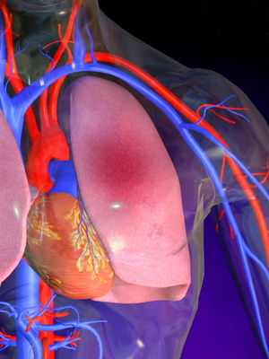
In the building human body Quite interesting is such an “anatomical structure” as the chest, where the bronchi and lungs, the heart and large vessels, as well as some other organs are located. This part of the body, formed by the ribs, sternum, spine and muscles, is designed to reliably protect the organ structures located inside it from external influence. Also, due to the respiratory muscles, the chest provides breathing, in which the lungs play one of the most important roles.
The human lungs, the anatomy of which will be discussed in this article, are very important organs, because it is thanks to them that the breathing process is carried out. They fill everything chest cavity, with the exception of the mediastinum, and are the main ones in the entire respiratory system.
In these organs, the oxygen contained in the air is absorbed by special blood cells (erythrocytes), and carbon dioxide is also released from the blood, which then breaks down into two components - carbon dioxide and water.
Where are the lungs in humans (with photo)
When approaching the question of where the lungs are located, you should first pay attention to one very fun fact regarding these organs: the location of the lungs in humans and their structure are presented in such a way that they very organically combine airways, blood and lymphatic vessels and nerves.
Externally, the anatomical structures considered are quite interesting. In their shape, each of them is similar to a vertically dissected cone, in which one convex and two concave surfaces can be distinguished. The convex one is called costal, due to its direct contact with the ribs. One of the concave surfaces is diaphragmatic (adjacent to the diaphragm), the other is medial, or in other words, median (i.e. located closer to the median longitudinal plane of the body). In addition, interlobar surfaces are also distinguished in these organs.
Using a diaphragm right part the anatomical structure we are considering is separated from the liver, and left side from the spleen, stomach, left kidney and transverse colon. The median surfaces of the organ border with large vessels and heart.
It is worth noting that the place where a person’s lungs are located also affects their shape. If a person has a narrow and long chest, then the lungs are correspondingly elongated and vice versa, these organs have a short and wide appearance with a similar shape chest.
Also in the structure of the described organ there is a base that lies on the dome of the diaphragm (this is the diaphragmatic surface) and an apex that protrudes into the neck area approximately 3-4 cm above the collarbone.
To form a clearer picture of what these anatomical structures look like, as well as to understand where the lungs are, the photo below is perhaps the best visual aid:

Anatomy of the right and left lung
Do not forget that anatomy right lung differs from the anatomy of the left lung. These differences lie primarily in the number of shares. On the right there are three (the bottom one, which is the largest, the top one, slightly smaller, and the smallest of the three - the middle one), while on the left there are only two (top and bottom). In addition, the left lung has a tongue located on its anterior edge, and also this organ, due to the lower position of the left dome of the diaphragm, is slightly longer in length than the right one.
Before entering the lungs, the air first passes through others, at least important departments respiratory tract, in particular the bronchi.
The anatomy of the lungs and bronchi overlaps, so much so that it is difficult to imagine the existence of these organs separately from each other. In particular, each lobe is divided into bronchopulmonary segments, which are sections of the organ, to one degree or another isolated from the same neighboring ones. Each of these areas has segmental bronchus. There are 18 such segments in total: 10 on the right and 8 on the left side of the organ.
The structure of each segment is represented by several lobules - areas within which the lobular bronchus branches. It is believed that a person has about 1,600 lobules in his main respiratory organ: approximately 800 on the right and left.
However, the conjugation of the location of the bronchi and lungs does not end there. The bronchi continue to branch, forming bronchioles of several orders, and they, in turn, give rise to alveolar ducts, dividing from 1 to 4 times and ultimately ending in alveolar sacs, into the lumen of which the alveoli open.


Such branching of the bronchi forms the so-called bronchial tree, otherwise called airways. In addition to them, there is also an alveolar tree.
Anatomy of the blood supply to the lungs in humans

Anatomy connects the blood supply to the lungs with the pulmonary and bronchial vessels. The former, entering the pulmonary circulation, are mainly responsible for the function of gas exchange. The latter, belonging to a large circle, provide nutrition to the lungs.
It is worth noting that the body’s nutrition largely depends on the extent to which the various lung areas are ventilated. This is also influenced by the relationship between blood flow rate and ventilation. A significant role is played by the degree of saturation of the blood with hemoglobin, as well as the rate of passage of gases through the membrane located between the alveoli and capillaries, and some other factors. When even one indicator changes, the physiology of breathing is disrupted, which negatively affects the entire body.
This article has been read 97,894 times.
The bronchi are one of the leading organs of the respiratory system, providing air flow into the acini (respiratory sections) with their moisturizing, warming and cleansing. With their help, a complete metabolism is ensured, air enriched with oxygen enters the lungs with its subsequent removal.
Location of the bronchi and their structure
The bronchi are in upper area chest, which provides their protection.
Location of the bronchiStructure of the bronchi
The internal and external structures of the bronchi are not the same, which is due to different mechanisms of action on their walls. The exoskeleton (outside the lung) consists of half rings of cartilage tissue, which are transformed into ligaments with thin lattice walls at the entrance to the lungs.

The bronchi of an adult, coming from the trachea, are no more than 18 mm in diameter. 2 partial bronchi extend from the main trunk to the left, and 3 to the right. Then they are divided into segments (10 pieces on each side). Their diameter decreases and division into small bronchioles occurs. In this case, the segmental cartilages disintegrate into plates, cartilage tissue they are completely absent. In an adult patient, there are about 23 alveolar ducts and branches.
The structure of the bronchi varies according to their order. As their diameter decreases, the shells soften, losing their cartilage. However, there are common characteristics in the form of 3 shells that form their walls.
- The mucosa is composed of several types of cells responsible for specific functions.
- Goblet - promotes mucus production.
- Intermediate and basal - restore the mucous membrane.
- Neuroendocrine - produce serotonin. On top, the mucosa is covered with several rows of ciliated epithelium.
- The fibromuscular cartilaginous sheath consists of cartilaginous (open) hyaline rings connected by fibrous tissue.
The adventitia consists of unformed, loose connective tissue.
Bronchial diseases
Pathologies of the bronchial system are mainly provoked by violations of their drainage function and patency. The most common violations are:
- bronchiectasis– characterized by dilation of the bronchi, which leads to an inflammatory process, dystrophy and sclerosis of the walls. Quite often, against the background of an inflammatory process, bronchiectasis develops, accompanied by the formation of a purulent process. The main symptom of this disease is a cough with the release of purulent contents. In particular severe cases possible pulmonary hemorrhage;
- Chronical bronchitis– this disease is characterized by the development of an inflammatory process, accompanied by hypertrophy of the mucous membrane and its sclerotic changes. The disease has a long-term, sluggish nature, there is a cough with sputum, as well as a tendency to exacerbations and remissions;
- bronchial asthma– this disease is accompanied by increased mucus secretion and suffocation, mainly at night.
In addition to these diseases, bronchospasm is quite often observed, accompanying chronic bronchitis, asthmatic syndrome and pulmonary emphysema.
Structure of the bronchi and lower respiratory system
The respiratory system refers to the lungs, but the human respiratory system is the upper (nasal cavity, including the paranasal sinuses and larynx) and lower (trachea and bronchial tree) respiratory tract. These components are unique in their functionality, but they are all interconnected and work as a whole.

Trachea
Trachea - The trachea carries air to the lungs. This is a kind of tube; it is formed by 18-20 cartilaginous (incomplete) rings, which are closed at the back by smooth muscle fibers. In the area of the 4th thoracic vertebra, a division occurs into 2 bronchi, which go to the lungs and form a tree, which is the basis of the lungs.
Bronchi
The diameter of the primary bronchi is no more than 2 cm. As they enter the lung, 5 branches are formed corresponding to the pulmonary lobes. Further branching continues, the lumen narrows, and segments are formed (10 on the right and 8 on the left). The inner bronchial surface is composed of mucous membranes with ciliated epithelium.
Bronchioles
Bronchioles are the smallest bronchi with a diameter of no more than 1 mm. They represent the final section of the airway on which the respiratory tract is located. lung tissue formed by alveoli. There are terminal and respiratory bronchioles, which is determined by the location of the branch relative to the edge of the bronchial tree.
Acinus
At the end of the bronchioles there are acini (microscopic pulmonary vesicles that facilitate gas exchange). Quite a lot of acini are present in lung tissue, which ensures the capture of a large area for oxygen supply.
Alveoli
Thanks to the alveoli, the blood is purified and distributes oxygen to organs and tissues, ensuring gas exchange. The alveolar walls are extremely thin. When air enters the alveoli, their walls stretch, and when air leaves the lungs, they collapse. The size of the alveoli is up to 0.3 mm, and their coverage area can be up to 80 square meters. m.
Bronchial walls
The bronchial walls are composed of cartilaginous rings and smooth muscle fibers. This structure provides support for the respiratory organs, the required expansion of the bronchial lumen and the prevention of their collapse. Inside, the walls are lined with mucous membrane, and the blood supply is provided by arteries - short branches that form vascular anastomoses (connections). In addition, they contain many lymph nodes that receive lymph from the lung tissues, which ensures not only the supply of air, but also its purification from harmful components.
Bronchial function
The physiological purpose of the bronchi is the delivery of air to the lungs and its subsequent removal to the outside, cleansing and drainage, due to which the respiratory tract is cleared of dust particles, bacteria and viruses. When small foreign particles enter the bronchi, they are removed by coughing. The air passing through the bronchi acquires the required humidity and temperature.
Prevention of bronchial diseases
To prevent the development of diseases associated with the respiratory system, compliance with preventive measures is required, including proper nutrition, quitting smoking, daily walks at a comfortable temperature.
Dosed doses are useful physical exercise, hardening procedures, breathing exercises, Spa treatment, strengthening the body's defenses and taking vitamin supplements.
All of the above measures help strengthen and optimize the functioning of the respiratory system, thereby providing positive influence for the whole body. To maintain the health of the bronchi, their position, structure, and distribution into segments and parts should be taken into account. Much depends on the timeliness of applying for medical care. As soon as the patient felt the slightest violations from the respiratory system, it is necessary to consult a doctor.


