How does donation work? Basic blood constants
Let us consider in more detail the composition of plasma and cellular elements of blood.
Plasma. After the separation of cellular elements suspended in the blood, an aqueous solution of complex composition remains, called plasma. As a rule, plasma is a clear or slightly opalescent liquid, the yellowish color of which is determined by the presence of small amounts of bile pigment and other colored organic substances.
However, after consumption fatty foods Many fat droplets (chylomicrons) enter the blood, causing the plasma to become cloudy and oily.
Plasma is involved in many vital processes of the body. It transports blood cells nutrients and metabolic products and serves as a link between all extravascular (i.e. located outside the blood vessels) fluids; the latter include, in particular, the intercellular fluid, and through it communication with the cells and their contents occurs. Thus, the plasma comes into contact with the kidneys, liver and other organs and thereby maintains the constant internal environment organism, i.e. homeostasis.
The main components of plasma and their concentrations are given in table. 1. Among the substances dissolved in plasma are low molecular weight organic compounds(urea, uric acid, amino acids, etc.); large and very complex protein molecules; partially ionized inorganic salts. The most important cations (positively charged ions) include sodium (Na +), potassium (K +), calcium (Ca 2+) and magnesium (Mg 2+) cations; The most important anions (negatively charged ions) are chloride anions (Cl –), bicarbonate (HCO 3 –) and phosphate (HPO 4 2– or H 2 PO 4 –). The main protein components of plasma are albumin, globulins and fibrinogen.
Plasma proteins
Of all proteins, albumin, synthesized in the liver, is present in the highest concentration in plasma. It is necessary to maintain osmotic balance, which ensures normal distribution of fluid between blood vessels and the extravascular space. During fasting or insufficient protein intake from food, the albumin content in plasma decreases, which can lead to increased accumulation of water in tissues (edema). This condition, associated with protein deficiency, is called starvation edema.
Plasma contains several types or classes of globulins, the most important of which are designated Greek letters a (alpha), b (beta) and g (gamma), and the corresponding proteins are a 1, a 2, b, g 1 and g 2. After separation of globulins (by electrophoresis), antibodies are detected only in fractions g 1, g 2 and b. Although antibodies are often called gamma globulins, the fact that some of them are also present in the b-fraction led to the introduction of the term “immunoglobulin”. The a- and b-fractions contain many different proteins that ensure the transport of iron, vitamin B12, steroids and other hormones in the blood. This same group of proteins also includes coagulation factors, which, along with fibrinogen, are involved in the process of blood clotting.
The main function of fibrinogen is to form blood clots (thrombi). During the process of blood clotting, whether in vivo (in a living body) or in vitro (outside the body), fibrinogen is converted into fibrin, which forms the basis blood clot; Plasma that does not contain fibrinogen, usually in the form of a clear, pale yellow liquid, is called blood serum.
Red blood cells.
Red blood cells, or erythrocytes, are round discs with a diameter of 7.2–7.9 µm and an average thickness of 2 µm (µm = micron = 1/10 6 m). 1 mm 3 of blood contains 5–6 million red blood cells. They make up 44–48% of the total blood volume.
Red blood cells have the shape of a biconcave disc, i.e. The flat sides of the disk are compressed, making it look like a donut without a hole. Mature red blood cells do not have nuclei. They contain mainly hemoglobin, the concentration of which in the intracellular aqueous medium is approx. 34%. [In terms of dry weight, the hemoglobin content in erythrocytes is 95%; per 100 ml of blood, the hemoglobin content is normally 12–16 g (12–16 g%), and in men it is slightly higher than in women.] In addition to hemoglobin, red blood cells contain dissolved inorganic ions (mainly K +) and various enzymes . The two concave sides provide the red blood cell with optimal surface area through which gases can be exchanged: carbon dioxide and oxygen. Thus, the shape of cells largely determines the efficiency of physiological processes. In humans, the area of surfaces through which gas exchange occurs averages 3820 m2, which is 2000 times the surface of the body.
In the fetus, primitive red blood cells are first formed in the liver, spleen and thymus. From the fifth month of intrauterine development, erythropoiesis gradually begins in the bone marrow - the formation of full-fledged red blood cells. In exceptional circumstances (for example, when normal bone marrow is replaced by cancerous tissue), the adult body can switch back to producing red blood cells in the liver and spleen. However, in normal conditions erythropoiesis in an adult occurs only in flat bones (ribs, sternum, pelvic bones, skull and spine).
Red blood cells develop from precursor cells, the source of which is the so-called. stem cells. In the early stages of red blood cell formation (in cells still in the bone marrow), the cell nucleus is clearly visible. As the cell matures, hemoglobin accumulates, formed during enzymatic reactions. Before entering the bloodstream, the cell loses its nucleus - due to extrusion (squeezing out) or destruction by cellular enzymes. With significant blood loss, red blood cells are formed faster than normal, and in this case, immature forms containing a nucleus may enter the bloodstream; This apparently occurs because the cells leave the bone marrow too quickly. The period of maturation of erythrocytes in the bone marrow - from the moment the youngest cell appears, recognizable as the precursor of an erythrocyte, until its full maturation - is 4-5 days. The lifespan of a mature erythrocyte in peripheral blood is on average 120 days. However, with certain abnormalities of the cells themselves, a number of diseases, or under the influence of certain medications, the lifespan of red blood cells can be shortened.
Most of the red blood cells are destroyed in the liver and spleen; in this case, hemoglobin is released and breaks down into its components heme and globin. Further fate globin was not traced; As for heme, iron ions are released from it (and returned to the bone marrow). Losing iron, heme turns into bilirubin, a red-brown bile pigment. After minor modifications occurring in the liver, bilirubin in bile is excreted through the gallbladder into the digestive tract. Based on the content of the final product of its transformations in feces, the rate of destruction of red blood cells can be calculated. On average, in an adult body, 200 billion red blood cells are destroyed and re-formed every day, which is approximately 0.8% of their total number (25 trillion).
Hemoglobin.
The main function of the red blood cell is to transport oxygen from the lungs to the tissues of the body. A key role in this process is played by hemoglobin, an organic red pigment consisting of heme (a porphyrin compound with iron) and globin protein. Hemoglobin has a high affinity for oxygen, due to which the blood is able to carry much more oxygen than a regular aqueous solution.
The degree of binding of oxygen to hemoglobin depends primarily on the concentration of oxygen dissolved in the plasma. In the lungs, where there is a lot of oxygen, it diffuses from the pulmonary alveoli through the walls of blood vessels and the aqueous medium of the plasma and enters the red blood cells; There it binds to hemoglobin - oxyhemoglobin is formed. In tissues where the oxygen concentration is low, oxygen molecules are separated from hemoglobin and penetrate into the tissue due to diffusion. Insufficiency of red blood cells or hemoglobin leads to a decrease in oxygen transport and thereby to disruption of biological processes in tissues.
In humans, a distinction is made between fetal hemoglobin (type F, from fetus) and adult hemoglobin (type A, from adult). There are many known genetic variants of hemoglobin, the formation of which leads to abnormalities of red blood cells or their function. Among them, the most famous is hemoglobin S, which causes sickle cell anemia.
Leukocytes.
White peripheral blood cells, or leukocytes, are divided into two classes depending on the presence or absence of special granules in their cytoplasm. Cells that do not contain granules (agranulocytes) are lymphocytes and monocytes; their kernels have a predominantly regular round shape. Cells with specific granules (granulocytes) are usually characterized by the presence of irregularly shaped nuclei with many lobes and are therefore called polymorphonuclear leukocytes. They are divided into three types: neutrophils, basophils and eosinophils. They differ from each other in the pattern of granules stained with various dyes.
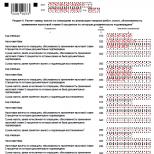
In a healthy person, 1 mm 3 of blood contains from 4,000 to 10,000 leukocytes (on average about 6,000), which is 0.5–1% of blood volume. The ratio of individual types of cells in the composition of leukocytes can vary significantly among different people and even from the same person in different time. Typical values are given in table. 2.
Polymorphonuclear leukocytes (neutrophils, eosinophils and basophils) are formed in the bone marrow from progenitor cells, which give rise to stem cells, probably the same ones that give rise to red blood cell precursors. As the nucleus matures, the cells develop granules that are typical for each cell type. In the bloodstream, these cells move along the walls of the capillaries primarily due to amoeboid movements. Neutrophils are able to leave the internal space of the vessel and accumulate at the site of infection. The lifespan of granulocytes appears to be approx. 10 days, after which they are destroyed in the spleen.
The diameter of neutrophils is 12–14 µm. Most dyes color their core purple; the nucleus of peripheral blood neutrophils can have from one to five lobes. The cytoplasm is stained pinkish; under a microscope, many intense pink granules can be distinguished in it. In women, approximately 1% of neutrophils carry sex chromatin (formed by one of the two X chromosomes), a drumstick-shaped body attached to one of the nuclear lobes. These so-called Barr bodies allow sex to be determined by examining blood samples.
Eosinophils are similar in size to neutrophils. Their nucleus rarely has more than three lobes, and the cytoplasm contains many large granules, which clearly stain bright red with eosin dye.
Unlike eosinophils, basophils have cytoplasmic granules stained blue with basic dyes.
Monocytes. The diameter of these non-granular leukocytes is 15–20 µm. The nucleus is oval or bean-shaped, and only in a small part of the cells is it divided into large lobes that overlap each other. When stained, the cytoplasm is bluish-gray and contains a small number of inclusions that are stained blue-violet with azure dye. Monocytes are formed both in the bone marrow and in the spleen and lymph nodes. Their main function is phagocytosis.
Lymphocytes. These are small mononuclear cells. Most peripheral blood lymphocytes have a diameter of less than 10 µm, but lymphocytes with a larger diameter (16 µm) are sometimes found. The cell nuclei are dense and round, the cytoplasm is bluish in color, with very sparse granules.
Although lymphocytes appear morphologically uniform, they differ clearly in their functions and cell membrane properties. They are divided into three broad categories: B cells, T cells, and O cells (null cells, or neither B nor T).
B lymphocytes mature in the human bone marrow and then migrate to the lymphoid organs. They serve as precursors to cells that form antibodies, the so-called. plasmatic. In order for B cells to transform into plasma cells, the presence of T cells is necessary.
T cell maturation begins in the bone marrow, where prothymocytes are formed, which then migrate to the thymus gland, an organ located in the chest behind the breastbone. There they differentiate into T lymphocytes, a highly heterogeneous population of cells immune system, performing various functions. Thus, they synthesize macrophage activation factors, B-cell growth factors and interferons. Among T cells there are inducer (helper) cells that stimulate the formation of antibodies by B cells. There are also suppressor cells that suppress the functions of B cells and synthesize the growth factor of T cells - interleukin-2 (one of the lymphokines).
O cells differ from B and T cells in that they do not have surface antigens. Some of them serve as “natural killers”, i.e. kill cancer cells and cells infected with a virus. However, the overall role of O cells is unclear.
1. Blood is the internal environment of the body. Blood functions. Composition of human blood. Hematocrit Amount of blood, circulating and deposited blood. Indicators of hematocrit and blood quantity in a newborn.
General properties of blood. Formed elements of blood.
Blood and lymph are the internal environment of the body. Blood and lymph directly surround all cells and tissues and provide vital functions. The entire amount of metabolism occurs between cells and blood. Blood is a type of connective tissue that includes blood plasma (55%) and blood cells or formed elements (45%). The formed elements are represented by - erythrocytes (red blood cells 4.5-5 * 10 in 12 l), leukocytes 4-9 * 10 in 9 l, platelets 180-320 * 10 in 9 l. The peculiarity is that the elements themselves are formed outside - in the hematopoietic organs, and why they enter the blood and live for some time. The destruction of blood cells also occurs outside this tissue. The scientist Lang introduced the concept of the blood system, in which he included the blood itself, the hematopoietic and blood-destructive organs and the apparatus for their regulation.
Features - the intercellular substance in this tissue is liquid. The bulk of the blood is in constant movement, due to which humoral connections are made in the body. The amount of blood is 6-8% of body weight, which corresponds to 4-6 liters. A newborn has more blood. Blood mass occupies 14% of body weight and by the end of the first year it decreases to 11%. Half of the blood is in circulation, the main part is located in the depot and represents deposited blood (spleen, liver, subcutaneous vascular systems, pulmonary vascular systems). Preserving blood is very important for the body. The loss of 1/3 can lead to death, and ½ of the blood is a condition incompatible with life. If blood is centrifuged, the blood is separated into plasma and formed elements. And the ratio of red blood cells to total blood volume is called hematocrit( in men 0.4-0.54 l/l, in women - 0.37-0.47 l/l ) .Sometimes expressed as a percentage.
Blood functions -
- Transport function - transfer of oxygen and carbon dioxide for nutrition. Blood carries antibodies, cofactors, vitamins, hormones, nutrients, water, salts, acids, bases.
- Protective (body's immune response)
- Stopping bleeding (hemostasis)
- Maintaining homeostasis (pH, osmolality, temperature, integrity vascular bed)
- Regulatory function (transport of hormones and other substances that change the activity of the organ)
Blood plasma
Organic
Inorganic
Inorganic substances in plasma- Sodium 135-155 mmol/l, chlorine 98-108 mmol/l, calcium 2.25-2.75 mmol/l, potassium 3.6-5 mmol/l, iron 14-32 µmol/l
2. Physico-chemical properties of blood, their features in children.
Physicochemical properties of blood
- Blood has a red color, which is determined by the content of hemoglobin in the blood.
- Viscosity - 4-5 units relative to the viscosity of water. In newborns, 10-14 due to the larger number of red blood cells, by the 1st year it decreases to an adult.
- Density - 1.052-1.063
- Osmotic pressure 7.6 atm.
- pH - 7.36(7.35-7.47)
The osmotic pressure of the blood is created by minerals and proteins. Moreover, 60% of the osmotic pressure comes from sodium chloride. Blood plasma proteins create an osmotic pressure of 25-40 mm. mercury column (0.02 atm). But despite its small size, it is very important for retaining water inside the vessels. A decrease in protein content in the cut will be accompanied by edema, because... water begins to enter the cell. It was observed during the Great Patriotic War during the famine. The value of osmotic pressure is determined by cryoscopy. Osmotic pressure temperatures are determined. A decrease in the freezing temperature below 0 - depression of the blood and the freezing temperature of the blood - 0.56 C. - osmotic pressure in this case is 7.6 atm. Osmotic pressure is maintained at a constant level. Proper kidney function is very important to maintain osmotic pressure. sweat glands and intestines. Osmotic pressure of solutions that have the same osmotic pressure. Like blood, they are called isotonic solutions. The most common is 0.9% sodium chloride solution, 5.5% glucose solution. Solutions with lower pressure are hypotonic, and higher ones are hypertonic.
Active blood reaction. Blood buffer system
- alkalosis
3. Blood plasma. Blood osmotic pressure.
Blood plasma- liquid opalescent liquid of yellowish color, which consists of 91-92% water, and 8-9% is a dense residue. It contains organic and inorganic substances.
Organic- proteins (7-8% or 60-82 g/l), residual nitrogen- as a result of protein metabolism (urea, uric acid, creatinine, creatine, ammonia) - 15-20 mmol/l. This indicator characterizes the functioning of the kidneys. The growth of this indicator indicates renal failure. Glucose - 3.33-6.1 mmol/l - diabetes mellitus is diagnosed.
Inorganic- salts (cations and anions) - 0.9%
Plasma is a yellowish, slightly opalescent liquid, and is a very complex biological medium, which includes proteins, various salts, carbohydrates, lipids, intermediate metabolic products, hormones, vitamins and dissolved gases. It includes both organic and inorganic substances (up to 9%) and water (91-92%). Blood plasma is in close connection with tissue fluids of the body. It enters the blood from tissues a large number of metabolic products, but, due to the complex activity of various physiological systems of the body, the composition of plasma does not normally undergo significant changes.
The amounts of proteins, glucose, all cations and bicarbonate are kept at a constant level and the slightest fluctuations in their composition lead to severe disorders in the normal functioning of the body. At the same time, the content of substances such as lipids, phosphorus, and urea can vary within significant limits without causing noticeable disorders in the body. The concentration of salts and hydrogen ions in the blood is very precisely regulated.
The composition of blood plasma has some fluctuations depending on age, gender, nutrition, geographical features of the place of residence, time and season of the year.
Functional osmotic pressure regulation system. The osmotic pressure of the blood of mammals and humans normally remains at a relatively constant level (Hamburger’s experiment with the introduction of 7 liters of 5% sodium sulfate solution into the blood of a horse). All this occurs due to the activity of the functional system for regulating osmotic pressure, which is closely linked with functional system regulation of water-salt homeostasis, since it uses the same executive organs.
The walls of blood vessels contain nerve endings that respond to changes in osmotic pressure ( osmoreceptors). Their irritation causes excitation of central regulatory formations in the medulla oblongata and diencephalon. From there, commands come, including certain organs, for example, kidneys, which remove excess water or salts. Of the other executive bodies of the FSOD, it is necessary to name the bodies digestive tract, in which both the removal of excess salts and water and the absorption of products necessary to restore OD occur; skin, the connective tissue of which absorbs excess water when the osmotic pressure decreases or releases it to the latter when the osmotic pressure increases. In the intestine, solutions of mineral substances are absorbed only in such concentrations that contribute to the establishment of normal osmotic pressure and ionic composition of the blood. Therefore, when taking hypertonic solutions ( Epsom salt, sea water) dehydration occurs due to the removal of water into the intestinal lumen. The laxative effect of salts is based on this.
A factor that can change the osmotic pressure of tissues, as well as blood, is metabolism, because body cells consume large-molecular nutrients and release significantly larger number molecules of low molecular weight products of their metabolism. This makes it clear why venous blood flowing from the liver, kidneys, and muscles has a higher osmotic pressure than arterial blood. It is no coincidence that these organs contain greatest number osmoreceptors.
Particularly significant shifts in osmotic pressure in the whole organism are caused by muscular work. During very intense work activities excretory organs may not be sufficient to maintain blood osmotic pressure at a constant level and, as a result, it may increase. The shift in blood osmotic pressure to 1.155% NaCl makes it impossible to further perform work (one of the components of fatigue).
4. Blood plasma proteins. Functions of the main protein fractions. The role of oncotic pressure in the distribution of water between plasma and intercellular fluid. Features of the protein composition of plasma in young children.
Blood plasma proteins are presented in several fractions that can be detected by electrophoresis. Albumin - 35-47 g/l (53-65%), globulins 22.5-32.5 g/l (30-54%), divided into alpha1, alpha 2 (alpha - transport proteins), beta and gamma ( protective bodies) globulins, fibrinogen 2.5 g/l (3%). Fibrinogen is a substrate for blood clotting. A blood clot forms from it. Gamma globulins are produced by plasma cells of lymphoid tissue, the rest in the liver. Plasma proteins take part in the creation of oncotic or colloid-osmotic pressure and are involved in the regulation of water metabolism. Protective function, transport function (transport of hormones, vitamins, fats). Participate in blood clotting. Blood clotting factors are formed by protein components. Possess buffer properties. In diseases, the level of protein in the blood plasma decreases.
The most complete separation of blood plasma proteins is carried out using electrophoresis. On the electropherogram, 6 fractions of plasma proteins can be distinguished:
Albumin. They are contained in the blood 4.5-6.7%, i.e. Albumin accounts for 60-65% of all plasma proteins. They perform mainly a nutritional and plastic function. The transport role of albumins is no less important, since they can bind and transport not only metabolites, but drugs. When there is a large accumulation of fat in the blood, some of it is also bound by albumin. Since albumins have very high osmotic activity, they account for up to 80% of the total colloid-osmotic (oncotic) blood pressure. Therefore, a decrease in the amount of albumin leads to disruption of water exchange between tissues and blood and the appearance of edema. Albumin synthesis occurs in the liver. Their molecular weight is 70-100 thousand, so some of them can pass through the renal barrier and be absorbed back into the blood.
Globulins usually accompany albumin everywhere and are the most abundant of all known proteins. The total amount of globulins in plasma is 2.0-3.5%, i.e. 35-40% of all plasma proteins. By faction, their contents are as follows:
alpha1 globulins - 0.22-0.55 g% (4-5%)
alpha2 globulins - 0.41-0.71g% (7-8%)
beta globulins - 0.51-0.90 g% (9-10%)
gamma globulins - 0.81-1.75 g% (14-15%)
The molecular weight of globulins is 150-190 thousand. The place of formation may vary. Most of it is synthesized in lymphoid and plasma cells of the reticuloendothelial system. Part is in the liver. The physiological role of globulins is diverse. Thus, gamma globulins are carriers of immune bodies. Alpha and beta globulins also have antigenic properties, but their specific function is to participate in coagulation processes (these are plasma coagulation factors). This also includes most of the blood enzymes, as well as transferrin, cerulloplasmin, haptoglobins and other proteins.
Fibrinogen. This protein makes up 0.2-0.4 g%, about 4% of all blood plasma proteins. It is directly related to coagulation, during which it precipitates after polymerization. Plasma devoid of fibrinogen (fibrin) is called blood serum.
In various diseases, especially those leading to disturbances in protein metabolism, sharp changes in the content and fractional composition of plasma proteins are observed. Therefore, the analysis of blood plasma proteins has diagnostic and prognostic significance and helps the doctor judge the degree of organ damage.
5. Blood buffer systems, their significance.
Blood buffer system(pH fluctuation by 0.2-0.4 is very serious stress)
- Bicarbonate (H2CO3 - NaHCO3) 1: 20. Bicarbonates are an alkaline reserve. During the exchange process, many acidic products are formed that need to be neutralized.
- Hemoglobin (reduced hemoglobin (a weaker acid than oxyhemoglobin. The release of oxygen by hemoglobin leads to the fact that reduced hemoglobin binds a hydrogen proton and prevents the reaction from shifting to the acidic side) - oxyhemoglobin, which binds oxygen)
- Protein (plasma proteins are amphoteric compounds and, unlike the medium, can bind hydrogen ions and hydroxyl ions)
- Phosphate (Na2HPO4 (alkaline salt) - NaH2PO4 (acid salt)). Phosphate formation occurs in the kidneys, so the phosphate system works best in the kidneys. The excretion of phosphates in the urine changes depending on the functioning of the kidneys. In the kidneys, ammonia is converted to ammonium NH3 to NH4. Impaired kidney function - acidosis - shift to the acidic side and alkalosis- shift of the reaction to the alkaline side. Accumulation of carbon dioxide during malfunction lungs. Metabolic and respiratory conditions (acidosis, alkalosis), compensated (without a transition to the acidic side) and uncompensated (alkaline reserves are exhausted, a shift in the reaction to the acidic side) (acidosis, alkalosis)
Any buffer system includes a weak acid and a salt formed by a strong base.
NaHCO3 + HСl = NaCl + H2CO3 (H2O and CO2 are removed through the lungs)
6. Red blood cells, their number, physiological role. Age-related fluctuations in the number of red blood cells.
red blood cells- the most numerous formed elements of blood, the content of which differs in men (4.5-6.5 * 10 in 12 l) and women (3.8-5.8). Nuclear-free highly specialized cells. They have the shape of a biconcave disk with a diameter of 7-8 microns and a thickness of 2.4 microns. This shape increases its surface area, increases the stability of the red blood cell membrane, and can fold when passing through capillaries. Red blood cells contain 60-65% water and 35-40% is dry residue. 95% of the dry residue is hemoglobin - a respiratory pigment. The remaining proteins and lipids account for 5%. Of the total mass of the red blood cell, the mass of hemoglobin is 34%. Red blood cell size (volume) is 76-96 femto/l (-15 degree), the average red blood cell volume can be calculated by dividing the hematocrit by the number of red blood cells per liter. The average hemoglobin content is determined by picograms - 27-32 pico/g - 10 in - 12. Outside, the erythrocyte is surrounded by a plasma membrane (a double lipid layer with integral proteins that penetrate this layer and these proteins are represented by glycophorin A, protein 3, ankyrin. C inside membranes - proteins spectrin and actin. These proteins strengthen the membrane). On the outside, the membrane has carbohydrates - polysaccharides (glycolipids and glycoproteins and polysaccharides carry antigens A, B and III). Transport function of integral proteins. There is sodium-potassium atphase, calcium-magnesium atphase. Inside, red blood cells have 20 times more potassium and 20 times less sodium than plasma. The packing density of hemoglobin is high. If the red blood cells in the blood have different sizes, this is called anisocytosis, if the form differs, it is called oikelocytosis. Red blood cells are formed in the red bone marrow and then enter the blood, where they live for an average of 120 days. Metabolism in red blood cells is aimed at maintaining the shape of the red blood cell and maintaining the affinity of hemoglobin for oxygen. 95% of glucose absorbed by red blood cells undergoes anaerobic glycolysis. 5% uses the pentose phosphate pathway. By-product glycolysis is the substance 2,3-diphosphoglycerate (2,3 - DPG). Under conditions of oxygen deficiency, more of this product is formed. When DPG accumulates, oxygen release from oxyhemoglobin is easier.
Functions of red blood cells
- Respiratory (O2, CO2 transport)
- Transfer of amino acids, proteins, carbohydrates, enzymes, cholesterol, prostaglandins, trace elements, leukotrienes
- Antigenic function (antibodies can be produced)
- Regulatory (pH, ionic composition, water exchange, erythropoiesis process)
- Formation of bile pigments (bilirubin)
An increase in red blood cells (physiological erythrocytosis) in the blood will be promoted by physical activity, food intake, and neuropsychic factors. The number of red blood cells increases in mountain residents (7-8 * 10 in 12). For blood diseases - erythrimymia. Anemia - a decrease in the content of red blood cells (due to lack of iron, failure to absorb folic acid (vitamin B12)).
Counting the number of red blood cells in the blood.
Produced in a special counting chamber. Chamber depth 0.1 mm. There is a gap of 0.1 mm under the cover stele and the chamber. On the middle part there is a grid - 225 squares. 16 small squares (side of a small square 1/10 mm, 1/400 - area, volume - 1/4000 mm3)
We dilute the blood 200 times with a 3% sodium chlorine solution. Red blood cells shrink. This diluted blood is fed under a cover glass into a counting chamber. Under a microscope, we count the number in 5 large squares (90 small ones), divided into small ones.
Number of red blood cells = A (number of red blood cells in five large squares) * 4000 * 200/80
7. Hemolysis of erythrocytes, its types. Osmotic resistance of erythrocytes in adults and children.
Destruction of the erythrocyte membrane with the release of hemoglobin into the blood. The blood becomes transparent. Depending on the causes of hemolysis, it is divided into osmotic hemolysis in hypotonic solutions. Hemolysis can be mechanical. When shaking the ampoules, they can be destroyed, thermal, chemical (alkali, gasoline, chloroform), biological (incompatibility of blood groups).
The resistance of erythrocytes to hypotonic solution changes in different diseases.
Maximum osmotic resistance is 0.48-044% NaCl.
Minimum osmotic resistance - 0.28 - 0.34% NaCl
Erythrocyte sedimentation rate. Red blood cells are kept suspended in the blood due to the small difference in density between red blood cells (1.03) and plasma (1.1). The presence of zeta potential on the red blood cell. Red blood cells are found in plasma, as in a colloidal solution. A zeta potential is formed at the boundary between the compact and diffuse layers. This ensures that red blood cells repel each other. Violation of this potential (due to the introduction of protein molecules into this layer) leads to the gluing of red blood cells (coin columns). The radius of the particle increases, and the segmentation speed increases. Continuous blood flow. The sedimentation rate of 1 erythrocyte is 0.2 mm per hour, and in fact in men (3-8 mm per hour), in women (4-12 mm), in newborns (0.5 - 2 mm per hour). The erythrocyte sedimentation rate obeys Stokes' law. Stokes studied the settling speed of particles. The settling rate of particles (V=2/9R in 2 * (g*(density 1 - density 2)/eta (viscosity in poise))) is observed in inflammatory diseases, when many coarse proteins are formed - gamma globulins. They reduce the zeta potential more and promote subsidence.
8. Erythrocyte sedimentation rate (ESR), mechanism, clinical significance. Age-related changes in ESR.
Blood is a stable suspension of small cells in a liquid (plasma). The property of blood as a stable suspension is disrupted when the blood transitions to a static state, which is accompanied by cell sedimentation and is most clearly manifested by erythrocytes. This phenomenon is used to assess the suspension stability of blood when determining the erythrocyte sedimentation rate (ESR).
If the blood is prevented from clotting, the formed elements can be separated from the plasma by simple settling. This is of practical clinical importance, since ESR changes markedly under certain conditions and diseases. Thus, ESR greatly accelerates in women during pregnancy, in patients with tuberculosis, and in inflammatory diseases. When blood stands, red blood cells stick together (agglutinate), forming so-called coin columns, and then conglomerates of coin columns (aggregation), which settle the faster the larger their size.
The aggregation of erythrocytes, their bonding depends on changes in the physical properties of the surface of erythrocytes (possibly with a change in the sign of the total charge of the cell from negative to positive), as well as on the nature of the interaction of erythrocytes with plasma proteins. The suspension properties of blood depend primarily on the protein composition of the plasma: an increase in the content of coarse proteins during inflammation is accompanied by a decrease in suspension stability and an acceleration of ESR. The value of ESR also depends on the quantitative ratio of plasma and erythrocytes. In newborns, ESR is 1-2 mm/hour, in men 4-8 mm/hour, in women 6-10 mm/hour. ESR is determined using the Panchenkov method (see workshop).
Accelerated ESR, caused by changes in plasma proteins, especially during inflammation, also corresponds to increased aggregation of erythrocytes in the capillaries. The predominant aggregation of erythrocytes in capillaries is associated with a physiological slowdown in blood flow in them. It has been proven that under conditions of slow blood flow, an increase in the content of coarse proteins in the blood leads to more pronounced cell aggregation. Red blood cell aggregation, reflecting the dynamic suspension properties of blood, is one of the oldest protective mechanisms. In invertebrates, erythrocyte aggregation plays a leading role in the processes of hemostasis; during an inflammatory reaction, this leads to the development of stasis (stopping blood flow in the border areas), helping to delineate the source of inflammation.
Recently, it has been proven that what matters in ESR is not so much the charge of erythrocytes, but the nature of its interaction with the hydrophobic complexes of the protein molecule. The theory of neutralization of the charge of erythrocytes by proteins has not been proven.
9. Hemoglobin, its types in the fetus and newborn. Compounds of hemoglobin with various gases. Spectral analysis of hemoglobin compounds.
Oxygen transfer. Hemoglobin attaches oxygen at high partial pressure (in the lungs). There are 4 hemes in the hemoglobin molecule, each of which can attach an oxygen molecule. Oxygenation is the addition of oxygen to hemoglobin, because There is no process of changing the valence of iron. In tissues where the partial pressure is low, hemoglobin releases oxygen - deoxykination. The combination of hemoglobin and oxygen is called oxyhemoglobin. The oxygenation process occurs in stages.
During oxygenation, the process of oxygen addition increases.
Cooperative effect - oxygen molecules at the end join 500 times faster. 1 g of hemoglobin adds 1.34 ml of O2.
100% blood saturation with hemoglobin - maximum percentage (volume) saturation
20ml per 100ml of blood. In fact, hemoglobin is saturated by 96-98%.
The addition of oxygen also depends on pH, on the amount of CO2, 2,3-diphosphoglycerate (a product of incomplete oxidation of glucose). As it accumulates, hemoglobin begins to release oxygen more easily.
Methemoglobin, in which iron becomes trivalent (under the action of strong oxidizing agents - potassium ferricyanide, nitrates, berthollet salt, phenacytin) It cannot give off oxygen. Methemoglobin is capable of binding hydrocyanic acid and other bonds, therefore, in case of poisoning with these substances, methemoglobin is injected into the body.
Carboxyhemoglobin (a compound of Hb with CO) carbon monoxide joins iron in hemoglobin, but the affinity of hemoglobin for carbon monoxide is 300 times higher than for oxygen. If there is more than 0.1% carbon monoxide in the air, then hemoglobin binds with carbon monoxide. 60% is due to carbon monoxide (death). Carbon monoxide is found in exhaust gases, in stoves, and is formed when smoking.
Help for victims - carbon monoxide poisoning begins unnoticed. The person himself cannot move; it is necessary to remove him from this room and provide breathing, preferably with a gas cylinder with 95% oxygen and 5% carbon dioxide. Hemoglobin can join carbon dioxide- carbhemoglobin. The connection occurs with the protein part. The acceptor is the amine parts (NH2) - R-NH2+CO2=RNHCOOH.
This compound is capable of removing carbon dioxide. The combination of hemoglobin with different gases has different absorption spectra. Reduced hemoglobin has one broad band in the yellow-green part of the spectrum. Oxyhemoglobin produces 2 bands in the yellow-green part of the spectrum. Methemoglobin has 4 bands - 2 yellow-green, red and blue. Carboxyhemoglobin has 2 bands in the yellow-green part of the spectrum, but this compound can be distinguished from oxyhemoglobin by the addition of a reducing agent. Since carboxyhemoglobin is a strong compound, adding a reducing agent does not add streaks.
Hemoglobin has an important function in maintaining normal pH levels. When releasing oxygen in tissues, hemoglobin attaches a proton. In the lungs, a hydrogen proton is given up to form carbonic acid. When hemoglobin is exposed to strong acids or alkalis, compounds with a crystalline form are formed and these compounds are the basis for confirming blood. Hemins, hemochromogens. Glycine and succinic acid. Globin is formed from amino acids through protein synthesis. In red blood cells that complete their life cycle hemoglobin also breaks down. In this case, the heme is separated from the protein part. Iron is calved from heme, and bile pigments are formed from heme residues (for example, bilirubin, which will then be captured by liver cells). Inside hepatocytes, hemoglobin combines with glucuronic acid. Bilirubin gyukuronite is excreted into the bile capillaries. It enters the intestine with the bile, where it undergoes oxidation, where it turns into urabillin, which is absorbed into the blood. Some remains in the intestines and is excreted in the feces (their color is stercobillin). Urrabillin colors the urine and is taken up again by the liver cells.
The content of hemoglobin in erythrocytes is judged by the so-called color index, or farb index (Fi, from farb - color, index - indicator) - a relative value characterizing the saturation of an average erythrocyte with hemoglobin. Fi is the percentage ratio of hemoglobin and red blood cells, while 100% (or units) of hemoglobin is conventionally taken to be 166.7 g/l, and 100% of red blood cells is 5*10 /l. If a person has a hemoglobin and red blood cell content of 100%, then the color index is 1. Normally, Fi ranges from 0.75-1.0 and very rarely can reach 1.1. In this case, the red blood cells are called normochromic. If Fi is less than 0.7, then such red blood cells are undersaturated with hemoglobin and are called hypochromic. When Fi is more than 1.1, red blood cells are called hyperchromic. In this case, the volume of the red blood cell increases significantly, which allows it to contain a higher concentration of hemoglobin. As a result, a false impression is created that the red blood cells are oversaturated with hemoglobin. Hypo- and hyperchromia occur only in anemia. Determining the color index is important for clinical practice, as it allows for a differential diagnosis for anemia of various etiologies.
10. Leukocytes, their number and physiological role.
White blood cells. These are nuclear cells without a polysaccharide shell
Dimensions - 9-16 microns
Normal quantity - 4-9 * 10 in 9l
Formation occurs in the red bone marrow, lymph nodes, and spleen.
Leukocytosis - increase in the number of white blood cells
Leukopenia - decrease in the number of leukocytes
Leukocyte count=B*4000*20/400. They count on Goryaev's grid. Blood is diluted with a 5% solution acetic acid tinted with methylene blue, diluted 20 times. In an acidic environment, hemolysis occurs. Next, the diluted blood is placed in a counting chamber. Count the number in 25 large squares. Counting can be done in undivided and divided squares. The total number of white blood cells counted will correspond to 400 small ones. Let's find out how many leukocytes there are on average per small square. Convert to cubic millimeters (multiply by 4000). We take into account the dilution of blood by 20 times. In newborns, the amount on the first day is increased (10-12*10 in 9 l). By the age of 5-6 years it reaches the level of an adult. An increase in leukocytes is caused by physical activity, food intake, pain, and stressful situations. The amount increases during pregnancy and during cooling. This is a physiological leukocytosis associated with the release of more leukocytes into the circulation. These are redistributive reactions. Daily fluctuations - in the morning there are fewer leukocytes, in the evening - more. In infectious inflammatory diseases, the number of leukocytes increases due to their participation in protective reactions. The number of white blood cells may increase in leukemia (leukemia)
General properties of leukocytes
- Independent mobility (formation of pseudopodia)
- Chemotaxis (approach to a focus with a changed chemical composition)
- Phagocytosis (absorption of foreign substances)
- Diapedesis - the ability to penetrate the vascular wall
11. Leukocyte formula, its clinical significance. B- and T-lymphocytes, their role.
Leukocyte formula
- Granulocytes
A. Neutrophils 47-72% (segmented (45-65%), band (1-4%), young (0-1%))
B. Eosinophils (1-5%)
B. Basophils (0-1%)
- Agranulocytes (no granularity)
A. Lymphocytes (20-40%)
B. Monocytes (3-11%)
The percentage of different forms of leukocytes is the leukocyte formula. Counting on a blood smear. Staining according to Romanovsky. Of 100 leukocytes, how many will be of these varieties. In the leukocyte formula, there is a shift to the left (an increase in young forms of leukocytes) and to the right (the disappearance of young forms and the predominance of segmented forms). A shift to the right characterizes the inhibition of the function of the red bone marrow, when new cells are not formed, but only mature forms are present. No longer favorable. Features of the functions of individual forms. All granulocytes have high cell membrane lability, adhesive properties, chemotaxis, phagocytosis, and free movement.
Neutrophil granulocytes are formed in the red bone marrow and live in the blood for 5-10 hours. Neutrophils contain lysosamal, peroxidase, hydrolytic, Nad-oxidase. These cells are our non-specific protectors from bacteria, viruses, and foreign particles. Their number at infection age. The source of infection is approached using chemotaxis. They are capable of capturing bacteria by phagocytosis. Phagocytosis was discovered by Mechnikov. Absonins, substances that enhance phagocytosis. Immune complexes, C-reactive protein, aggregated proteins, fibronectins. These substances coat foreign agents and make them “tasty” to leukocytes. Upon contact with a foreign object - protrusion. This bubble then separates. Then inside, it fuses with lysosomes. Further, under the influence of enzymes (peroxidase, adoxidase), neutralization occurs. Enzymes break down the foreign agent, but the neutrophils themselves die.
Eosinophils. They phagocytose histamine and destroy it with the enzyme histaminase. Contains a protein that destroys heparin. These cells are necessary to neutralize toxins and capture immune complexes. Eosinophils destroy histamine during allergic reactions.
Basophils - contain heparin (anti-clotting effect) and histamine (dilate blood vessels). Mast cells, which contain receptors for immunoglobulins E on their surface. Active substances are derivatives of arachidonic acid - platelet activation factors, thromboxanes, leukotrienes, prostaglandins. The number of basophils increases in the final stage of the inflammatory reaction (in this case, basophils dilate blood vessels, and heparin facilitates the resorption of the inflammatory focus).
Agranulocytes. Lymphocytes are divided into -
- 0-lymphocytes(10-20%)
- T-lymphocytes (40-70%). Development is completed in the thymus. Formed in the red bone marrow
- B lymphocytes (20%). Place of formation - red bone marrow. The final stage of this group of lymphocytes occurs in the lymphoepithelial cells along the small intestine. In birds, they complete development in a special bursa in the stomach.
12. Age-related changes in the child’s leukocyte formula. The first and second “crossovers” of neutrophils and lymphocytes.
The leukocyte formula, like the number of leukocytes, undergoes significant changes during the first years of a person’s life. If in the first hours a predominance of granulocytes is noted in a newborn, then by the end of the first week after birth the number of granulocytes decreases significantly and their bulk consists of lymphocytes and monocytes. Starting from the second year of life, there is a gradual increase in the relative and absolute number of granulocytes and a decrease in mononuclear cells, mainly lymphocytes. The intersection points of the agranulocyte and granulocyte curves are 5 months and 5 years. In persons aged 14-15 years, the leukocyte formula is practically no different from that of adults.
When assessing leukograms, great importance should be attached not only to the percentage of leukocytes, but also to their absolute values (“leukocyte profile” according to Moshkovsky). It is understandable that a decrease in the absolute number of certain types of leukocytes leads to an apparent increase in the relative number of other forms of leukocytes. Therefore, only the determination of absolute values can indicate changes that are actually taking place.
13. Platelets, their number, physiological role.
platelets, or blood platelets, are formed from giant cells of the red bone marrow - megakaryocytes. In the bone marrow, megakaryocytes are tightly pressed into the spaces between fibroblasts and endothelial cells, through which their cytoplasm protrudes out and serves as material for the formation of platelets. In the bloodstream, platelets have a round or slightly oval shape, their diameter does not exceed 2-3 microns. The platelet does not have a nucleus, but has a large number of granules (up to 200) of various structures. Upon contact with a surface that differs in its properties from the endothelium, the platelet is activated, spreads out, and up to 10 notches and processes appear, which can be 5-10 times the diameter of the platelet. The presence of these processes is important for stopping bleeding.
Normally, the number of platelets in a healthy person is 2-4-1011 / l, or 200-400 thousand in 1 μl. An increase in the number of platelets is called "thrombocytosis" decrease - "thrombocytopenia". Under natural conditions, the number of platelets is subject to significant fluctuations (their number increases with painful stimulation, physical activity, stress), but rarely goes beyond normal limits. As a rule, thrombocytopenia is a sign of pathology and is observed when radiation sickness, congenital and acquired diseases of the blood system.
The main purpose of platelets is to participate in the process of hemostasis (see section 6.4). An important role in this reaction belongs to the so-called platelet factors, which are concentrated mainly in the granules and platelet membrane. Some of them are designated by the letter P (from the word platelet - plate) and an Arabic numeral (P 1, P 2, etc.). The most important are P 3, or partial (incomplete) thromboplastin, representing a fragment of cell membrane; P 4, or antiheparin factor; P 5, or platelet fibrinogen; ADF; contractile protein thrombastenin (resembling actomyosin), vasoconstrictor factors - serotonin, adrenaline, norepinephrine, etc. Plays a significant role in hemostasis thromboxane A 2 (TxA 2), which is synthesized from arachidonic acid, which is part of cell membranes (including platelets) under the influence of the enzyme thromboxane synthetase.
On the surface of platelets there are glycoprotein formations that perform the functions of receptors. Some of them are “masked” and are expressed after platelet activation by stimulating agents - ADP, adrenaline, collagen, microfibrils, etc.
Platelets take part in protecting the body from foreign agents. They have phagocytic activity, contain IgG, are a source of lysozyme and β -lysines, which can destroy the membrane of some bacteria. In addition, peptide factors were found in their composition that cause the transformation of “zero” lymphocytes (0-lymphocytes) into T- and B-lymphocytes. These compounds are released into the blood during platelet activation and, in the event of vascular injury, protect the body from pathogenic microorganisms.
Regulators of thrombocytopoiesis are short-term and long acting. They are formed in the bone marrow, spleen, liver, and are also part of megakaryocytes and platelets. Short-acting plateletpoietins enhance the detachment of blood platelets from megakaryocytes and accelerate their entry into the blood; long-acting thrombocytopoietins promote the transition of bone marrow giant cell precursors to mature megakaryocytes. The activity of thrombocytopoietins is directly influenced by IL-6 and IL-11.
14. Regulation of erythropoiesis, leukopoiesis and thrombopoiesis. Hemopoietins.
The continuous loss of blood cells requires their replenishment. They are formed from undifferentiated stem cells in the red bone marrow. From which arise the so-called colony-stimulating (CFU), which are the precursors of all hematopoietic lines. Both bi and unipotent cells can arise from them. From them, differentiation and formation of various forms of erythrocytes and leukocytes occurs.
1. Proerythroblast
2. Erythroblast -
Basophilic
Polychromatic
Orthochromatic (loses the nucleus and becomes a reticulocyte)
3. Reticulocyte (contains remnants of RNA and ribosomes, hemoglobin formation continues) 25-65 * 10 * 9 l turn into mature red blood cells in 1-2 days.
4. Erythrocyte - every minute 2.5 million mature red blood cells are formed.
Factors accelerating erythropoiesis
1. Erythropoietins (formed in the kidneys, 10% in the liver). Accelerate the processes of mitosis, stimulate the transition of reticulocytes to mature forms.
2. Hormones - somatotropic, ACTH, androgenic, hormones of the adrenal cortex, inhibit erythropoiesis - estrogens
3. Vitamins - B6, B12 (external factor of hematopoiesis, but absorption occurs if it combines with the internal factor of Castle, which is formed in the stomach), folic acid.
You also need iron. The formation of leukocytes is stimulated by leukopoietin substances, which accelerate the maturation of granulocytes and promote their release from the red bone marrow. These substances are formed during the breakdown of tissue, in areas of inflammation, which enhances the maturation of leukocytes. There are interleukins, which also stimulate the formation of leukzoites. Growth hormone and adrenal hormones cause leukocytosis (increase in the number of hormones). Thymosin is necessary for the maturation of T lymphocytes. The body has 2 reserves of leukocytes - vascular - accumulation along the walls of blood vessels and bone marrow reserve. In pathological conditions, leukocytes are released from the bone marrow (30-50 times more).
15. Blood coagulation and its biological significance. Rate of coagulation in adults and newborns. Blood clotting factors.
If the blood released from the blood vessel is left for some time, then from the liquid it first turns into jelly, and then a more or less dense clot is organized in the blood, which, by contracting, squeezes out a liquid called blood serum. This is plasma devoid of fibrin. The described process is called blood clotting (by hemocoagulation). Its essence lies in the fact that the fibrinogen protein dissolved in plasma under certain conditions becomes insoluble and precipitates in the form of long fibrin filaments. In the cells of these threads, as in a mesh, cells get stuck and the colloidal state of the blood as a whole changes. The significance of this process is that coagulated blood does not flow out of the wounded vessel, preventing the body from dying from blood loss.
Blood coagulation system. Enzymatic theory of coagulation.
The first theory explaining the process of blood clotting by the work of special enzymes was developed in 1902 by the Russian scientist Schmidt. He believed that coagulation occurs in two phases. First, one of the plasma proteins prothrombin under the influence of enzymes released from blood cells destroyed during injury, especially platelets ( thrombokinase) And Ca ions goes into enzyme thrombin. At the second stage, under the influence of the enzyme thrombin, fibrinogen dissolved in the blood is converted into insoluble fibrin, which causes the blood to clot. IN last years life, Schmidt began to distinguish 3 phases in the process of hemocoagulation: 1- formation of thrombokinase, 2- formation of thrombin. 3- formation of fibrin.
Further study of the coagulation mechanisms showed that this representation is very schematic and does not fully reflect the entire process. The main thing is that there is no active thrombokinase in the body, i.e. an enzyme capable of converting prothrombin into thrombin (according to the new nomenclature of enzymes, this should be called prothrombinase). It turned out that the process of prothrombinase formation is very complex; a number of so-called proteins are involved in it. thrombogenic enzyme proteins, or thrombogenic factors, which, interacting in a cascade process, are all necessary for blood clotting to occur normally. In addition, it was discovered that the coagulation process does not end with the formation of fibrin, because its destruction begins at the same time. Thus, the modern blood coagulation scheme is much more complicated than Schmidt’s.
The modern blood coagulation scheme includes 5 phases, successively replacing each other. These phases are as follows:
1. Formation of prothrombinase.
2. Thrombin formation.
3. Fibrin formation.
4. Fibrin polymerization and clot organization.
5. Fibrinolysis.
Over the past 50 years, many substances involved in blood clotting, proteins, the absence of which in the body leads to hemophilia (non-clotting of blood), have been discovered. Having considered all these substances, the international conference of hemocoagulologists decided to designate all plasma coagulation factors in Roman numerals, and cellular coagulation factors in Arabic numerals. This was done in order to eliminate confusion in names. And now in any country, after the generally accepted name of the factor (they can be different), the number of this factor according to the international nomenclature must be indicated. In order that we may consider the folding scheme further, let us first give brief description these factors.
A. Plasma clotting factors .
I. Fibrin and fibrinogen . Fibrin is the end product of the blood clotting reaction. The coagulation of fibrinogen, which is its biological feature, occurs not only under the influence of a specific enzyme - thrombin, but can be caused by the venoms of some snakes, papain and other chemicals. Plasma contains 2-4 g/l. Place of formation: reticuloendothelial system, liver, bone marrow.
II. Thrombin and prothrombin . Only traces of thrombin are normally found in circulating blood. Its molecular weight is half the molecular weight of prothrombin and is equal to 30 thousand. The inactive precursor of thrombin - prothrombin - is always present in the circulating blood. This is a glycoprotein consisting of 18 amino acids. Some researchers believe that prothrombin is a complex compound of thrombin and heparin. Whole blood contains 15-20 mg% prothrombin. This content in excess is enough to convert all fibrinogen in the blood into fibrin.
The level of prothrombin in the blood is a relatively constant value. Of the moments causing vibrations of this level, one should indicate menstruation (increases), acidosis (decreases). Taking 40% alcohol increases the prothrombin content by 65-175% after 0.5-1 hour, which explains the tendency to thrombosis in people who regularly drink alcohol.
In the body, prothrombin is constantly used and synthesized at the same time. Antihemorrhagic vitamin K plays an important role in its formation in the liver. It stimulates the activity of liver cells that synthesize prothrombin.
III.Thromboplastin . This factor is not present in active form in the blood. It is formed when blood cells and tissues are damaged and can be, respectively, blood, tissue, erythrocyte, platelet. Its structure is a phospholipid, similar to the phospholipids of cell membranes. According to thromboplastic activity, tissues of various organs are arranged in descending order: lungs, muscles, heart, kidneys, spleen, brain, liver. Sources of thromboplastin are also human milk and amniotic fluid. Thromboplastin is involved as an essential component in the first phase of blood coagulation.
IV. Ionized calcium, Ca++. The role of calcium in the process of blood clotting was known to Schmidt. It was then that they were offered sodium citrate as a blood preservative - a solution that bound Ca++ ions in the blood and prevented its clotting. Calcium is necessary not only for the conversion of prothrombin to thrombin, but for other intermediate stages of hemostasis, in all phases of coagulation. The content of calcium ions in the blood is 9-12 mg%.
V and VI.Proaccelerin and accelerin (AS-globulin ). Formed in the liver. Participates in the first and second phases of coagulation, while the amount of proaccelerin decreases and accelerin increases. Essentially V is a precursor to factor VI. Activated by thrombin and Ca++. It is an accelerator of many enzymatic coagulation reactions.
VII.Proconvertin and convertin . This factor is a protein found in the beta globulin fraction of normal plasma or serum. Activates tissue prothrombinase. Vitamin K is required for the synthesis of proconvertin in the liver. The enzyme itself becomes active upon contact with damaged tissues.
VIII.Antihemophilic globulin A (AGG-A ). Participates in the formation of blood prothrombinase. Capable of providing clotting of blood that has not had contact with tissues. The absence of this protein in the blood causes the development of genetically determined hemophilia. It has now been obtained in dry form and is used in the clinic for its treatment.
IX.Antihemophilic globulin B (AGG-B, Christmas factor , plasma component of thromboplastin). Participates in the coagulation process as a catalyst, and is also part of the blood thromboplastic complex. Promotes activation of X factor.
X.Koller factor, Steward-Prower factor . The biological role is reduced to participation in the formation of prothrombinase, since it is its main component. When rolled up it is disposed of. Named (like all other factors) after the names of patients in whom a form of hemophilia was first discovered, associated with the absence of the specified factor in their blood.
XI.Rosenthal factor, plasma thromboplastin precursor (PPT) ). Participates as an accelerator in the formation of active prothrombinase. Refers to beta globulins in the blood. Reacts in the first stages of phase 1. Formed in the liver with the participation of vitamin K.
XII.Contact factor, Hageman factor . Plays the role of a trigger in blood clotting. Contact of this globulin with a foreign surface (roughness of the vessel wall, damaged cells, etc.) leads to activation of the factor and initiates the entire chain of coagulation processes. The factor itself is adsorbed on the damaged surface and does not enter the bloodstream, thereby preventing the generalization of the coagulation process. Under the influence of adrenaline (under stress), it is partially able to activate directly in the bloodstream.
XIII.Fibrin stabilizer Lucky-Loranda . Necessary for the formation of terminally insoluble fibrin. This is a transpeptidase that cross-links individual fibrin strands with peptide bonds, promoting its polymerization. Activated by thrombin and Ca++. In addition to plasma, it is found in formed elements and tissues.
The 13 factors described are the generally accepted basic components necessary for normal process blood clotting. Caused by their absence various shapes bleeding disorders belong to different types of hemophilia.
IN. Cellular factors coagulation.
Along with plasma factors, cellular factors released from blood cells also play a primary role in blood coagulation. Most of them are found in platelets, but they are also found in other cells. It’s just that during hemocoagulation, platelets are destroyed in greater quantities than, say, erythrocytes or leukocytes, so platelet factors are of greatest importance in coagulation. These include:
1f.AC platelet globulin . Similar to V-VI blood factors, performs the same functions, accelerating the formation of prothrombinase.
2f.Thrombin accelerator . Accelerates the action of thrombin.
3f.Thromboplastic or phospholipid factor . It is found in granules in an inactive state and can only be used after platelets have been destroyed. Activated upon contact with blood, necessary for the formation of prothrombinase.
4f.Antiheparin factor . Binds heparin and delays its anticoagulant effect.
5f.Platelet fibrinogen . Necessary for the aggregation of blood platelets, their viscous metamorphosis and the consolidation of the platelet plug. Found both inside and outside the platelet. promotes their gluing.
6f.Retractozyme . Provides compaction of the blood clot. Several substances are determined in its composition, for example thrombostenin + ATP + glucose.
7f.Antifibinosilin . Inhibits fibrinolysis.
8f.Serotonin . Vasoconstrictor. Exogenous factor, 90% is synthesized in the gastrointestinal mucosa, the remaining 10% in platelets and the central nervous system. Released from cells when they are destroyed, promotes spasm small vessels, thereby helping to prevent bleeding.
In total, up to 14 factors are found in platelets, such as antithromboplastin, fibrinase, plasminogen activator, AC globulin stabilizer, platelet aggregation factor, etc.
Other blood cells contain mainly these same factors, but normally they do not play a significant role in hemocoagulation.
WITH.Tissue coagulation factors
Participate in all phases. These include active thromboplastic factors like plasma factors III, VII, IX, XII, and XIII. Tissues contain activators of factors V and VI. There is a lot of heparin, especially in the lungs, prostate gland, and kidneys. There are also antiheparin substances. In inflammatory and cancerous diseases, their activity increases. There are many activators (kinins) and inhibitors of fibrinolysis in tissues. The substances contained in the vascular wall are especially important. All these compounds constantly flow from the walls of blood vessels into the blood and regulate coagulation. The tissues also ensure the removal of coagulation products from the vessels.
16. Blood coagulation system, blood coagulation factors (plasma and platelet) Factors that maintain the fluid state of blood.
The function of blood is possible when it is transported through blood vessels. Damage to the blood vessels could cause bleeding. Blood can perform its functions in a liquid state. Blood can form a clot. This will block blood flow and lead to blockage of blood vessels. Causes their necrosis - heart attack, necrosis - consequences of intravascular thrombus. For normal function of the circulatory system, it should have liquid properties, but if damaged, it should clot. Hemostasis is a series of sequential reactions that stop or reduce bleeding. These reactions include -
- Compression and narrowing of damaged vessels
- Lamellar thrombus formation
- Blood coagulation, blood clot formation.
- Thrombus retraction and lysis (dissolution)
The first reaction - compression and narrowing - occurs due to contraction of muscle elements, due to the release chemical substances. Endothelial cells (in capillaries) stick together and close the lumen. In larger cells with smooth muscle elements, depolarization occurs. The tissues themselves can react and compress the vessel. The area around the eyes has very faint elements. They compress the vessel very well during childbirth. Vasoconstriction is caused by serotonin, adrenaline, fibrinopeptide B, thromboxane A2. This primary reaction improves bleeding. Formation of a plate thrombus (associated with the function of platelets) Platelets are non-nuclear elements and have a flat shape. Diameter - 2-4 microns, thickness - 0.6-1.2 microns, volume 6-9 femtol. Quantity 150-400*10 in 9 l. They are formed from megakaryocytes by detachment. Life expectancy is 8-10 days. Electron microscopy of platelets made it possible to establish that these cells have a complex structure, despite their small size. On the outside, the platelet is covered with a thrombotic membrane containing glycoproteins. Glycoproteins form receptors that can interact with each other. The platelet membrane has invaginations that increase the area. These membranes contain tubules for secreting substances from the inside. Phosphomembranes are very important. Lamellar factor from membrane phospholipids. Under the membrane there are dense tubes - the remains of the sarcoplasmic reticulum with calcium. Under the membrane there are also microtubules and filaments of actin and myosin, which maintain the shape of platelets. Inside platelets there are mitochondria and dense dark granules and alpha granules - light. In platelets, there are 2 types of granules containing bodies.
In dense - ADP, serotonin, calcium ions
Light (alpha) - fibrinogen, von Willebrand factor, plasma factor 5, antiheparin factor, lamellar factor, beta-thromboglobulin, thrombospondin and lamellar growth factor.
The plates also have lysosomes and glycogen granules.
When vessels are damaged, the plates take part in aggregation processes and the formation of a plate thrombus. This reaction is due to a number of properties inherent in the plate - When the vessels are damaged, subendothelial proteins are exposed - adhesion (the ability to stick to these proteins due to receptors on the plate. Von Willebranca factor also contributes to adhesion). In addition to the property of adhesion, platelets have the ability to change their shape and - secrete active substances(Thromboxane A2, serotonin, ADP, membrane phospholipids - lamellar factor 3, thrombin is released - coagulation - thrombin), aggregation (sticking to each other) is also characteristic. These processes lead to the formation of a plate thrombus, which can stop bleeding. The formation of prostaglandins plays an important role in these reactions. From membrane phospholipyls - arachidonic acid is formed (under the action of phospholipase A2), - Prostaglandins 1 and 2 (under the action of cyclooxygenase). First formed in the prostate gland in men. - They are converted into thromboxane A2, which suppresses adenylate cyclase and increases the content of calcium ions - aggregation occurs (lamella sticking together). Prostocyclin is formed in the vascular endothelium - it activates adenylate cyclase, reduces calcium, and this inhibits aggregation. The use of aspirin reduces the formation of thromboxane A2 without affecting prostacyclin.
Clotting factors that lead to the formation of a blood clot. The essence of the blood coagulation process is the conversion of soluble plasma protein fibrinogen into insoluble fibrin under the action of the protease thrombin. This is the final stage of blood clotting. In order for this to happen, the action of the blood coagulation system is necessary, which includes blood coagulation factors and they are divided into plasma (13 factors) and plate factors. The coagulation system also includes antifactors. All factors are in an inactive state. In addition to the coagulation system, there is a fibrinolytic system - dissolution of the formed blood clot .
Plasma coagulation factors -
1. Fibrinogen, is a unit of fibrin polymer with a concentration of 3000 mg/l
2. Prothrombin 1000 - Protease
3. Tissue thromboplastin - cofactor (released when cells are damaged)
4. Ionized calcium 100 - cofactor
5. Proaccelerin 10 - cofactor (active form - accelerin)
7. Proconvertin 0.5 - protease
8. Antihemophilic globulin A 0.1 - cofactor. Connected to the Willibring factor
9. Christmas factor 5 - protease
10. Stewart-Prover factor 10 - protease
11. Plasma precursor of thromboplastin (Rosenthal factor) 5 - protease. Its absence leads to hemophilia type C
12. Hageman factor 40 - proteases. This is where the coagulation process begins.
13. Fibrin stabilizing factor 10 - transamidase
Without numbers
Prekallikrein (Fletcher factor) 35 - protease
Kininogen with a high MB factor (Fitzgerald factor.) - 80 - cofactor
Platelet phospholipids
These factors include clotting factor inhibitors, which prevent the onset of a blood clotting reaction. The smooth wall of blood vessels is of great importance; the endothelium of blood vessels is covered with a thin film of heparin, which is an anticoagulant. Inactivation of products that are formed during blood clotting - thrombin (10 ml is enough to clot all the blood in the body). There are mechanisms in the blood that prevent this action of thrombin. Phagocytic function of the liver and some other organs that are capable of absorbing thromboplastin 9, 10 and 11 factors. The concentration of blood clotting factors is reduced by constant blood flow. All this inhibits the formation of thrombin. The already formed thrombin is absorbed by fibrin threads, which are formed during blood clotting (they absorb thrombin). Fibrin is antithrombin 1. Another antithrombin 3 inactivates the formed thrombin and its activity increases with the combined action of heparin. This complex inactivates factors 9, 10, 11, 12. The resulting thrombin binds to thrombomodulin (located on endothelial cells). As a result, the thrombomodulin-thrombin complex promotes the conversion of protein C into the active protein form. Protein S acts together with protein C. They inactivate blood clotting factors 5 and 8. For their formation, these proteins (C and S) require the supply of vitamin K. Through the activation of protein C, the fibrinolytic system opens in the blood, which is designed to dissolve a blood clot that has formed and completed its task. The fibrinolytic system includes factors that activate and inhibit this system. In order for the process of blood dissolution to take place, activation of plasminogen is necessary. Plasminogen activators are tissue plasminogen activator, which is also in an inactive state and plasminogen can activate active factor 12, kallikrein, high molecular weight kininogen and the enzymes urokinase and streptokinase.
To activate tissue plasminogen activator, the interaction of thrombin with thrombomodulin, which is an activator of protein C, is necessary, and activated protein C activates tissue plasminogen activator and it converts plasminogen into plasmin. Plasmin ensures fibrin lysis (transforms insoluble filaments into soluble ones)
Physical activity and emotional factors lead to the activation of plasminogen. During childbirth, sometimes a large amount of thrombin can also be activated in the uterus; this condition can lead to threatening uterine bleeding. Large amounts of plasmin can act on fibrinogen, reducing its content in plasma. Increased plasmin content in venous blood, which also promotes blood flow. IN venous vessels there are conditions for the dissolution of the blood clot. Currently, plasminogen activator drugs are used. This is important in case of myocardial infarction, which will prevent necrosis of the area. In clinical practice, drugs are used that are prescribed to prevent blood clotting - anticoagulants, and anticoagulants are divided into the group direct action and indirect action. The first group (direct) includes salts of citric and oxalic acids - sodium citrate and sodium oxalate, which bind calcium ions. You can restore it by adding potassium chloride. Hirudin (leeches) is an antithrombin, capable of inactivating thrombin, so leeches are widely used for medicinal purposes. Heparin is also prescribed as a drug to prevent blood clotting. Heparin is also included in numerous ointments and creams.
Indirect anticoagulants include vitamin K antagonists (in particular, drugs obtained from clover - dicoumarin). When dicoumarin is introduced into the body, the synthesis of vitamin K-dependent factors is disrupted (2,7,9,10). In children, when the microflora is not sufficiently developed, blood clotting processes occur.
17. Stopping bleeding in small vessels. Primary (vascular-platelet) hemostasis, its characteristics.
Vascular-platelet hemostasis is reduced to the formation of a platelet plug, or platelet thrombus. Conventionally, it is divided into three stages: 1) temporary (primary) vasospasm; 2) formation of a platelet plug due to adhesion (attachment to the damaged surface) and aggregation (sticking together) of platelets; 3) retraction (contraction and compaction) of the platelet plug.
Immediately after injury there is primary spasm of blood vessels, due to which bleeding may not occur in the first seconds or may be limited. Primary vascular spasm is caused by the release of adrenaline and norepinephrine into the blood in response to painful stimulation and lasts no more than 10-15 seconds. In the future comes secondary spasm caused by the activation of platelets and the release of vasoconstrictor agents into the blood - serotonin, TxA 2, adrenaline, etc.
Damage to blood vessels is accompanied by immediate activation of platelets, which is due to the appearance of high concentrations of ADP (from collapsing red blood cells and injured vessels), as well as exposure of the subendothelium, collagen and fibrillar structures. As a result, secondary receptors “open” and optimal conditions are created for adhesion, aggregation and formation of a platelet plug.
Adhesion is due to the presence in plasma and platelets of a special protein - von Willebrand factor (FW), which has three active centers, two of which bind to expressed platelet receptors, and one to receptors of the subendothelium and collagen fibers. Thus, with the help of FW, the platelet becomes “suspended” to the injured surface of the vessel.
Simultaneously with adhesion, platelet aggregation occurs, carried out with the help of fibrinogen, a protein contained in plasma and platelets and forming connecting bridges between them, which leads to the appearance of a platelet plug.
A complex of proteins and polypeptides called “integrins” plays an important role in adhesion and aggregation. The latter serve as binding agents between individual platelets (when sticking to each other) and structures damaged vessel. Platelet aggregation can be reversible (following aggregation comes disaggregation, i.e., disintegration of aggregates), which depends on an insufficient dose of the aggregating (activating) agent.
From platelets that have undergone adhesion and aggregation, granules and the biologically active compounds they contain are intensively secreted - ADP, adrenaline, norepinephrine, factor P4, TxA2, etc. (this process is called the release reaction), which leads to secondary, irreversible aggregation. Simultaneously with the release of platelet factors, thrombin is formed, which sharply increases aggregation and leads to the appearance of a fibrin network in which individual erythrocytes and leukocytes get stuck.
Thanks to the contractile protein thrombostenin, platelets are pulled towards each other, the platelet plug contracts and thickens, i.e. retraction.
Normally, stopping bleeding from small vessels takes 2-4 minutes.
An important role for vascular platelet hemostasis is played by arachidonic acid derivatives - prostaglandin I 2 (PgI 2), or prostacyclin, and TxA 2. While maintaining the integrity of the endothelial cover, the action of Pgl prevails over TxA 2, due to which adhesion and aggregation of platelets is not observed in the vascular bed. When the endothelium is damaged at the site of injury, Pgl synthesis does not occur, and then the influence of TxA 2 manifests itself, leading to the formation of a platelet plug.
18. Secondary hemostasis, hemocoagulation. Phases of hemocoagulation. External and internal pathways for activating the blood coagulation process. Composition of a blood clot.
Let us now try to combine all coagulation factors into one common system and analyze the modern hemostasis scheme.
The chain reaction of blood coagulation begins from the moment blood comes into contact with the rough surface of a wounded vessel or tissue. This causes activation of plasma thromboplastic factors and then the gradual formation of two prothrombinases, clearly different in their properties - blood and tissue - occurs.
However, before the chain reaction of prothrombinase formation ends, processes associated with the participation of platelets (the so-called vascular-platelet hemostasis). Due to their ability to adhesion, platelets stick to the damaged area of the vessel, stick to each other, sticking together with platelet fibrinogen. All this leads to the formation of the so-called. lamellar thrombus (“Gayem’s platelet hemostatic nail”). Platelet adhesion occurs due to ADP released from the endothelium and erythrocytes. This process is activated by wall collagen, serotonin, factor XIII and contact activation products. At first (within 1-2 minutes) blood still passes through this loose plug, but then the so-called viscose degeneration of the blood clot, it thickens and the bleeding stops. It is clear that such an end to events is only possible when small vessels are injured, where blood pressure is not able to squeeze out this “nail”.
1st coagulation phase . During the first phase of coagulation, education phase prothrombinase, there are two processes that occur at different speeds and have different meanings. This is the process of formation of blood prothrombinase, and the process of formation of tissue prothrombinase. The duration of phase 1 is 3-4 minutes. however, the formation of tissue prothrombinase takes only 3-6 seconds. The amount of tissue prothrombinase produced is very small, it is not enough to convert prothrombin into thrombin, however, tissue prothrombinase acts as an activator of a number of factors necessary for the rapid formation of blood prothrombinase. In particular, tissue prothrombinase leads to the formation of a small amount of thrombin, which converts internal coagulation factors V and VIII into an active state. A cascade of reactions ending in the formation of tissue prothrombinase ( external mechanism of hemocoagulation), as follows:
1. Contact of destroyed tissues with blood and activation of factor III - thromboplastin.
2. III factor translates VII to VIIa(proconvertin to convertin).
3. A complex is formed (Ca++ + III + VIIIa)
4. This complex activates a small amount of X factor - X goes to Ha.
5. (Ha + III + Va + Ca) form a complex that has all the properties of tissue prothrombinase. The presence of Va (VI) is due to the fact that there are always traces of thrombin in the blood, which activates V factor.
6. The resulting small amount of tissue prothrombinase converts a small amount of prothrombin into thrombin.
7. Thrombin activates a sufficient amount of V and VIII factors necessary for the formation of blood prothrombinase.
If this cascade is turned off (for example, if, with all precautions using paraffin needles, you take blood from a vein, preventing its contact with tissues and with a rough surface, and place it in a paraffin tube), the blood clots very slowly, within 20-25 minutes or longer.
Well, normally, simultaneously with the process already described, another cascade of reactions associated with the action of plasma factors is launched, ending with the formation of blood prothrombinase in an amount sufficient to convert a large amount of prothrombin from thrombin. These reactions are as follows ( interior mechanism of hemocoagulation):
1. Contact with a rough or foreign surface leads to the activation of factor XII: XII - XIIa. At the same time, a Gayem hemostatic nail begins to form (vascular-platelet hemostasis).
2. Active factor XII converts factor XI into an active state and a new complex is formed XIIa +Ca++ +XIa+ III(f3)
3. Under the influence of the specified complex, factor IX is activated and a complex is formed IXa + Va + Ca++ +III(f3).
4. Under the influence of this complex, a significant amount of X factor is activated, after which the last complex of factors is formed in large quantities: Xa + Va + Ca++ + III(ph3), which is called blood prothrombinase.
This entire process normally takes about 4-5 minutes, after which the coagulation moves into the next phase.
2 coagulation phase - thrombin generation phase lies in the fact that under the influence of the enzyme prothrombinase, factor II (prothrombin) goes into an active state (IIa). This is a proteolytic process, the prothrombin molecule is split into two halves. The resulting thrombin goes to the implementation of the next phase, and is also used in the blood to activate more and more accelerin (V and VI factors). This is an example of a positive feedback system. The thrombin generation phase lasts several seconds.
3rd phase of coagulation - fibrin formation phase- also an enzymatic process, as a result of which a piece of several amino acids is split off from fibrinogen due to the action of the proteolytic enzyme thrombin, and the remainder is called fibrin monomer, which in its properties differs sharply from fibrinogen. In particular, it is capable of polymerization. This connection is designated as Im.
4 coagulation phase - fibrin polymerization and clot organization. It also has several stages. Initially, in a few seconds, under the influence of blood pH, temperature, and ionic composition of the plasma, long fibrin polymer filaments are formed Is which, however, is not yet very stable, since it can dissolve in urea solutions. Therefore, at the next stage, under the influence of the fibrin stabilizer Lucky-Loranda ( XIII factor) fibrin is finally stabilized and converted into fibrin Ij. It falls out of solution in the form of long threads that form a network in the blood, in the cells of which cells get stuck. Blood changes from a liquid state to a jelly-like state (coagulates). The next stage of this phase is the retraction (compaction) of the clot, which lasts quite a long time (several minutes), which occurs due to the contraction of fibrin threads under the influence of retractozyme (thrombostenin). As a result, the clot becomes dense, the serum is squeezed out of it, and the clot itself turns into a dense plug that blocks the vessel - a thrombus.
5 coagulation phase - fibrinolysis. Although it is not actually associated with the formation of a blood clot, it is considered the last phase of hemocoagulation, since during this phase the thrombus is limited to only the area where it is actually needed. If the thrombus has completely closed the lumen of the vessel, then during this phase this lumen is restored (there is thrombus recanalization). In practice, fibrinolysis always occurs in parallel with the formation of fibrin, preventing the generalization of coagulation and limiting the process. Fibrin dissolution is ensured by a proteolytic enzyme plasmin (fibrinolysin) which is contained in plasma in an inactive state in the form plasminogen (profibrinolysine). The transition of plasminogen to the active state is carried out by a special activator, which in turn is formed from inactive precursors ( proactivators), released from tissues, vessel walls, blood cells, especially platelets. In the processes of transferring proactivators and plasminogen activators into an active state, acid and alkaline blood phosphatases, cell trypsin, tissue lysokinases, kinins, environmental reaction, and factor XII play an important role. Plasmin breaks down fibrin into individual polypeptides, which are then utilized by the body.
Normally, a person’s blood begins to clot within 3-4 minutes after leaving the body. After 5-6 minutes it completely turns into a jelly-like clot. You will learn how to determine bleeding time, blood clotting rate and prothrombin time in practical classes. All of them have important clinical significance.
19. Fibrinolytic system of the blood, its significance. Retraction of a blood clot.
Prevents blood clotting and fibrinolytic blood system. According to modern ideas, it consists of profibrinolysin (plasminogen), proactivator and plasma and tissue systems plasminogen activators. Under the influence of activators, plasminogen transforms into plasmin, which dissolves the fibrin clot.
Under natural conditions, the fibrinolytic activity of the blood depends on the plasminogen depot, the plasma activator, on the conditions that ensure activation processes, and on the entry of these substances into the blood. Spontaneous activity of plasminogen in healthy body observed during a state of excitement, after an injection of adrenaline, during physical stress and in conditions associated with shock. Among artificial blockers of fibrinolytic activity of the blood, gamma aminocaproic acid (GABA) occupies a special place. Normally, plasma contains an amount of plasmin inhibitors that is 10 times greater than the level of plasminogen reserves in the blood.
The state of hemocoagulation processes and the relative constancy or dynamic balance of coagulation and anticoagulation factors is associated with the functional state of the organs of the hemocoagulation system (bone marrow, liver, spleen, lungs, vascular wall). The activity of the latter, and consequently the state of the hemocoagulation process, is regulated by neurohumoral mechanisms. Blood vessels have special receptors that sense the concentration of thrombin and plasmin. These two substances program the activity of these systems.
20. Anticoagulants of direct and indirect action, primary and secondary.
Despite the fact that the circulating blood contains all the factors necessary for the formation of a blood clot, under natural conditions, in the presence of vascular integrity, the blood remains liquid. This is due to the presence in the bloodstream of anticoagulants, called natural anticoagulants, or the fibrinolytic component of the hemostasis system.
Natural anticoagulants are divided into primary and secondary. Primary anticoagulants are always present in the circulating blood, secondary anticoagulants are formed as a result of proteolytic cleavage of blood coagulation factors during the formation and dissolution of a fibrin clot.
Primary anticoagulants can be divided into three main groups: 1) antithromboplastins - having antithromboplastic and antiprothrombinase effects; 2) antithrombins - binding thrombin; 3) inhibitors of fibrin self-assembly - allowing the transition of fibrinogen to fibrin.
It should be noted that when the concentration of primary natural anticoagulants decreases, favorable conditions are created for the development of thrombosis and disseminated intravascular coagulation syndrome.
MAIN NATURAL ANTICOAGULANTS (according to Barkagan 3.S. and Bishevsky K.M.)
|
Primary |
|
|
Antithrombin III |
γ 2 -Globulin. Synthesized in the liver. A progressive inhibitor of thrombin, factors Xa, IXa, XIa, XIIa, kallikrein and, to a lesser extent, plasmin and trypsin. Plasma cofactor of heparin |
|
Sulfated polysaccharide. Transforms antithrombin III from a progressive anticoagulant to an immediate anticoagulant, significantly increasing its activity. Forms complexes with thrombogenic proteins and hormones that have anticoagulant and non-enzymatic fibrinolytic effects |
|
|
α 2 -Antiplasma |
Protein. Inhibits the action of plasmin, trypsin, chymotrypsin, kallikrein, factor Xa, urokinase |
|
α 2 -Macroglobulin |
Progressive inhibitor of thrombin, kallikrein, plasmin and trypsin |
|
α 2 -Antitrypsin |
Thrombin, trypsin and plasmin inhibitor |
|
C1-esterase inhibitor |
α 2 -Neuroaminoglycoprotein. Inactivates kallikrein, preventing its effect on kininogen, factors XIIa, IXa, XIa and plasmin |
|
Lipoprotein-associated coagulation inhibitor (LACI) |
Inhibits the thromboplastin-factor VII complex, inactivates factor Xa |
|
Apolipoprotein A-11 |
Inhibits thromboplastin-factor VII complex |
|
Placental anticoagulant protein |
Formed in the placenta. Inhibits the thromboplastin–factor VII complex |
|
Protein C |
Vitamin K-dependent protein. Formed in the liver and endothelium. It has the properties of a serine protease. Together with protein S, it binds factors Va and VIIIa and activates fibrinolysis |
|
Protein S |
Vitamin K-dependent protein is formed by endothelial cells. Enhances the effect of protein C |
|
Thrombomodulin |
Protein C cofactor, binds to factor IIa Produced by endothelial cells |
|
Fibrin self-assembly inhibitor |
A polypeptide produced in various tissues. Acts on fibrin monomer and polymer |
|
"Floating" receptors |
Glycoproteins bind factors IIa and Xa, and possibly other serine proteases |
|
Autoantibodies to active factor am folding |
Found in plasma, they inhibit factors IIa, Xa, etc. |
|
Secondary (formed during the process of proteolysis - during blood clotting, fibrinolysis, etc.) |
|
|
Antithrombin I |
Fibrin. Adsorbs and inactivates thrombin |
|
Derivatives (degradation products) of prothrombin P, R, Q, etc. |
Inhibit factors Xa, Va |
|
Metafactor Va |
Factor Xa inhibitor |
|
Metafactor XIa |
XIIa+X1a complex inhibitor |
|
Fibrinopeptides |
Products of fibrinogen proteolysis by thrombin; inhibit factor IIa |
|
Degradation products of fibrinogen and fibrin (usually the latter) (PDF) |
They disrupt the polymerization of fibrin monomer, block fibrinogen and fibrin monomer (form complexes with them), inhibit factors XIa, IIa, fibrinolysis and platelet aggregation |
To secondary anticoagulants include “spent” blood coagulation factors (participated in coagulation) and degradation products of fibrinogen and fibrin (FDP), which have a powerful antiaggregation and anticoagulation effect, as well as stimulating fibrinolysis. The role of secondary anticoagulants is reduced to limiting intravascular coagulation and the spread of thrombus through the vessels.
21. Blood groups, their classification, significance in blood transfusion.
The doctrine of blood groups arose from the needs of clinical medicine. When transfusing blood from animals to humans or from humans to humans, doctors often observed severe complications, sometimes ending in the death of the recipient (the person to whom the blood was transfused).
With the discovery of blood groups by the Viennese physician K. Landsteiner (1901), it became clear why in some cases blood transfusions are successful, while in others they end tragically for the patient. K. Landsteiner was the first to discover that the plasma, or serum, of some people is capable of agglutinating (gluing together) the red blood cells of other people. This phenomenon is called isohemagglutination. It is based on the presence in erythrocytes of antigens called agglutinogens and designated by the letters A and B, and in plasma - natural antibodies, or agglutinins, called α And β . Agglutination of erythrocytes is observed only if the same agglutinogen and agglutinin are found: A and α , In and β .
It has been established that agglutinins, being natural antibodies (AT), have two binding centers, and therefore one agglutinin molecule is able to form a bridge between two erythrocytes. In this case, each of the erythrocytes can, with the participation of agglutinins, contact the neighboring one, due to which a conglomerate (agglutinate) of erythrocytes appears.
There cannot be agglutinogens and agglutinins of the same name in the blood of the same person, since otherwise there would be a massive gluing of red blood cells, which is incompatible with life. Only four combinations are possible in which the same agglutinogens and agglutinins, or four blood groups, do not occur: I - αβ , II - Aβ , III - B α , IV - AB.
In addition to agglutinins, plasma or serum of blood contains hemolysins: there are also two types of them and they are designated, like agglutinins, by the letters α And β . When the same agglutinogen and hemolysin meet, hemolysis of red blood cells occurs. The effect of hemolysins manifests itself at a temperature of 37-40 o WITH. That is why when transfusing, do not compatible blood in humans already after 30-40 s. hemolysis of red blood cells occurs. At room temperature, if agglutinogens and agglutinins of the same name occur, agglutination occurs, but hemolysis is not observed.
In the plasma of people with blood groups II, III, IV there are antiagglutinogens that have left the erythrocyte and tissues. They are designated, like agglutinogens, by the letters A and B (Table 6.4).
Table 6.4. Serological composition of the main blood groups (ABO system)
As can be seen from the table below, blood group I does not have agglutinogens, and therefore, according to the international classification, it is designated as group 0, II is called A, III - B, IV - AB.
To resolve the issue of blood group compatibility, the following rule is used: the recipient’s environment must be suitable for the life of the donor’s red blood cells (the person who donates blood). Plasma is such a medium; therefore, the recipient must take into account the agglutinins and hemolysins found in the plasma, and the donor must take into account the agglutinogens contained in erythrocytes. To resolve the issue of blood group compatibility, the blood being tested is mixed with serum obtained from people with different blood groups (Table 6.5).
Table 6.5. Compatibility various groups blood
|
Serum group |
Red blood cell group |
|||
|
I(ABOUT) |
II(A) |
III(IN) |
IV(AB) |
|
|
Iαβ |
||||
|
II β |
||||
|
III α |
||||
|
IV |
||||
Note. “+” - presence of agglutination (groups are incompatible); “--” – absence of agglutination (groups are compatible.
The table shows that agglutination occurs when group I serum is mixed with erythrocytes of groups II, III and IV, group II serum is mixed with erythrocytes of groups III and IV, group III serum is mixed with erythrocytes of groups II and IV.
Consequently, blood group I is compatible with all other blood groups, therefore a person with blood group I is called universal donor. On the other hand, red blood cells of blood group IV should not give an agglutination reaction when mixed with plasma (serum) of people with any blood group, therefore people with blood group IV are called universal recipients.
Why, when deciding on compatibility, do they not take into account the donor’s agglutinins and hemolysins? This is explained by the fact that agglutinins and hemolysins, when transfused with small doses of blood (200-300 ml), are diluted in a large volume of plasma (2500-2800 ml) of the recipient and are bound by its antiagglutinins, and therefore should not pose a danger to red blood cells.
In everyday practice, to decide on the type of blood to be transfused, a different rule is used: blood of the same type should be transfused and only for health reasons, when a person has lost a lot of blood. Only in the absence of single-group blood can a small amount of compatible blood of a different group be transfused with great care. This is explained by the fact that approximately 10-20% of people have a high concentration of very active agglutinins and hemolysins, which cannot be bound by antiagglutinins even in the case of transfusion of a small amount of blood of a different group.
Post-transfusion complications sometimes arise due to errors in determining blood groups. It has been established that agglutinogens A and B exist in different options, differing in their structure and antigenic activity. Most of them received a digital designation (A 1, A, 2, A 3, etc., B 1, B 2, etc.). The higher the serial number of the agglutinogen, the less activity it exhibits. Although agglutinogen types A and B are relatively rare, they may not be detected when determining blood groups, which can lead to transfusion of incompatible blood.
It should also be taken into account that the majority of human erythrocytes carry antigen H. This antigen is always found on the surface of cell membranes in people with blood group 0, and is also present as a latent determinant on the cells of people with blood groups A, B and AB. H is the antigen from which antigens A and B are formed. In people with blood group I, the antigen is accessible to the action of anti-H antibodies, which are quite common in people with blood groups II and IV and relatively rare in people with group III. This circumstance can cause blood transfusion complications when blood of group 1 is transfused to people with other blood groups.
The concentration of agglutinogens on the surface of the erythrocyte membrane is extremely high. Thus, one erythrocyte of blood group A 1 contains on average 900,000-1,700,000 antigenic determinants, or receptors, for agglutinins of the same name. With an increase in the serial number of the agglutinogen, the number of such determinants decreases. Group A 2 erythrocyte has only 250,000-260,000 antigenic determinants, which also explains the lower activity of this agglutinogen.
Currently, the AB0 system is often referred to as AVN, and the terms “antigens” and “antibodies” are used instead of the terms “agglutinogens” and “agglutinins” (for example, AVN antigens and AVN antibodies).
22. Rh factor, its significance.
K. Landsteiner and A. Wiener (1940) discovered rhesus AG in the erythrocytes of the rhesus macaque monkey, which they called Rh factor. It later turned out that approximately 85% of people of the white race also have this hypertension. Such people are called Rh positive (Rh +). About 15% of people do not have this hypertension and are called Rh negative (Rh).
It is known that the Rh factor is a complex system, including more than 40 antigens, designated by numbers, letters and symbols. The most common Rh antigens are type D (85%), C (70%), E (30%), e (80%) - they also have the most pronounced antigenicity. The Rh system does not normally have the same ag-glutinins, but they can appear if Rh-positive blood is transfused into a Rh-negative person.
The Rh factor is inherited. If the woman is Rh and the man is Rh +, then the fetus in 50-100% of cases will inherit the Rh factor from the father, and then the mother and the fetus will be incompatible for the Rh factor. It has been established that during such a pregnancy the placenta has increased permeability to the red blood cells of the fetus. The latter, penetrating into the mother’s blood, lead to the formation of antibodies (anti-resus agglutinins). Penetrating into the blood of the fetus, antibodies cause agglutination and hemolysis of its red blood cells.
The most severe complications that arise from transfusion of incompatible blood and Rh conflict are caused not only by the formation of erythrocyte conglomerates and their hemolysis, but also by intense intravascular coagulation, since erythrocytes contain a set of factors that cause platelet aggregation and formation of fibrin clots. In this case, all organs suffer, but the kidneys are especially severely damaged, since clots clog the “wonderful network” of the glomerulus of the kidney, preventing the formation of urine, which may be incompatible with life.
According to modern concepts, the erythrocyte membrane is considered as a set of very different antigens, of which there are more than 500. More than 400 million combinations, or group characteristics of blood, can be made from these antigens alone. If we take into account all the other antigens found in the blood, then the number of combinations will reach 700 billion, i.e., significantly more than there are people on the globe. Of course, not all hypertension are important for clinical practice. However, when blood is transfused with relatively rare hypertension, severe transfusion complications and even death of the patient can occur.
Serious complications often occur during pregnancy, including severe anemia, which can be explained by the incompatibility of blood groups according to the systems of little-studied antigens of the mother and fetus. In this case, not only the pregnant woman suffers, but the unborn child is also in unfavorable conditions. Incompatibility of mother and fetus by blood groups can cause miscarriages and premature births.
Hematologists identify the most important antigenic systems: ABO, Rh, MNSs, P, Lutheran (Lu), Kell-Kellano (Kk), Lewis (Le), Duffy (Fy) and Kid (Jk). These antigen systems are taken into account in forensic medicine to establish paternity and sometimes during organ and tissue transplantation.
Currently, whole blood transfusions are performed relatively rarely, since they use the transfusion of various blood components, i.e., they transfuse what the body needs most: plasma or serum, red blood cells, leukocytes or platelets. In such a situation, a smaller amount of antigens is introduced, which reduces the risk of post-transfusion complications.
23. Formation, lifespan and destruction shaped elements blood, Erythropoiesis,. leukopoiesis, thrombocytopoiesis. Regulation of hematopoiesis.
Hematopoiesis (hematopoiesis) - difficult process formation, development and maturation of blood cells. Hematopoiesis occurs in special hematopoietic organs. The part of the body's hematopoietic system that is directly involved in the production of red blood cells is called erythron. Erythron is not a single organ, but is scattered throughout the hematopoietic tissue of the bone marrow.
According to modern concepts, the single mother cell of hematopoiesis is a precursor cell (stem cell), from which erythrocytes, leukocytes, lymphocytes, and platelets are formed through a series of intermediate stages.
Red blood cells are formed intravascularly (inside the vessel) in the sinuses of the red bone marrow. Red blood cells entering the blood from the bone marrow contain a basophilic substance that is stained with basic dyes. These cells are called reticulocytes. The content of reticulocytes in the blood of a healthy person is 0.2-1.2%. The lifespan of red blood cells is 100-120 days. Red blood cells in the cells of the macrophage system are destroyed.
Leukocytes are formed extravascularly (outside the vessel). In this case, granulocytes and monocytes mature in the red bone marrow, and lymphocytes in the thymus gland, lymph nodes, tonsils, adenoids, lymphatic formations of the gastrointestinal tract, and spleen. The lifespan of leukocytes is up to 15-20 days. Leukocytes die off in the cells of the macrophage system.
Platelets are formed from megakaryocyte giant cells in the red bone marrow and lungs. Like leukocytes, platelets develop outside the vessel. The penetration of blood platelets into the vascular bed is ensured by amoeboid mobility and the activity of their proteolytic enzymes. The lifespan of platelets is 2-5 days, and according to some data up to 10-11 days. Blood platelets in the cells of the macrophage system are destroyed.
The formation of blood cells occurs under the control of humoral and nervous regulatory mechanisms.
The humoral components of the regulation of hematopoiesis, in turn, can be divided into two groups: exogenous and endogenous factors.
Exogenous factors include biologically active substances - B vitamins, vitamin C, folic acid, as well as microelements: iron, cobalt, copper, manganese. These substances, influencing enzymatic processes in the hematopoietic organs, promote the maturation and differentiation of formed elements, the synthesis of their structural (component) parts.
Endogenous factors regulating hematopoiesis include: Castle factor, hemopoietins, erythropoietins, thrombocytopoietins, leukopoietins, some gland hormones internal secretion. Hemopoietins are products of the breakdown of formed elements (leukocytes, platelets, erythrocytes) and have a pronounced stimulating effect on the formation of blood formed elements.
24. Lymph, its composition and properties. Formations and movement of lymph.
Lymph is the fluid contained in vertebrate animals and humans in the lymphatic capillaries and vessels. The lymphatic system begins with lymphatic capillaries, which drain all tissue intercellular spaces. The movement of lymph is in one direction - towards the large veins. On this path, small capillaries merge into large ones lymphatic vessels, which gradually, increasing in size, form the right lymphatic and thoracic ducts. Not all lymph flows into the bloodstream through the thoracic duct, since some lymphatic trunks (right lymphatic duct, jugular, subclavian and bronchomediastinal) independently flow into the veins.
Along the lymphatic vessels there are lymph nodes, after which the lymph is again collected into slightly larger lymphatic vessels.
In fasting people, lymph is a clear or slightly opalescent fluid. The average specific gravity is 1016, the reaction is alkaline, pH - 9. The chemical composition is close to the composition of plasma, tissue fluid, as well as other biological fluids (spinal, synovial), but there are some differences and depend on the permeability of the membranes separating them from each other. The most important difference in the composition of lymph from blood plasma is its lower protein content. The total protein content on average is about half of the content in the blood.
During the digestion period, the concentration of substances absorbed from the intestines in the lymph increases sharply. In the chyle (lymph of the mesenteric vessels), the concentration of fat, to a lesser extent carbohydrates, and slightly proteins increases sharply.
The cellular composition of lymph is not exactly the same depending on whether it has passed through one or all lymph nodes or has not been in contact with them. Accordingly, they distinguish between peripheral and central (taken from thoracic duct) lymph. Peripheral lymph is much poorer in cellular elements. So, 2 mm. cube A dog's peripheral lymph contains an average of 550 leukocytes, and the central lymph contains 7800 leukocytes. In a person, in the central lymph there can be up to 20,000 leukocytes per 1 mm3. Along with lymphocytes, which make up 88%, lymph includes small amounts of erythrocytes, macrophages, eosinophils, and neutrophils.
The total production of lymphocytes in human lymph nodes is 3 million per 1 kg of mass/hour.
Basic functions of the lymphatic system are very diverse and mainly consist of:
Return of protein to the blood from tissue spaces;
Participating in the redistribution of fluid in the body;
In protective reactions, both by removing and destroying various bacteria, and by participating in immune reactions;
Participating in the transport of nutrients, especially fats.
The third physiological compound of hemoglobin is carbohemoglobin - a compound of hemoglobin with carbon dioxide. Thus, hemoglobin is involved in the transfer of carbon dioxide from tissues to the lungs. Carbohemoglobin is found in venous blood.
When hemoglobin is exposed to strong oxidizing agents (berthollet salt, potassium permanganate, nitrobenzene, aniline, phenacetin, etc.), iron is oxidized and becomes trivalent. In this case, hemoglobin is converted into methemoglobin and acquires a brown color. Being a product of true oxidation of hemoglobin, the latter firmly retains oxygen and therefore cannot serve as its carrier. The formation of a significant amount of methemoglobin sharply worsens the respiratory functions of the blood. This can happen after the introduction of drugs with oxidizing properties into the body. Methemoglobin is a pathological compound of hemoglobin.
Hemoglobin very easily combines with carbon monoxide to form carboxyhemoglobin (HbCO). The chemical affinity of carbon monoxide for hemoglobin is approximately 200 times greater than that of oxygen. Therefore, it is enough to add a small amount of CO to the air to form a significant number of molecules of this compound. It is very strong, and hemoglobin, blocked by CO, cannot be an oxygen carrier. Therefore, carbon monoxide is very poisonous. When inhaling air containing 0.1% CO, severe consequences of oxygen starvation (vomiting, loss of consciousness) develop after 30-60 minutes. When the air contains 1% CO, death occurs within a few minutes. Affected people and animals must be taken out into clean air or allowed to breathe oxygen. Under the influence of high oxygen pressure, carboxyhemoglobin is slowly broken down.
When hydrochloric acid acts on hemoglobin, hemin is formed. In this compound, iron is in the oxidized trivalent form. To obtain it, a drop of dried blood is heated on a glass slide with crystals of table salt and 1-2 drops of glacial acetic acid. Brown orthorhombic crystals of hemin are examined under a microscope. Hemin crystals of different animal species differ in their shape. This is due to species differences in the structure of globin. This reaction, called the hemin test, can be used to detect traces of blood.
When viewing a diluted solution of oxyhemoglobin through a spectroscope, two characteristic dark absorption bands are visible in the yellow-green part of the spectrum, between the Fraunhofer lines D and E. Reduced hemoglobin is characterized by one wide absorption band in the yellow-green part of the spectrum. The spectrum of carboxyhemoglobin is very similar to that of oxyhemoglobin. They can be distinguished by the addition of a reducing agent. Carboxyhemoglobin and after that has two absorption bands. Methemoglobin has a characteristic spectrum: one narrow absorption band is on the left, on the border of the red and yellow parts of the spectrum, another narrow band on the border of the yellow and green zones, and a wide dark band in the green part.
The amount of hemoglobin is determined by the colorimetric method and expressed as a gram percent (g%), and then using the International System of Units (SI) conversion factor, which is equal to 10, the amount of hemoglobin is found in grams per liter (g/l). It depends on the type of animal. The content of red blood cells and hemoglobin is affected by age, gender, breed, altitude, work, feeding. Thus, newborn animals have a higher content of red blood cells and hemoglobin than adults; In males, the number of red blood cells is 5-10% higher than in females.
The number of erythrocytes in racehorses is greater than in draft horses, and reaches 10-10.5 million/µl of blood, or according to the SI system 10-10.5.1012 l, and in draft horses it is 7.4-7.6 million/µl
The decrease in oxygen pressure at high altitudes stimulates the formation of red blood cells. Therefore, in sheep and cows on mountain pastures the number of red blood cells and hemoglobin is increased. Intense physical activity produces the same effect. The amount of hemoglobin in the blood of trotters, equal to an average of 12.6 g% (126 g/l) before running, increases to 16-18 g% (160-180 g/l) after running. Deterioration of feeding leads to a decrease in the content of red blood cells and hemoglobin. The lack of microelements and vitamins (cyanocobalamin, folic acid, etc.) has a particularly large impact.
To determine the saturation of each red blood cell with hemoglobin, a color indicator or index I is used
Normally, the color index is 1. If it is less than 1, the hemoglobin content in red blood cells is reduced (hypochromia), if more than 1, it is increased (hyperchromia).
Myoglobin. Skeletal and cardiac muscles contain muscle hemoglobin (myoglobin). It has similarities and differences with blood hemoglobin. The similarity of these two substances is expressed by the presence of the same prosthetic group, the same amount of iron, and the ability to form reversible compounds with O and CO. However, the mass of myoglobin is much smaller, and it has a much greater affinity for oxygen than blood hemoglobin, and is therefore adapted to the function of storing (binding) oxygen, which is of great importance for supplying oxygen to contracting muscles. When muscles contract, their blood supply is temporarily reduced due to constriction of the capillaries. And at this moment, myoglobin serves as an important source of oxygen. It “stores” oxygen during relaxation and releases it during contraction. Myoglobin content increases under the influence of muscle loads.
Erythrocyte sedimentation rate (ESR). To determine ESR, blood is mixed with a solution of sodium citrate, and a test tube with millimeter graduations is collected in a glass tube. After some time, the height of the upper transparent layer is counted. ESR varies among animals of different species. Horse erythrocytes settle very quickly, and ruminants settle very slowly. The value of ESR is influenced by the physiological state of the body. Strenuous training slows down this reaction. In sports horses selected for Olympic competition, with an average load, the ESR in the first 15 minutes was 9.6 mm. After 2 months of intense training, in the same first 15 minutes it was 2.6 mm.
ESR increases greatly during pregnancy, as well as during chronic inflammatory processes, infectious diseases, and malignant tumors. This is associated with an increase in the amount of large molecular proteins in the plasma - globulins and especially fibrinogen. It is likely that large molecular proteins reduce the electrical charge and electrical repulsion of erythrocytes, which contributes to a greater rate of sedimentation.
Lifespan of red blood cells. It is different for different animals. Erythrocytes in a horse remain in the vascular bed for an average of 100 days, in cattle - 120-16, in a sheep - 130, in a reindeer - in a rabbit - 45-60 days.
In 1951, A.L. Chizhevsky, as a result of experimental studies and mathematical calculations, came to the conclusion that in the arterial vessels of healthy people and animals, red blood cells move in a system consisting of coin columns.
Moreover, coin columns of large diameter erythrocytes are adjacent to the slow parietal layer of blood, and coin columns of small diameter erythrocytes are carried in the fast axial blood flow. In addition to translational movement, red blood cells also perform rotational movements around their own axis. In diseases, the spatial arrangement of red blood cells in the vessels is disrupted.
Leukocytes. White blood cells have a cytoplasm and a nucleus. They are divided into two large groups: granular (granulocytes) and non-granular (agranulocytes). The cytoplasm of granular leukocytes contains grains (granules), while the cytoplasm of non-granular leukocytes contains no granules.
Granular leukocytes, depending on the color of the granules, are distinguished as eosinophilic (granules are painted pink with acidic dyes, such as eosin), basophilic (blue with basic dyes) and neutrophilic with both pink-violet dyes). In young granulocytes, the nucleus is round, in young it is in the form of a horseshoe or stick (rod); As it develops, the core is laced and divided into several segments. Segmented neutrophils make up the bulk of granulocytes.
In birds, instead of segmented neutrophils, there are pseudoeosinophils, the cytoplasm of which contains rod-shaped and spindle-shaped granules.
Non-granular leukocytes are divided into lymphocytes and monocytes. Lymphocytes have a large nucleus surrounded by a narrow belt of cytoplasm. Depending on the size, large, medium and small lymphocytes are distinguished. Lymphocytes make up the majority of white blood cells: in cattle
50-60% of all leukocytes, In pigs - 45-60, in sheep - 55-65, in goats - 40-50, in rabbits - 50-65, in chickens - 45-65%. These types of animals are characterized by the so-called lymphocytic blood profile. In horses and carnivores, segmented neutrophils predominate - the neutrophil profile of the blood. However, even in these animals the number of lymphocytes is significant - 20-40% of all leukocytes. Monocytes are the largest blood cells, mostly round in shape, with a well-defined cytoplasm.
In addition, the blood of birds contains Turkic cells - large, with an eccentrically located nucleus and a significant amount of cytoplasm.
The total number of leukocytes in the blood is significantly less than that of red blood cells. In mammals it is about 0.1--0.2% of the number of red blood cells, in birds it is slightly more (about 0.5--1%).
An increase in the number of leukocytes is called leukocytosis, and a decrease is called leukopenia.
There are two types of leukocytosis: physiological and reactive. Physiological, in turn, is divided into:
digestive (a significant increase in the number of leukocytes occurs after eating food; especially pronounced in horses, pigs, dogs and rabbits);
myogenic (develops after heavy muscular work);
emotional;
with painful effects;
during pregnancy.
Physiological leukocytoses are redistributive in nature, that is, leukocytes in these cases leave the depot (spleen, bone marrow, lymph nodes). They are characterized by rapid development, short duration, and absence of changes in the leukocyte formula.
Reactive, or true, leukocytosis occurs in inflammatory processes and infectious diseases. At the same time, the formation of white blood cells in the hematopoietic organs sharply increases and the number of leukocytes in the blood increases more significantly than with redistributive leukocytosis. But the main difference is that with reactive leukocytosis, the leukocyte formula changes: the number of young forms of neutrophils in the blood - myelocytes, young, and stab - increases. The severity of the disease and the reactivity of the body are assessed by the nuclear shift to the left.
Recently, leukopenias are more common than before. This is due to an increase in background radioactivity and other reasons related to technological progress. Particularly severe leukopenia caused by bone marrow damage is observed with radiation sickness. Leukopenia is also detected in some infectious diseases (calf paratyphoid fever, swine fever).
Functions of leukocytes. Leukocytes play an important role in the protective and regenerative processes of the body. Monocytes and neutrophils are capable of amoeboid movement. The speed of movement of the latter can reach up to 40 μm/m, which is equal to a distance 3-4 times greater than the diameter of these cells. These types of leukocytes pass through the endothelium of the capillaries and move in the tissues to the place of accumulation of microbes, foreign particles or decaying cells of the body itself. One neutrophil can capture up to 20-30 bacteria, and a monocyte phagocytoses up to 100 microbes. In addition to proteolytic enzymes, these forms of leukocytes secrete, also adsorb on their surface and transport substances that neutralize microbes and foreign proteins - antibodies.
Basophils have a weak ability for phagocytosis or do not show it at all. Like mast cells of connective tissue, they synthesize heparin, a substance that prevents blood clotting. In addition, basophils are capable of producing histamine. Heparin prevents blood clotting, and histamine expands the capillaries at the site of inflammation, which accelerates the process of resorption and healing.
Lymphocytes take part in the production of antibodies, therefore they are of great importance in creating immunity to infectious diseases (infectious immunity), and are also responsible for reactions to the introduction of foreign proteins and rejection of foreign tissue during organ transplantation (transplantation immunity).
The leading role in immunity, especially transplantation, is played by the so-called T-lymphocytes. They are formed from precursor cells in the bone marrow, undergo differentiation in the thymus (thymus gland), and then move to the lymph nodes, spleen or circulating blood, where they account for 40-70% of all lymphocytes. T lymphocytes are heterogeneous. Among them there are several groups:
1) helpers (assistants) interact with B lymphocytes and transform them into plasma cells that synthesize antibodies;
2) suppressors - suppress excessive reactions of B-lymphocytes and maintain a constant ratio of different forms of lymphocytes;
H) killers (killers) - interact with foreign cells and destroy them;
4) amplifiers - activate killers;
5) immune memory cells
B lymphocytes are formed in the bone marrow, differentiated in mammals in the lymphoid tissue of the intestine, appendix, pharyngeal and palatine tonsils. In birds, differentiation takes place in the bursa of Fabricius. The Latin word for bursa is bursa, hence B lymphocytes. They account for 20-30% of circulating lymphocytes. The main function of B lymphocytes is to produce antibodies and create humoral immunity. After meeting the antigen, B lymphocytes move to the bone marrow, spleen, and lymph nodes, where they multiply and turn into plasma cells that form antibodies and immune globulins. B lymphocytes are specific: each group of them reacts with only one antigen and is responsible for the production of antibodies only against it.
There are also so-called zero lymphocytes, which do not undergo differentiation in the organs of the immune system, but, if necessary, can turn into T and B lymphocytes. They make up 10-20% of lymphocytes.
Lifespan of leukocytes. Most of them live relatively short lives. Using the technique of labeled atoms, it was established that granulocytes live a maximum of 8-10 days, often much less - hours and even minutes. The average lifespan of neutrophils in a calf is 5 hours. Among lymphocytes, short-lived and long-lived forms are distinguished. The first (B-lymphocytes) live from several hours to a week, the second (T-lymphocytes) can live for months and even years.
Blood platelets (platelets). In mammals, these blood cells do not have nuclei; in birds and all lower vertebrates there are nuclei. Blood plates have the amazing property of changing shape and size depending on location. Thus, in the blood stream they have the shape of a ball with a diameter of half a micron (at the resolution limit of an optical microscope). But once on the wall of a blood vessel or on a glass slide, they spread out, from round to star-shaped, increasing their area by 5-10 times, their diameter becomes from 2 to 5 microns. The number of blood platelets depends on the type of animal. It increases with heavy muscular work, digestion, and during pregnancy. Daily fluctuations were also noted: there are more of them during the day than at night. The number of blood platelets decreases in acute infectious diseases and anaphylactic shock.
In 1882, the Russian scientist V.P. Obraztsov first proved that platelets are independent elements of the blood, originating from red bone marrow cells - megakaryocytes (diameter up to 140 microns). Megakaryocyte- a cell with a huge nucleus. For a long time, the “explosion theory” was accepted, according to which a “mature” megakaryocyte seems to explode, disintegrating into small particles - platelets. Moreover, the megakaryocyte nucleus also disintegrates, transferring a certain supply of the substance of heredity - DNA - to platelets. However, careful studies under an electron microscope did not confirm this hypothesis. It turned out that in the cytoplasm of a megakaryocyte, under the control of its giant nucleus, the conception and development of 3-4 thousand platelets occurs. The megakaryocyte then releases its cytoplasmic processes through the walls of the blood vessels. The processes contain mature blood platelets, they tear off, enter the bloodstream and begin to perform their functions. But the megakaryocyte does not cease to exist. Its nucleus grows new cytoplasm, in which a new cycle of birth, maturation and “birth” of plates takes place. Thus, the “explosion theory” was replaced by the “birth theory”. Each megakaryocyte produces 8-10 generations of platelets during its existence in the bone marrow. The plates are released into the blood from the bone marrow in a mature state with a full set of organelles, but without a nucleus and nuclear hereditary material (DNA). They exist, but do not develop, spend themselves, but do not recover. In the absence of a nucleus in the blood stream, only synthesis is possible due to the reserves of substances and energy received from the megakaryocyte. That is why each platelet does not live long in the bloodstream (3-5 days).
In a light microscope, the plates look like pieces of cytoplasm with a small number of grains inside. Using an electron microscope, it was shown that behind the apparent simplicity there is hidden a unique and complex organization. The chemical composition of blood platelets also turned out to be very complex. They contain the enzymes adrenaline, norepinephrine, lysozyme, ATP, serotonin granules and a number of other substances.
Functions of platelets. Platelets perform various functions. First of all, they are involved in the process of blood clotting.
Having a very sticky surface, they are able to quickly stick to the surface of a foreign object. When they come into contact with foreign bodies or a rough surface, platelets stick together, and then break up into small fragments, and at the same time substances lying in the mitochondria are released - the so-called lamellar, or platelet factors, which are usually denoted by Arabic numerals. They take part in all phases of blood coagulation.
Platelets serve building material for primary thrombus. When blood clots, the blood platelets release tiny processes - star-shaped tendrils, then interlock with them, forming a frame on which a blood clot is formed - a thrombus.
Platelets also secrete substances necessary for the compaction of a blood clot - retractozymes. The most important of them is thrombostenin, which in its properties resembles skeletal muscle actomyosin.
Platelet-derived growth factor (TGF) is released from blood platelets into wounded tissue, which stimulates cell division, so the wound heals quickly.
Platelets strengthen the walls of blood vessels. The inner wall of the vessel is formed by epithelial cells, but its strength is determined by the adhesion of parietal platelets. And they are always located along the walls of blood vessels, serving as a kind of barrier. When the strength of the vessel wall is increased, the vast majority of wall platelets have a dendritic, most “tenacious” form, and many of them are at different stages of penetration into epithelial cells. Without interaction with platelets, the vascular endothelium begins to allow red blood cells to pass through.
Platelets carry various substances. For example, serotonin, which is adsorbed by platelets from the blood. This substance constricts blood vessels and reduces bleeding. Platelets also carry the so-called creative substances necessary to preserve the structure of the vascular wall. About 15% of platelets circulating in the blood are used for these purposes.
Platelets have the ability to phagocytose. They absorb and digest foreign particles, including viruses.
BLOOD CLOTTING
When a blood vessel is injured, the blood clots and a blood clot forms, which clogs the defect and prevents further bleeding. Blood clotting, or hemocoagulation, protects the body from blood loss and is the body’s most important protective reaction. With reduced blood clotting, even a minor injury can lead to death.
The rate of blood clotting varies among animals of different species. Blood clotting can occur inside blood vessels when their inner lining (intima) is damaged or due to increased blood clotting. In these cases, vascular blood clots form inside, which pose a danger to the body.
Blood coagulation is caused by a change in the physicochemical state of the plasma protein fibrinogen, which at the same time changes from a soluble to an insoluble form, turning into fibrin. Thin and long fibrin filaments form a network, in the loops of which there are formed elements. If you continuously stir the blood released from the vessel with a whisk, fibrin fibers will settle on it. Blood from which fibrin has been removed is called defibrinated. It consists of formed elements and serum. Blood serum- this is plasma in which there is no fibrinogen and some other substances involved in coagulation.
Not only whole blood, but also plasma can clot.
Modern theory of blood coagulation. It is based on the enzymatic theory of A. Schmidt (1872). According to the latest data, blood coagulation occurs in three phases: 1 - formation of prothrombinase, 2 - formation of thrombin, 3 - formation of fibrin.
In addition, the prephase and postphase of blood coagulation are distinguished. In the prephase, the so-called vascular-platelet, or microcirculatory, hemostasis is carried out. The post-phase includes two parallel processes: retraction (compaction) And fibrinolysis (dissolution) blood clot.
Hemostasis is a set of physiological processes that culminate in stopping bleeding when blood vessels are damaged. Vascular-platelet, or microcirculatory, hemostasis - stopping bleeding from small vessels with low blood pressure. It consists of two sequential processes: vasospasm and the formation of a platelet plug.
In case of injury, a reflex decrease in the lumen (spasm) of small blood vessels occurs. The reflex spasm is short-term. Longer vascular spasm is maintained by vasoconstrictor substances (serotonin, norepinephrine, adrenaline), which are secreted by platelets and damaged tissue cells. Vasospasm leads only to a temporary stop of bleeding.
The formation of a platelet plug is of primary importance for stopping bleeding in small vessels. A platelet plug is formed due to the ability of platelets to stick to a foreign surface (platelet adhesion) and stick together (platelet aggregation). The resulting platelet clot is then compacted as a result of the contraction of a special protein, thrombostenin, contained in platelets.
Stopping bleeding when small vessels are injured occurs in animals within 4 minutes. This hemostasis in vessels with low pressure is called primary. It is caused by prolonged spasm of blood vessels and mechanical blockage of platelet aggregates.
Secondary hemostasis ensures tight closure of damaged vessels by a thrombus. It protects against resumption of bleeding from small vessels and serves as the main mechanism of protection against bleeding when muscle-type vessels are damaged. In this case, irreversible platelet aggregation and clot formation occur.
In large vessels, hemostasis also begins with the formation of a platelet plug, but it cannot withstand high pressure and is washed out. In these vessels, coagulation (enzymatic) hemostasis takes place, carried out in three phases.
First phase. The formation of prothrombinase is the most complex and lengthy. There are tissue and blood and tissue prothrombinases.
The formation of tissue prothrombinase occurs in 5-10 s, and blood prothrombinase in 5-10 minutes.
The process of formation of tissue prothrombinase begins with damage to the walls of blood vessels and surrounding tissues and the release of tissue thrombin, which is fragments of cell membranes (phospholipids), into the blood. Substances contained in plasma, the so-called plasma factors, also take part in this process: VII - convertin, V - globulin - accelerator, X - thrombotropin and IV - calcium cations. The formation of tissue prothrombinase serves as a trigger for subsequent reactions.
The process of formation of blood prothrombinase begins with the activation of a special plasma substance - factor XII, or Hageman factor. In the circulating blood it is in an inactive state, which is due to the presence of an antifactor in the plasma that prevents its activation. Upon contact with a rough surface, the antifactor is destroyed, and then the Hageman factor is activated. The rough surface is the collagen fibers exposed when a blood vessel is damaged. With the activation of the Hageman factor, a chain reaction begins. Factor XII activates factor XI, a precursor of plasma thromboplastin, and forms a complex with it called contact factor. Under the influence of the contact factor, factor IX - antihemophilic globulin B is activated, which reacts with factor VIII antihemophilic globulin A - and calcium ions, forming a calcium complex.
The latter has a strong effect on blood platelets. They stick together, swell and secrete granules containing platelet factor Z. Contact factor, calcium complex and platelet factor form an intermediate product that activates factor X. The latter factor forms a complex on fragments of cell membranes, platelets and erythrocytes (blood thromboplastin) by combining with the factor V and calcium ions. This completes the formation of blood prothrombinase. The main link here is the active factor X.
It is important for patients with pathologies of the hematopoietic system to know what the lifespan of red blood cells is, how aging and destruction of red cells occurs, and what factors reduce their lifespan.
The article discusses these and other aspects of the functioning of red blood cells.
The single circulatory system in the human body is formed by blood and organs involved in the production and destruction of blood cells.
The main purpose of blood is transportation, maintaining the water balance of tissues (adjusting the ratio of salt and protein, ensuring the permeability of the walls of blood vessels), protection (supporting human immunity).
The ability to clot is the most important property of blood, necessary to prevent heavy blood loss in the event of damage to body tissues.
The total blood volume in an adult depends on body weight and is approximately 1/13 (8%), that is, up to 6 liters.
IN children's body the blood volume is relatively larger: in children under one year of age - up to 15%, after one year - up to 11% of body weight.
The total volume of blood is maintained at a constant level, while not all of the available blood moves through the blood vessels, some part is stored in blood depots - the liver, spleen, lungs, and skin vessels.
Blood consists of two main parts - liquid (plasma) and formed elements (erythrocytes, leukocytes, platelets). Plasma occupies 52–58% of the total, blood cells account for up to 48%.
The formed elements of blood include erythrocytes, leukocytes and platelets. The fractions play their role, and in a healthy body the number of cells in each fraction does not exceed certain acceptable limits.
Platelets, together with plasma proteins, help clot blood and stop bleeding, preventing excessive blood loss.
Leukocytes - white blood cells - are part of the human immune system. Leukocytes protect the human body from the effects of foreign bodies, recognize and destroy viruses and toxins.
Due to their shape and size, white bodies leave the bloodstream and penetrate the tissues, where they perform their main function.
Erythrocytes are red blood cells that transport gases (mostly oxygen) due to the protein hemoglobin they contain.
Blood is a rapidly regenerating tissue type. Renewal of blood cells occurs due to the breakdown of old elements and the synthesis of new cells, which occurs in one of the hematopoietic organs.
In the human body, the bone marrow is responsible for the production of blood cells, and the spleen is the blood filter.
The role and properties of red blood cells
Erythrocytes are red blood cells that perform a transport function. Thanks to the hemoglobin they contain (up to 95% of the cell mass), blood bodies deliver oxygen from the lungs to the tissues and carbon dioxide in the opposite direction.
Although the cell diameter is from 7 to 8 microns, they easily pass through capillaries whose diameter is less than 3 microns due to the ability to deform their cytoskeleton.
Red blood cells perform several functions: nutritional, enzymatic, respiratory and protective.
Red cells carry amino acids from the digestive organs to the cells, transport enzymes, carry out gas exchange between the lungs and tissues, bind toxins and help remove them from the body.
The total volume of red cells in the blood is enormous; red blood cells are the most numerous type of blood element.
When conducting general analysis blood in the laboratory, the concentration of bodies is calculated in a small volume of material - 1 mm 3.
Valid values red blood cells in the blood vary for different patients and depend on their age, gender and even place of residence.
The increased number of red blood cells in infants in the first days after birth is explained by high content oxygen in the blood of children during intrauterine development.
An increase in the concentration of red blood cells helps protect the child’s body from hypoxia when there is insufficient oxygen supply from the mother’s blood.
Residents of the highlands are characterized by a change normal indicators red cells to the larger side.
Moreover, when changing place of residence to flat terrain, the values of erythrocyte volume return to general norms.
Both an increase and a decrease in the number of red bodies in the blood is considered one of the symptoms of the development of pathologies of internal organs.
An increase in the concentration of red blood cells is observed in kidney diseases, COPD, heart defects, and malignant tumors.
A decrease in the number of red blood cells is typical for patients with anemia of various origins and cancer patients.
Red cell formation
The common material of the hematopoietic system for the formed elements of blood are considered to be pluripotent undifferentiated cells, from which erythrocytes, leukocytes, lymphocytes and platelets are produced at various stages of synthesis.
When these cells divide, only a small part remains in the form of stem cells, which remain in the bone marrow, and with age, the number of original mother cells decreases naturally.
Most of the resulting bodies differentiate, and new types of cells are formed. Red blood cells are produced inside the blood vessels of the red bone marrow.
The process of creating blood cells is regulated by vitamins and microelements (iron, copper, manganese, etc.). These substances accelerate the production and differentiation of blood components and participate in the synthesis of their components.
Hematopoiesis is also regulated by internal factors. The breakdown products of blood elements become a stimulator for the synthesis of new blood cells.
Erythropoietin plays the role of the main regulator of erythropoiesis. The hormone stimulates the formation of red blood cells from previous cells and increases the rate of release of reticulocytes from the bone marrow.
Erythropoietin is produced in the adult body by the kidneys, and a small amount is produced by the liver. The increase in red blood cell volume is explained by a lack of oxygen in the body. The kidneys and liver more actively produce the hormone in case of oxygen starvation.
The average lifespan of red blood cells is 100 – 120 days. In the human body, the depot of red blood cells is constantly renewed, which is replenished at a rate of up to 2.3 million per second.
The process of red blood cell differentiation is strictly monitored to maintain a constant number of circulating red cells.

The key factor influencing the time and rate of red blood cell production is the concentration of oxygen in the blood.
The red blood cell differentiation system is highly sensitive to changes in oxygen levels in the body.
Aging and death of red blood cells
The lifespan of red blood cells is 3-4 months. After this, red blood cells are removed from the circulatory system to prevent their excessive accumulation in the vessels.
It happens that red cells die immediately after formation in the bone marrow. Mechanical damage can lead to the destruction of red blood cells at an early stage of formation (trauma leads to damage to blood vessels and the formation of a hematoma, where red blood cells are destroyed).
The absence of mechanical resistance to blood flow affects the lifespan of red blood cells and increases their service life.
Theoretically, if deformation is excluded, red blood cells can circulate through the blood indefinitely, but such conditions are impossible for human vessels.
During their existence, red blood cells receive multiple damage, as a result of which the diffusion of gases through the cell membrane deteriorates.
The efficiency of gas exchange is sharply reduced, so these red blood cells must be removed from the body and replaced with new ones.
If damaged red blood cells are not destroyed in time, their membrane begins to collapse in the blood, releasing hemoglobin.
A process that should normally take place in the spleen occurs directly in the bloodstream, which can lead to protein entering the kidneys and causing kidney failure.
Obsolete red blood cells are removed from the bloodstream by the spleen, bone marrow and liver. Macrophages recognize cells that have been circulating in the blood for a long time.
Such cells contain a low number of receptors or are significantly damaged. The red blood cell is engulfed by the macrophage and iron ions are released in the process.
In modern medicine, in the treatment of diabetes mellitus, data on red blood cells (what is their life expectancy, which affects the production of blood cells) plays an important role, since they help determine the content of glycated hemoglobin.
Based on this information, doctors can understand how much the blood sugar concentration has increased over the past 90 days.