Anatomy of a kitten. Anatomy of a cat: structure of the skeleton and skull. Schematic representation of the liver and gallbladder
The cat, like humans, belongs to the class of mammals. But evolution has taken us very far apart, which is noticeable in our anatomy and external morphology. Cats have unusual structural features that largely determine the animal’s lifestyle. Knowing about the external and internal structure of their pets is useful for every owner, as this information helps to understand their pet and not make mistakes in caring for it.
Features of the external structure of cats
On average, the length of an adult cat excluding the tail is 50–60 cm, with a tail - 75–85 cm. Sexual dimorphism is weakly expressed - females are only 5–7 cm smaller than males. The height of cats at the withers is 25–28 cm.
The largest cat, according to the Guinness Book of Records, is a Maine Coon from Melbourne named Omar, its length is 121.9 cm.
A pet weighs on average from 2.5 to 6.5 kg, but there are breeds whose representatives are considered real heavyweights. For example, the jungle cat, Siberian cat and Maine Coon can gain weight up to 13 kg.
Head
Cats have an elongated or rounded head. Relative to the entire body, it is small in size. For example, wild relatives like the tiger and lion have a larger muzzle due to a more massive jaw and pronounced fangs.
The surface pattern of a cat's nose is unique, like a human fingerprint.
The cat can rightfully be called a big-eyed animal. And it's not just a matter of keen eyesight. Cats are among the ten animals with the largest eye sizes relative to the size of their muzzle. Thanks to this feature, cats can immediately see a picture with a 200° field of view without moving their head (for comparison, a person’s visual range is only 180°).
Each cat’s ear is controlled by more than 10 muscles, thanks to which cats can change the position of the ears on the head - press them, bend them, turn them towards the sound, etc.
A special feature of the cat is the presence of very sensitive whiskers on its face. These are hard whiskers, which at the base are penetrated by a large number of nerve endings. Under no circumstances should you pull, let alone tear out, these whiskers - this causes pain to the animal.
With the help of whiskers, the cat receives information about everything that surrounds him - about objects, about the weather, approaching enemies and even the temperature of food
Torso
The body of a cat is divided into the back, chest, and abdomen. According to the relationship of the body to the head and paws, cats have three body types:
- Heavy - these cats have a wide body, a large head and rather short but dense legs and tail.
- Lungs - the body is slender and elongated, the head seems miniature in comparison.
- Medium - in this case, there is maximum harmony between the sizes of the body, head and tail. As a rule, outbred animals have an average body type.
Hair coat is of great importance for a cat. There are no wild naked cats (sphynxes are the result of artificial selection, they are not able to survive in nature). Wool protects the animal from cold, direct rays of the sun, and injuries. Miniature muscles located at the roots of the hairs can raise them on end - at such moments, cats seem larger than usual. This is a defense mechanism designed to scare off the enemy.
Cats love to climb higher - their long tail allows them to maintain balance.
Limbs
Some people mistakenly consider only the pads on which the animal steps when walking and running to be a cat's foot. In fact, it is longer and reaches the outgrowth, which is a vestigial finger (it can be easily felt, since the claw never retracts into it). It turns out that the cat moves “on tiptoe” all the time.
The photo shows that cats have five fingers - 4 on one side of the pad and one, rudimentary, located away from their “comrades”, on the opposite side
Anatomy of a cat
The internal structure of cats is a set of all vital systems inherent in any other representatives of mammals. But there are some peculiarities in the structure of individual organs.
Musculoskeletal system
The cat's skeleton consists of 230 bones, which is 24 more bones than in humans. But cats have fewer muscles - 517 muscles versus our 650.
The cat's musculoskeletal system allows it to accelerate to 50 km/h
10% of all bones in the skeleton in cats are in the tail (of course, this does not apply to breeds with short tails or their complete absence). The skull has pronounced facial and cerebral sections. This tells us that our pets' brains are well developed.
Ulna, radius, femur and tibia - these bones are the most vulnerable and most often break in cats
An interesting feature of the musculoskeletal system of cats is that the bones of the paws are not directly connected to the skeleton, but are held in place only by muscles and tendons. The collarbones are atrophied. This makes the animal more flexible and maneuverable, allowing it to crawl into narrow crevices.
Cat skeleton video
The cardiovascular system
The structure of the cardiovascular system in cats is standard, like in all mammals. But they still have their own characteristics. For example, there are many leukocytes in the blood of cats, which explains the strong natural immunity of these animals. In addition, cats' blood can clot twice as fast as that of humans.
The cat's heart is four-chambered and weighs from 16 to 30 grams, which is much less than that of other warm-blooded animals leading an active lifestyle. The “motor” beats twice as often as ours - in a calm state, when the animal is not sick, it makes 120–140 beats per minute.
Cats have a faster heart rate than cats, but the reason for this is unknown
Respiratory system
When inhaled, air enters the nasal cavity, which is lined with mucous membrane. It contains many glands that produce mucus and hairs-cilia - this is a protective barrier that holds dust and germs. After the nasal cavity, the air passes through the pharynx, larynx, trachea and lungs. The latter organs are large in a cat - they occupy the largest space in the chest.
Cats breathe on average 30–40 times per minute, with kittens under 3 weeks of age and pregnant and lactating cats breathing more rapidly than other cats when at rest.
Nervous system
Kittens are born with an incompletely formed nervous system, which explains the cubs' inhibited reflexes. The brain, spinal cord, and associated nerves are present but are unable to transmit electrical impulses adequately and in a coordinated manner. By the second week, the system comes into order, which is noticeable in how the baby begins to react to stimuli, learn, and move.
The weight of the brain of an adult cat is 30 grams, the spinal cord is 8–9 grams
Under the skin of the withers, cats have nerve endings that cause a specific behavior - the “neck reflex.” When a mother cat takes her kitten for this place, he automatically relaxes, stops twitching, and tucks his tail and paws to his stomach. In adult cats, this reflex remains.
Digestive system
Cats have a single-chamber stomach and are not suitable for digesting large amounts of plant food. You can verify this if you remember why pets eat grass - to induce vomiting and cleanse themselves. The approximate volume of a cat's (adult) stomach is 300–350 ml, which is equal to one large tea cup. In newborn kittens, the stomach can hold only 2 ml; by three weeks it can already hold 14 ml. The intestines are three times longer than the body of cats (it is approximately 1.6–1.7 meters). There is no appendix, so pets are not at risk of appendicitis.
The peculiarity of the digestive tract of cats is that it can digest fairly large pieces of food - this is important, since the animal is not inclined to chew food thoroughly
Genitourinary system
Among the features of the urinary system of cats, it is worth noting the structure of the urethra. In males, it is long and narrow - it is because of this that the male animal is prone to urolithiasis (the canal quickly becomes clogged with solid particles). Females are less susceptible to this pathology, since their urethra is shortened and wide.
The genital organs of cats are represented by the testes with appendages, vas deferens, spermatic cord, penis and prepuce (a fold of skin that hides the cat's penis when the animal is not aroused). Sperm formation begins when the cat reaches 6–7 months of age. The cat's reproductive system consists of the ovaries, fallopian tubes, uterus, vagina and external genitalia. The reproductive system of a female is fully formed only by the age of 1.5 years, which is why it is not recommended to breed an animal before this age.
The genital organ in cats is small and hidden by a fold of skin - this structure makes it difficult to determine the sex of small kittens
Deviations in the internal and external structure of cats
Sometimes kittens are born with anomalies of external or internal structure. The reason is disturbances in intrauterine development (for example, due to exposure to toxins on the embryo) or genetic failures. There are thousands of types of deviations - it is impossible to list them all in one article. Here are the most common:
- Polydactyly is a pathology in which a kitten is born with 6 or more toes. There are cases of oligodactyly, when one or more fingers are missing.
- Micromelia - the front legs are too short, the pathology is also called “kangaroo disease”.
- Syndrome of flattened chest, in which it is 3–5 times shorter than normal in length (but wider). The pathology is dangerous because it interferes with the cat’s breathing.
- Transposition of the heart is the location of the organ on the wrong side. As a rule, this pathology is not accompanied by any complications and does not in any way affect the cat’s well-being.
- Pituitary dwarfism is a delay in growth and physical development caused by underdevelopment of the organs of the endocrine system and, as a consequence, insufficiency of produced hormones.
- Hip dysplasia is underdevelopment of the joints, which leads to shortened paws and their weakness (the animal constantly limps and is prone to dislocations and fractures).
- Megaesophagus is a pathology of the digestive system in which a kitten is born with an enlarged esophagus.
- Neuroaxonal dystrophy is an anomaly of the nervous system associated with underdevelopment of the brain.
An example of polydactyly is a cat with 7 toes on its front paws, whereas normally there should be 5
Important: many deviations in the external structure that arose unintentionally (without human participation) are initially considered deviations, but later become the basis for a new breed and are recognized as the norm. Example: curled ears, lack of tail or fur, too short legs or body, etc.
A cat is an animal with an interesting internal and external structure. It has something in common with human physiology and anatomy, but there are still more differences. The entire structure of its body is the result of evolution: nature has endowed the animal with the ability to hunt, run quickly, nimbly climb, jump high and easily adapt to changing conditions.
Surely every owner of a mustachioed four-legged pet will be interested and useful to know “what his miracle consists of” and how much the cat’s anatomy differs from that of a human. Cats, as you know, belong to the class of mammals, just like us, and therefore we should have a lot in common. But to learn more about what a cat’s skeleton is and how all vital processes take place in the body of our beloved purrs, our educational article will help you!
[Hide]
Cat skeleton
Since ancient times, the cat has been considered the standard of grace and elegance. It is unlikely that anyone can compare with her in the ability to climb trees, in agility and the ability to land on her soft cat paws. Nature created our beloved purrs as ideal dexterous predators, but we turned them into pampered pets. However, in necessary situations, the cat quickly “remembers” its purpose, and the cat’s skeleton and its muscles help her in this.
Scull
The cat's skull has almost equally pronounced facial and brain sections. This tells us that the intelligence of domestic predators is very well developed. The bite of the purr is straight and pincer-like, and the size of the jaw is impressive compared to the small dimensions of the animal, which makes the predator dangerous and unpredictable. Cats' teeth tend to change, but by seven months the animal should acquire 30 permanent teeth.
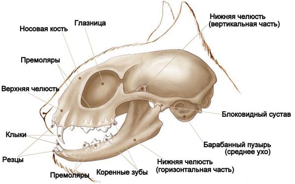
The main role is played by the fangs, which are quite long and sharp, and the auxiliary role is played by the incisors. The cat's skull has very large eye sockets, because keen-sighted cat eyes are impressive in size.
Torso bones
Cats have an unusually flexible spine. This flexibility is created due to the fact that it consists of small movable bones that have a high density. The larger bones make up the cervical region; it has 7 vertebrae, two of which have poetic names - the atlas and the epistropheus. These vertebrae have the property of rotating 180 0 .
The thoracic region consists of 13 vertebrae, to which 12 pairs of ribs are attached on both sides. 8 pairs of them end up attached to the sternum, and 5 pairs are not attached to anything. This ensures the flexibility of the cat's body and its ability to turn around even in very limited space.

Next comes the lumbar region, which consists of 7 vertebrae, becoming larger as they approach the tail. The vertebrae of the lumbar region have many strong protrusions, because the muscles and tendons that hold all the organs of the abdominal cavity are attached to them. In the sacral region there are 3 powerful fused vertebrae. And the longest is the caudal section, 21-23 decreasing towards the end of the vertebrae; some breeds, which are characterized by a shortened tail, have fewer caudal vertebrae.
One of the leading features of the cat's skeleton is the structure of its collarbones. The fact is that they are in a rudimentary state and do not limit the animal’s movements, as, for example, happens in dogs. Thanks to the “underdeveloped” collarbones, a cat can fit into any gap, as long as its head fits through.
Limb bones
Our fellow cats walk on their toes, and the lower back of their paw was once a foot. The cat's front paws have 5 toes, the outer phalanx of which forms the basis for the claw. The first finger is a rudiment and the claw cannot be removed from it.
The hind limbs of cats are longer, and the joints there are stronger, this allows the cat to withstand sudden strong loads. In addition, this structure of the limbs allows the cat to develop tremendous speed both horizontally and vertically. This is why cats are such good poison dart climbers.

The cat's hind legs have fewer toes - 4, and the fifth is also a rudiment. Based on how many fingers a cat has, she may have polydactyly (more fingers than normal) or oligodactyly (not enough fingers).
Internal organs
The internal structure of a cat is a set of all the same vital systems that are inherent in other mammals. Let's look at them in order.
Circulatory and respiratory systems
The circulatory system of a cat is not particularly different; the animal's pulse at rest ranges from 100 to 150 beats per minute and can be measured by pressing the femoral artery. Normally, blood in an animal’s body should be approximately 7% of its mass; cat blood is specific and clots faster than human blood.
With each beat, a cat's heart pumps about 3 ml of blood. The blood circulation of a cat occurs similarly to that of a person: in the lungs the blood is saturated with oxygen, and in the digestive organs with useful substances. After which the heart carries fresh blood through the arteries to all organs. And through the veins, blood flows back to the heart so that it again sends it to the lungs to be enriched with the necessary oxygen.

The respiratory system, in addition to supplying the blood with oxygen, is also involved in thermoregulation. The breathing rate in cats is 20-30 breaths per minute, in kittens about 40 breaths, and inhalation is through the nose. The air inhaled by a cat through the nose is first warmed and filtered, then passes through the pharynx into the larynx, trachea and lungs of the animal. There is an assumption that a cat makes purring sounds using pocket-like folds that are located in the larynx.
Excretory and digestive systems
The cat's digestive system also has much in common with humans. It begins with the mouth and ends with the rectum and sphincter. Between them are the pharynx, esophagus, stomach, small and large intestines. The pancreas and liver are also considered components of the digestive system.
It is noteworthy that the cat's stomach can digest fairly large pieces of food, which the cat bites off thanks to its strong and sharp incisors and fangs. The cat's intestine is approximately 3 times longer than the animal's body and is 1-1.8 m. The cat also has a cecum, but the murka does not have an appendix.

Fluid is removed from the cat's body through the urinary system - the kidneys, bladder and ureters. Urine formation begins in the kidneys, which also regulate blood chemistry. Urine then moves into the bladder through the ureters, from where it is removed from the body. The process of urination occurs under the control of the closing muscle, which prevents spontaneous urination. The following video will help you understand the structure of a cat by literally looking inside the cat's body!
Reproductive system
The purpose of the reproductive system is obvious - it is the continuation of the cat family. A cat's reproductive organs are the gonads, testicles, vas deferens and penis. In a cat, these are the ovaries, uterus, oviducts and external genitalia. The time for male cats to reach puberty is 6-8 months, however, the safe age for mating, when full-fledged offspring can be expected, is at least 10 months. During puberty, the behavior of cats changes greatly and they show in every possible way their readiness to reproduce.

Sense organs
Nature has endowed our smaller brothers with unusually developed sense organs. Our pets see, hear and smell much more keenly than we do.
Eye
A cat's eyes are much larger in relation to the size of its body than those of a human. The cornea of the cat's eye is more convex, which means that the quality of the image perceived by the cat's eye is higher. Cats can distinguish colors, it is believed that they see at least 3 colors - red, green and blue. The pupil of a cat's eye, just like a human's, is able to expand and contract thanks to a special constrictor muscle. Our mustachioed friends have unusually acute vision, but they don’t see what’s going on under their noses; the optimal distance for the perception of information by a cat’s eye is 2-6 m.
The structure of a cat's eye is distinguished by the presence of a special vascular layer called tapetum, thanks to which the cat's eyes can see in the dark and glow mystically. In addition, our pets may have differently pigmented irises, which is why they are so “different-eyed.”

Ear
The structure of a cat's ear gives it the ability to perceive sounds in the range from 30 hertz to 45 kilohertz, and purrs can also detect ultrasound. Almost all cats have erect ears, with the exception of some breeds. Cats, unlike humans, can actively move their ears; 27 muscles help them do this. All purrs have a fold of skin on the inside of the ear, which some call the “third ear.” It is noteworthy that sometimes completely white cats are born deaf due to gene mutations.
Nose
A cat's nose is considered one of the most vulnerable organs of a purr's body, especially its tip. By the way, the tip of the nose is completely devoid of vegetation and can be of different colors depending on the breed of the cat. Cats' sense of smell is quite well developed because they have a larger number of olfactory receptors compared to us.

In terms of their ability to recognize odors, cats are macrosomatic animals, while humans are microsomatic creatures and the number of odors they perceive is very limited. However, compared to dogs, cats still have weaker olfactory abilities.
Photo gallery
Video “Cats from a scientific point of view”
A very interesting and educational video with a selection of little-known facts about our furry pets to complete our tour of cat anatomy!
Sorry, there are no surveys available at this time.Cats, like all mammals, have a complex internal structure, with their own distinctive features. Given this fact, today we will take a detailed look at the internal structure of a cat and talk about each of its components.
The cat's digestive system consists of:
- esophagus;
- stomach;
- small intestine;
- duodenum;
- jejunum;
- liver;
- large intestine.
Esophagus It has a hose-like shape of a relatively small size, and connects the animal’s mouth and its stomach. The esophagus originates from the inner base of the mouth, extends through the neck and chest, passes close to the heart, extends through the muscles of the diaphragm, and connects to the stomach. It is important to note that the esophagus is equipped with special muscles that push food into the stomach, producing synchronous movements similar to waves. The esophagus is one of the most difficult organs in terms of surgical treatment, as it is difficult to access and extremely difficult to heal.
Feline stomach It is single-chamber, and differs in the location of the mucous membrane on its inner walls. The cat's stomach is adapted to accommodate a large amount of food, but it is almost never completely filled, since cats are not prone to gluttony (the vast majority). Also, the inner surface of the stomach is dotted with folds, which have an additional mechanical effect on the process of breaking down food. Food processed by gastric juice enters the duodenum through the pyloric sphincter. Most often, eaten food remains in the stomach for about 12 hours.
Small intestine It is a tubular organ connecting the stomach and large intestine. Often the length of a cat's small intestine is about 1.5-2 meters, and includes the duodenum, jejunum, and ileum.
Duodenum It is small in size and serves to mix food with liver and pancreatic enzymes, which is extremely important for digestion.
Jejunum is the longest part of the small intestine, and its inner walls are dotted with thin hairs, which, when in contact with food that comes into contact with them, penetrate it and suck out all the useful substances. It is here that the final extraction of all useful substances from food occurs, after which it enters the ileum, and then the large intestine, where it turns into feces.
Colon in cats it works as in all mammals: it serves for temporary storage of feces, as well as its removal from the anus. Also here, the walls of the large intestine absorb moisture from the feces stored in it, in order, if necessary, to maintain the necessary water balance in the body.
Liver is the largest gland in the cat’s body, and breaks down nutrients obtained from the stomach and intestines into elements necessary for the body. It is important to note that in order to fully produce the required complex of amino acids, the cat must receive 90% protein in its diet, otherwise the animal will die, because the liver will not be able to provide the body with the necessary substances from plant foods.
 General diagram of the structure of the internal organs of cats
 Respiratory system
The anatomy of the respiratory system of cats is similar to other carnivorous mammals and consists of the nose, nasopharynx, larynx, trachea, bronchi and, of course, lungs. The respiratory system is designed to carry out gas exchange in any environmental conditions (if there is oxygen), as well as saturate the body with this oxygen through its processing by the lungs. The structure, functions and principle of operation of the lungs are similar to other animals, and have no distinctive characteristics.
Circulatory system
The circulatory system in cats works the same way as in other mammals: the heart pushes blood through arteries that have elastic walls and rhythmically carry out contraction and relaxation movements. It is thanks to such movements that arteries located close to the skin can be felt, and this is called the pulse. The cat's pulse is easiest to detect on the inside of the thigh, and in a healthy animal it should range between 100-150 beats per minute.
The cat's brain absorbs 15-20% of the blood, the muscular system absorbs up to 40% of the total blood, and about 25-30% of the blood goes to the internal organs. During physical activity, muscles can absorb up to 90% of the blood, which is why cats get tired so quickly, but can focus maximum strength for a short period of time.
The animal's heart is a hollow organ located in the chest, just behind the breastbone. An important nuance is the fact that the weight of a cat’s heart depends on their weight, and does not have clearly established standards. Most often, the heart of an animal weighs 0.6% of the total body weight. The cat's heart consists of 2 ventricles and 2 atria.
The cat has a double circulation. The main blood circulation is provided by capillaries and arteries connected to the heart, which connect to all internal organs. The second circle of blood circulation is provided by veins, which pump blood into the right ventricle of the heart, straight through the lungs and their arteries.
Cat blood has high clotting rates compared to humans, and it cannot be replaced with the blood of other animals, as this can lead to the death of the cat. The bulk of the blood is yellow plasma, 30-45% are red blood cells, and the rest is allocated to white blood cells and platelets. Cat blood has 3 groups: A, B, AB. Cat blood type AB is extremely rare, which should be taken into account by owners of such animals.

urinary system
The excretory system is represented by the bladder, kidneys and ureters. Urine is formed in the kidneys; a cat produces about 100 ml of urine per day. Next, urine enters the ureters and goes to the bladder, where it is released into the external environment through urination.
Reproductive system
The reproductive system of cats has the following internal organs:
- vulva;
- vagina;
- Cervix;
- uterus;
- fallopian tubes and ovaries;
- mammary gland;
- oviduct.

The reproductive system of cats has the following organs:
- testicles;
- penis;
- prostate;
- genital tract that carries sperm from the testicles to the penis.

Endocrine system
The endocrine system is primarily responsible for hormones and their production in the corresponding organs. Thus, the cat’s brain produces antidiuretic hormone, oxytocin, corticotropic hormone, adrenocorticotropic hormone, cortisol and growth hormone.
The adrenal glands produce a lot of other hormones, the main purpose of which is to regulate metabolism, and are also responsible for behavioral characteristics. The adrenal glands also produce cortisol, a small portion of testosterone, as well as epinephrine and norepinephrine.
There are a number of other glands of external and internal secretion, the principle of operation of which is common to all mammals.
Nervous system
The nervous system of cats is divided into central and peripheral. Each of these systems in a cat performs functions that are standard for most mammals.
The central nervous system consists of the brain, brain stem and the so-called spinal cord. The central nervous system is the most important in the body of any living creature, and simple and complex reactions, as well as some reflexes, depend on it. In addition, the central nervous system interacts with the peripheral and autonomic ones, ensuring their functioning and control.
The peripheral nervous system is responsible for the cat's conscious motor abilities. So, thanks to this system, a cat can move its paws, extend its claws, run, and generally lead the lifestyle that it leads. Also, the peripheral nervous system transmits pain impulses to the central nervous system from any part of the body where peripheral nerve endings are present.

Musculoskeletal system
The cat's body has two main types of muscles: smooth muscle and striated muscle. Smooth muscles are found in all the internal organs of the cat, and are directly connected to the autonomic nervous system, thereby ensuring the work and unconscious functioning of the internal organs, an excellent example of which would be the esophagus and the heart.
The striated muscles are attached to the skeleton and provide the cat with physical strength, the ability to move, hunt and fight. The striated muscles are familiar muscles that we can feel on the limbs and body of the pet.
An important part of the musculoskeletal system of a cat are tendons, ligaments and joints, which in all cats are distinguished by strength, flexibility and enviable elasticity until old age.
The cat's shoulder girdle, which has a unique structure, deserves special mention. Thus, in almost all mammals, the bones of the forelegs are connected to the body with the help of the collarbone, but in cats, the bones of the limbs are connected to the body exclusively with the help of muscles, which provides them with incredible mobility.
1. Oral cavity (Cavum oris)
Food, entering the initial section of the digestive apparatus through the oral opening, enters the oral cavity, the skeleton of which is the upper and lower jaws, palatine and incisive bones. The hyoid bone, located inside the oral cavity, serves as a place of fixation for the muscles of the tongue, pharynx and larynx. The oral cavity extends orally from the lips, and aborally ends with the pharynx and passes into the pharynx. The dental edge of the closed jaws and lips form the vestibule of the oral cavity. Behind the vestibule is the oral cavity itself. The vestibule communicates with the external environment through the oral fissure. The oral fissure begins at the junction of the upper and lower lips, called the angle of the mouth.
Appearance of the oral cavity
Lips- upper and lower muscle-skin folds, covered on the outside with hair and on the inside with mucous membrane. Outside, the upper lip is sagittally divided by a deep groove - a filter, passing towards the nasal septum. On the upper lip there are hard whiskers, collected in 2 side tufts - mustaches.
Cheeks are a continuation of the lips behind their commissure and form the lateral walls of the oral cavity. The cheeks of cats are relatively small, thin, and covered with hair on the outside. Their inner surface is smooth, and the ducts of the salivary glands open on it.
Teeth- durable organs of the oral cavity that serve to capture and hold food, bite it, crush and grind it, as well as defend and attack.


Adult cats have 30 teeth, 16 of which are in the upper jaw and 14 in the lower jaw. Cats are carnivores by nature, which largely reflects the arrangement of their teeth. Cats have six front teeth and two canines in each jaw. These teeth are involved in the process of biting into meat and then tearing it. Cats have only 6 premolars and 2 molars in the upper jaw and 4 premolars and 2 molars in the lower jaw. Cats are also characterized by an increased size of the upper 4th molar (also called the “carnivore tooth”) and the 1st lower incisor. Due to the arrangement of these “carnivorous teeth”, eating food occurs according to the “scissor principle”, which is extremely effective when cutting raw meat.
STRUCTURE OF TEETH
A tooth consists of dentin, enamels And cement.
Schematic representation of the cutter:

Dentine- tissue that forms the basis of the tooth. Dentin consists of a calcified matrix penetrated by dentinal tubules containing processes of odontoblast cells lining the tooth cavity. The intercellular substance contains organic (collagen fibers) and mineral components (hydroxyapatite crystals). Dentin has different zones that differ in microstructure and color.
Enamel- a substance covering dentin in the crown area. It consists of crystals of mineral salts, oriented in a special way to form enamel prisms. Enamel does not contain cellular elements and is not tissue. The normal color of enamel is from white to cream with a yellowish tint (distinguishable from plaque).
Cement- tissue covering dentin in the root area. The structure of cement is close to bone tissue. Consists of cementocyte and cementoblast cells and a calcified matrix. Nutrition of cement occurs diffusely from the periodontium.
Inside there is tooth cavity, which is divided into coronalcavity And root canal, opening with the above hole in the apex of the tooth. Fills the dental cavity dental pulp, consisting of nerves and blood vessels immersed in loose connective tissue and providing metabolism in the tooth. Distinguish coronal And root pulp.
Gum- mucous membrane that covers the dental edges of the corresponding bones, tightly fused with their periosteum.
The gum covers the tooth in the neck area. It is abundantly supplied with blood (tendency to bleeding), but relatively poorly innervated. The grooved depression located between the tooth and the free edge of the gum is called the gingival sulcus.
The periodontium, alveolar wall and gums form supporting apparatus of the tooth - periodontium.
Periodontium- provides attachment of the tooth to the dental alveolus. It consists of the periodontium, the wall of the dental alveoli and the gums. The periodontium performs the following functions: supporting and shock-absorbing, barrier, trophic and reflex.
The teeth are distributed as follows: 12 incisors (I), 4 canines (C), 10 premolars (P) and 4 molars (M). Thus, the dental formula is as follows:
![]()
All teeth are of pronounced short-crown type.
There are 4 types of teeth: incisors, fangs And permanent teeth: preradical(false, small molars), or premolars And truly indigenous, or molars that do not have milk precursors.
Teeth arranged in order in a row form top
and lower dental arches (arcades)
.
Incisors- small, with uneven edges and 3 protruding points. Each root is single. The lateral incisors are larger than the medial ones, and the incisors of the upper jaw are larger than those of the lower jaw.
Schematic representation of the incisors:

Behind the incisors are located fangs. These are long, strong, deep-set teeth with a simple root and a rounded crown. When the jaws are closed, the lower canines lie laterocaudal to the upper ones. Behind the fangs on each jaw there is an edge free from teeth.
Schematic representation of fangs:

Molars of the upper dental arch.
Premolars are located behind the diastema; there are 3 pairs of them on the upper jaw
and 2 pairs on the bottom. The first premolar of the upper jaw is small,
with a simple crown and a simple root. The second premolar is larger, it has 4 projections - a large central one, a small cranial one
and 2 small caudal ones. The most massive tooth is the third premolar: it has 3 large protrusions located along the length
and small projections lying on the medial side of the first; the root of the tooth has 3 processes.
Schematic representation of premolars:

Upper dental arcade of a seven-month-old cat:

Molars located caudal to the last premolar in the upper jaw. These are small teeth with 2 projections and 2 roots.
Schematic arrangement of molars:

Molars of the lower dental arch.
In the lower arcade 2 premolar; they are identical in size and shape. The crown of each premolar bears 4 projections - one large, one small in front and two more behind. Each premolar has
2 roots.
Molar the lower jaw is the most massive in the arcade and has
2 protrusions and 2 roots. The molars sit obliquely in the sockets, so that when the jaws are closed, the teeth of the upper jaw adjoin the lower teeth from the inside.
The lower dental arcade of a seven-month-old cat:

Baby teeth appear in kittens soon after birth.
They are smaller in size than the permanent ones and less developed. Their color
milky white. There are fewer primary teeth than permanent teeth because molars have no predecessors.
The dental formula of primary teeth is as follows: ![]()
MECHANICAL DIGESTION
Digestion in the oral cavity occurs mainly mechanically; when chewing, large fragments of food are broken into pieces and mixed with saliva.
Mechanical digestion also increases the area exposed to digestive enzymes. The arrangement of teeth is closely related to the natural diet of various animal species and indicates their natural feeding behavior and preferred feeding patterns.
ORAL CAVITY
The oral cavity itself is separated from above, from the side of the nasal cavity, by the hard palate, from the pharynx by the soft palate, and is limited in front and on the sides by dental arcades.
Solid sky arched like a vault. Its mucous membrane forms 7 - 8 caudally concave transverse ridges - palatine ridges, between which the papillae are located. In the anterior part behind the incisors there is a small incisive papilla;
to the right and left of it lie the slit-like nasopalatine canals, which are the excretory ducts of the nasopharynx organ.
In the aboral direction, in the area of the choanae, the hard palate passes into the soft palate without a visible boundary.
Soft palate or velum- is a continuation of the hard palate and is a fold of the mucous membrane that covers the entrance to the choanae and pharynx. The soft palate is based on special muscles: the levator velum palatine, the tensor velum palatine, and the palatine muscle that shortens it after the act of swallowing. The velum palatine hangs from the end of the bony palate and, in a calm state, its free edge touches the root of the tongue, covering the pharynx, the exit from the oral cavity into the pharynx.
The free edge of the velum is called the arch of the palate. The palatine arch, together with the pharynx, forms the velopharyngeal arches, and with the root of the tongue - the palatoglossus arches. Aborally on the sides of the root of the tongue, in the tonsil sinuses there is one palatine tonsil.
SALIVARY GLANDS
Cats have 5 pairs of salivary glands: parotid, submandibular, sublingual, molar and infraorbital.
Layout of the salivary glands of a cat:

1 - parotid
2 - submandibular
3 - sublingual
4 - radical
5 – infraorbital
Parotid salivary gland located ventral to the external auditory canal under the cutaneous muscles. It is flat, has a lobular structure, and borders orally with the large masseter muscle. The excretory ducts of the individual lobules of the gland merge to form the common parotid (stenon) duct. It passes cranially as part of the fascia covering the large masticatory muscle, at the cranial edge of the muscle it turns inward, goes under the mucous membrane and opens into the buccal vestibule of the mouth opposite the last premolar with the salivary papilla. Along the duct there are one or more small accessory parotid salivary glands.
Submandibular gland rounded, lies ventral to the previous one near the large masseter muscle and consists of individual glandular lobules connected by connective tissue. The excretory duct of the submandibular gland is located on its inner surface, it stretches forward under the base of the tongue and opens at the bottom of the oral cavity with a sublingual wart, next to which the duct of the sublingual gland opens.
Sublingual gland elongated, conical, its base is adjacent to the submandibular gland, stretching 1-1.5 cm along its duct. The excretory duct of the sublingual gland is located on the ventral side; in its course it accompanies the duct of the submandibular gland, following first dorsally and then ventrally from it.
Indigenous salivary gland, absent in other domestic animals, in the cat it is located at the cranial edge of the large masseter muscle, between the mucous membrane of the lower lip and the orbicularis oris muscle. It is a flat formation that widens caudally and tapers orally. The anterior edge of the gland is visualized at the level of the canine. It has several ducts that open directly into the oral mucosa.
Orbital or zygomatic gland Of all domestic animals, only dogs and cats have it. It has a round shape and reaches a length of 1.5 cm. It is located medial to the zygomatic arch in the lower part of the orbit. The ventral edge is located behind the molar. Its large excretory duct and additional small ducts open into the oral cavity 3 - 4 mm caudal to the upper molar.
ENZYMATIVE DIGESTION
Saliva is secreted into the oral cavity by five pairs of salivary glands. Typically, a small amount of saliva is present in the mouth, but its flow may increase if the animal sees or smells food.
Salivation continues as food enters the oral cavity, and its effect is enhanced by the chewing process.
Saliva is 99% water, while the remaining 1% is mucus, inorganic salts and enzymes. Mucus acts as an effective lubricant and promotes swallowing, especially dry food. Unlike humans, cats lack the starch-digesting enzyme amylase in their saliva, which prevents the rapid absorption of starch in the mouth. The absence of this enzyme is consistent with the observed carnivorous behavior of cats that tend to consume foods low in starch.
Language- a muscular, movable organ lying at the bottom of the oral cavity.
Tongue and dorsally open pharynx:

Language in cats it is elongated, flat, widened in the middle and slightly narrowed at the end. When the oral cavity is closed, the tongue completely fills it. In terms of external shape, the tongue of cats is long, wide and thin.
The root of the tongue extends from the molars to the epiglottis and is closely connected to the hyoid bone.
The body of the tongue is almost twice as long as the root; it is located between the molars and has a dorsal back and 2 lateral surfaces. At the border with the apex below, the body forms a median fold containing parts of both geniohyoid muscles, this is the frenulum of the tongue. Folds are directed from the caudal end of the body to the epiglottis. The tip of the tongue rests with its free end against the incisor teeth.
On the back of the tongue and in the region of its apex, the mucous membrane is dotted with many rough, keratinized filiform papillae; their apices are directed caudally. Fungiform papillae are located on the surface of the dorsum, the largest of them lie along the edges of the tongue. Large ridge-shaped, or grooved, papillae in two caudally converging rows of 2-3 in each are located at the root of the tongue. The ventral surface and lateral edges of the tongue are smooth, soft, and free of papillae.
The muscles of the tongue consist of longitudinal, transverse and perpendicular bundles. The first ones go from the root of the tongue to its apex, the second ones - from the middle connective tissue septum of the tongue to the sides, the third ones run vertically from the back of the tongue to the bottom surface. These are the actual muscles of the tongue, located in its thickness;
with their help, the tongue can be shortened, thickened and flattened. In addition, there are muscles that connect the tongue to the bones of the oral cavity.
Genioglossus muscle passes from the symphysis of the mandible, where it originates on the medial surface; its fibers pass dorsally, located above the geniohyoid muscle, diverge; of these, the cranial ones reach the tip of the tongue, the caudal ones end at the root of the tongue. Dorsally, the muscle is mixed with the muscle of the same name on the opposite side.
Function: pulls the root of the tongue forward and the top of it to the side.
Lingual lateral muscle arises from the mastoid process of the temporal bone, from the ligament connecting the edge of the external auditory canal and the angular process of the mandible, and from the proximal part of the cranial horns of the hyoid bone. It passes into the lateral part of the tongue between the digastric and lingual main muscles, then, diverging, goes forward to the tip of the tongue, where it ends.
Function: pulls the tongue back with bilateral action, shortening it when swallowing; with unilateral action, turns the tongue to the side.
2. Pharynx (Pharynx)
Pharynx a mobile muscular-cavitary organ in which the digestive tract crosses, going through the pharynx from the oral cavity to the pharynx and further to the esophagus and the respiratory tract - through the choanae to the pharynx and further to the larynx.
Appearance of the pharynx:

Due to the fact that the cross-section of the digestive and respiratory tracts occurs in the pharynx, its mucous membrane, with the help of folds - the velopharyngeal arches, is divided into the upper, respiratory, and lower, digestive parts. The respiratory part is a continuation of the choanae, and is therefore called the nasal part of the pharynx, or nasopharynx. Near the choanae, a paired opening of the auditory tubes opens into the lateral wall of the pharynx. The digestive, or laryngeal, part in front borders the pharynx, being separated from it by the velum palatine, and is a caudal continuation of the oral cavity, rests against the epiglottis at the back and then, located on top of the larynx, follows towards the esophagus, which lies in this area above the trachea.
The muscles of the pharynx are striated, represented constrictors And dilators.
Cranial constrictor The pharynx consists of 2 paired muscles - the pterygopharyngeal and glossopharyngeal.
Pterygopharyngealmuscle flat, triangular, begins at the apex of the uncinate process of the pterygoid bone. Heading caudally, the muscle diverges under the medial constrictor. Some of the fibers are attached to the median suture of the pharynx, the dorsal fibers are attached to the base of the pterygoid bone, the ventral ones run along the length of the pharynx and end on the larynx.
Glossopharyngeal muscle begins on the geniohyoid muscle, passes as a thin ribbon outside the cranial horns of the hyoid bone, turns dorsally and attaches to the middorsal suture of the pharynx.
Middle, or sublingual, constrictor pharynx - a thin muscle covering the middle part of the lateral surface of the pharynx. It begins with two heads - on the cranial horns and the free caudal horn of the hyoid bone; attaches to the dorsal suture of the pharynx and the base of the sphenoid bone.
Caudal or laryngeal constrictor The pharynx begins on the lateral side of the thyroid and cricoid cartilages. The fibers run dorsally and cranially and attach to the pharyngeal suture.
Stylopharyngeal muscle begins at the apex of the mastoid process of the temporal bone. The ribbon-shaped abdomen extends ventrocaudally and is attached to the dorsal wall of the pharynx and larynx. Laterally, the muscle is covered by the middle and caudal constrictors. Contraction of the pharyngeal muscles underlies the complex act of swallowing, which also involves the soft palate, tongue, esophagus and larynx. At the same time, the pharyngeal levators pull it upward, and the compressors successively backwards narrow its cavity, pushing the food bolus into the esophagus. At the same time, the larynx rises and tightly covers the epiglottis, due to pressure on it with the root of the tongue. In this case, the muscles of the soft palate pull it upward and caudally so that the velum palatine rests on the palatopharyngeal arches, separating the nasopharynx. During breathing, the shortened velum palatine hangs obliquely downward, covering the pharynx, while the epiglottis, built of elastic cartilage and directed upward and forward, provides access to a stream of air into the larynx.
3. Esophagus (Oesophagus)
Esophagus It is a cylindrical tube following the pharynx, flattened at the top and bottom.
Esophageal endoscopy:


It is the initial section of the foregut and in structure is a typical tube-shaped organ. The esophagus is a direct continuation of the laryngeal part of the pharynx.
Usually the esophagus is in a collapsed state. The mucous membrane of the esophagus along its entire length is collected in longitudinal folds that straighten out as the food coma passes.
The submucosal layer contains many mucous glands that improve the sliding of food. The muscular layer of the esophagus is a complex multi-level striated layer. The outer membrane of the cervical and thoracic parts of the esophagus is connective tissue adventitia, and the abdominal part is covered with visceral peritoneum. The attachment points of the muscle layers are: laterally - the arytenoid cartilages of the larynx, ventrally - the annular cartilage, and dorsally - the tendon suture of the larynx.
The diameter of the esophagus is relatively constant throughout its entire length and reaches 1 cm during the passage of the food bolus. The esophagus is divided into cervical, thoracic and abdominal sections. Upon exiting the pharynx, the esophagus is located dorsal to the larynx and trachea, covering the bodies of the cervical vertebrae from below, then descends to the left side of the trachea and in the area of its bifurcation returns again to the midline. In the chest cavity, it lies in the mediastinum, passing over the base of the heart and under the aorta. It enters the abdominal cavity through the esophageal opening of the diaphragm, which lies approximately 2 cm ventral to the vertebral column. The abdominal region is very short.

1 - language
2 - pharynx and larynx
3 - esophagus in a collapsed state
4 – stomach
During the process of swallowing, a lump of unchewed food formed by the tongue enters the esophagus. The esophagus does not secrete digestive enzymes, but the cells of the esophagus secrete mucus, which serves to lubricate peristalsis, the automatic wave-like muscle contractions that are stimulated by the presence of food in the esophagus and allow it to move through the gastrointestinal tract. The process of moving food from the mouth to the stomach takes only a few seconds.
4. Stomach (Ventriculus)
Stomach is the organ of the digestive tract where food is retained and subjected to chemical processing. The cat's stomach is single-chamber, intestinal type. It is an extension of the digestive tube behind the diaphragm.

1 - pyloric part of the stomach
2 - cardiac part of the stomach
3 - fundic part of the stomach
4 - exit of the duodenum
5 - cardial opening (entrance of the esophagus)
Appearance of the opened stomach:

TOPOGRAPHY OF THE CAT'S STOMACH
The stomach is located in the anterior part of the abdominal cavity to the left of the midline, in the plane of the IX-XI intercostal space and in the region of the xiphoid process. The anterior, or diaphragmatic, wall is adjacent to the diaphragm only dorsally; the cardiac part of the stomach does not touch the diaphragm, so a small segment of the esophagus passes into the abdominal cavity. The posterior, visceral wall is adjacent to the intestinal loops.
Contrast radiograph of a cat's stomach:

STRUCTURE OF A CAT'S STOMACH
Diagram of the cross section of the stomach indicating the anatomical and functional elements:

In the enlarged initial part of the stomach, which lies on the left, there is the inlet of the esophagus. In the narrowed-elongated part lying to the right and below there is a second opening leading to the duodenum, the pyloric opening, and the pylorus.
In accordance with this, the cardiac and pyloric parts of the stomach are distinguished. The concave and convex sections located between them are called the lesser and greater curvature. The concave lesser curvature faces cranially and to the right. The convex greater curvature is directed caudally and to the left. The middle part of the stomach on the side of the greater curvature is called the fundus of the stomach.


In an empty stomach mucous membrane collected in longitudinal folds running parallel to each other. The surface of the gastric mucosa is about 1/5 - 1/6 of the total surface of the intestinal mucosa.
Muscularis The stomach is well developed and is represented by three layers.
Ultrasound image of the wall of a healthy stomach:

The superficial thin longitudinal layer is directed from the esophagus to the pylorus. In the area where the bottom and pyloric glands are located, the circular, or circular, layer of fibers reaches its greatest expression. In the left part of the stomach, the internal oblique layer predominates. As they approach the pylorus, the muscle walls thicken and, at the border with the duodenum, break off in the form of a thickened annular ridge. This strong muscular sphincter is called the sphincter muscle, or constrictor pylorus. In the constrictor area, the mucous membrane is also collected in longitudinal folds.
The outside of the stomach is covered serosa, which at the lesser curvature passes into the lesser omentum, in the area of greater curvature into the greater omentum. The first connects the stomach to the liver through the hepatogastric ligament. This ligament on the left merges with the ligament of the liver and esophagus and on the right - with the ligament of the liver and duodenum. The greater omentum, from the stomach to the lower back, forms the omental sac.
On the right, near the kidney, at the caudal vena cava and portal vein, there is an entrance to the omental sac. The spleen, located between the layers of the greater omentum, connects to the stomach through the gastrosplenic ligament.
During embryonic development, the stomach, as part of a straight digestive tube, undergoes two 180° rotations. One in the frontal plane counterclockwise and the other in the segmental plane.
FUNCTIONS OF THE STOMACH
The stomach has several functions: it serves to temporarily store food and controls the rate at which food enters the small intestine.
The stomach also secretes enzymes necessary for the digestion of macromolecules.
The stomach muscles regulate motility, allowing food to move aborally (away from the mouth), and aid digestion by mixing and grinding food.
PHASES OF STOMACH SECRETION
Gastric secretion is regulated by complex processes of nervous and hormonal interaction, due to which secretion is produced at the right time and in the required volume. The secretion process is divided into three phases: cerebral, gastric and intestinal.
Brain phase
The medullary phase of secretion is initiated by the anticipation of food intake and the sight, smell and taste of food, which stimulates the secretion of pepsinogen, although gastrin and hydrochloric acid are also released in small quantities.
Gastric phase
The gastric phase is initiated by mechanical stretching of the gastric mucosa, a decrease in acidity, as well as products of protein digestion. In the gastric phase, the main secretion product is gastrin, which also stimulates the secretion of hydrochloric acid, pepsinogen and mucus. Gastrin secretion slows down sharply if the pH drops below 3.0 and may also be controlled by peptic hormones such as secretin.
or enteroglucagon.
Intestinal phase
The intestinal phase is initiated by both mechanical distension of the intestinal tract and chemical stimulation with amino acids and peptides.
5. Small intestine (Intestinum tenue)
Small intestine is a narrowed section of the intestinal tube and consists of many loops that occupy most of the space of the abdominal cavity. The total length of the intestine is almost 4 times the length of the body and is about 1.98 m, with the small intestine accounting for 1.68 m and the large intestine 0.30 m. The mucous membrane of the small intestine is velvety due to the presence of villi. The muscular coat is represented by a longitudinal and circular layer of smooth muscle fibers. The serous membrane passes to the intestine from the mesentery.
According to its position, the small intestine is divided into duodenum, jejunum and ileum. Their length is, respectively, 0.16; 1.45; 0.07 m.

Ultrasound of the small intestine:

The wall of the thin section is richly vascularized. Arterial blood flows through the branches of the cranial mesenteric artery, and to the duodenum also through the hepatic artery. Venous drainage occurs in the cranial mesenteric vein, which is one of the roots of the portal vein of the liver.
Lymphotok from the intestinal wall comes from the lymphatic sinuses of the villi and intraorgan vessels through the mesenteric (intestinal) lymph nodes into the intestinal trunk, which flows into the lumbar cistern, then into the thoracic lymphatic duct and the cranial vena cava.
Nervous support The thin section is represented by branches of the vagus nerve and postganglionic fibers of the solar plexus from the semilunar ganglion, which form two plexuses in the intestinal wall: intermuscular (Auerbach) between the layers of the muscular layer and submucosal (Meissner) in the submucosal layer.
Control of intestinal activity by the nervous system is carried out both through local reflexes and through vagal reflexes involving the submucosal nerve plexus and intermuscular nerve plexus.
Bowel function is regulated by the parasympathetic nervous system. Control is directed from the medullary portion of the vagus nerve to the small intestine. The sympathetic nervous system (control directed from the ganglia in the paravertebral sympathetic trunk) plays a less important role. The processes of local control and coordination of motility and secretion of the intestine and associated glands are of a more complex nature; nerves, paracrine and endocrine chemicals take part in them.
TOPOGRAPHY
The thin section begins from the pylorus of the stomach at the level of the 12th rib, is covered ventrally by the leaves of the greater omentum, and is limited dorsolaterally by the thick section. There are no clear boundaries between the sections of the small intestine, and the identification of individual sections is mainly topographical in nature. Only the duodenum is most clearly distinguished, which is distinguished by its large diameter and topographic proximity to the pancreas.
MININGS OF THE INTESTINAL
The functional features of the small intestine leave an imprint on its anatomical structure.
There are mucous membrane and submucosal layer, muscular (external longitudinal and internal transverse muscles) and serous membranes of the intestine.

Mucous membrane forms numerous devices that significantly increase the suction surface.
These devices include circular folds, or Kirkring folds, in the formation of which not only the mucous membrane is involved, but also the submucosal layer and villi, which give the mucous membrane a velvety appearance.
The folds cover 1/3 or 1/2 of the circumference of the intestine. The villi are covered with a special bordered epithelium, which carries out parietal digestion and absorption. The villi, contracting and relaxing, perform rhythmic movements with a frequency of 6 times per minute, due to which they act as a kind of pumps during suction.
In the center of the villus there is a lymphatic sinus, which receives fat processing products.
Each villus from the submucosal plexus contains 1-2 arterioles, which break up into capillaries. Arterioles anastomose with each other and during absorption all capillaries function, while during a pause there are short anastomoses. Villi are thread-like outgrowths of the mucous membrane, formed by loose connective tissue rich in smooth myocytes, reticulin fibers and immunocompetent cellular elements, and covered with epithelium. The length of the villi is 0.95-1.0 mm, their length and density decreases in the caudal direction, that is, in the ileum the size and number of villi are much smaller than in the duodenum and jejunum.
The mucous membrane of the thin section and villi is covered with a single-layer columnar epithelium, which contains three types of cells: columnar epithelial cells with a striated border, goblet exocrinocytes (secrete mucus) and gastrointestinal endocrinocytes.
Mucous membrane of the thin section is replete with numerous parietal glands - common intestinal, or Lieberkühn glands (Lieberkühn's crypts), which open into the lumen between the villi. The number of glands averages about 150 million (in the duodenum and jejunum there are 10 thousand glands per 1 cm 2 of surface, and 8 thousand in the ileum). The crypts are lined with five types of cells: epithelial cells with a striated border, goblet glandulocytes, gastrointestinal endocrinocytes, small borderless cells of the crypt bottom (stem cells of the intestinal epithelium) and enterocytes with acidophilic granules (Paneth cells). The latter secrete an enzyme involved in the breakdown of peptides and lysozyme.
The duodenum is characterized by tubular-alveolar duodenal, or Bruner's glands, which open into crypts. These glands are a continuation of the pyloric glands of the stomach and are located only on the first 1.5-2 cm of the duodenum.
The final segment of the thin section (ileum) is rich in lymphoid elements, which lie in the mucous membrane at different depths on the side opposite to the attachment of the mesentery, and are represented by both single (solitary) follicles and their clusters in the form Peyer'splaques. Plaques begin in the final part of the duodenum.
The total number of plaques is from 11 to 25, they are round or oval in shape, length from 7 to 85 mm, and width from 4 to 15 mm. The lymphoid apparatus takes part in the digestive processes. As a result of the constant emigration of lymphocytes into the intestinal lumen and their destruction, interleukins are released, which have a selective effect on the intestinal microflora, regulating its composition and distribution between the thin and thick sections. In young organisms, the lymphoid apparatus is well developed, and the plaques are large. With age, a gradual reduction of lymphoid elements occurs, which is expressed in a decrease in the number and size of lymphatic structures.
Muscularis represented by two layers of smooth muscle tissue: longitudinal And circular, and the circular layer is better developed than the longitudinal one. The muscular layer provides peristaltic movements, pendulum-like movements
and rhythmic segmentation, which moves and mixes the intestinal contents.
Serosa forms the mesentery on which the entire thin section is suspended. At the same time, the mesentery of the jejunum and ileum is better expressed, and therefore they are combined under the name mesenteric colon.
INTESTINAL FUNCTIONS
Digestion of food is completed in the small intestine under the action of enzymes produced by the wall ( liver and pancreas) and wall ( Lieberkühnand Brunner's) glands, absorption of digested products into the blood and lymph, and biological disinfection of incoming substances.
The latter occurs due to the presence of numerous lymphoid elements enclosed in the wall of the intestinal tube.
The endocrine function of the thin section is also great, which consists in the production of some biologically active substances by intestinal endocrinocytes (secretin, serotonin, motilin, gastrin, pancreozymin-cholecystokinin, etc.).
PARTS OF THE SMALL INTESTINE
It is customary to distinguish three sections of the thin section: the initial segment or duodenum, middle segment or jejunum and the end segment or ileum.
DUODENUM
Structure
Duodenum- the initial section of the thin section, which is connected to the pancreas and the common bile duct and has the form of a loop facing caudally and located under the lumbar spine.
The duodenum accounts for 10% of the total length of the small intestine. This section of the thin section is characterized by the presence of duodenal (Bruner's) glands and a short mesentery, as a result of which the intestine does not form loops, but forms 4 distinct convolutions.

Topography
The duodenum, leaving the stomach, rotates so that it forms an acute angle (cranial bend). Initially, it is directed caudally and slightly to the right, but soon acquires a caudal direction, located in the right hypochondrium. Approximately 10 cm caudal to the pylorus, the intestine makes a U-shaped bend, passing 4 - 5 cm forward and to the left, then passes into the jejunum without pronounced boundaries. Between the branches of the U-shaped bend is the duodenum of the pancreas. Approximately 3 cm from the pylorus, the intestine receives the common bile and pancreatic duct. At the confluence of the duct on the mucous membrane there is a small papilla, the apex of which bears an oval opening. The confluence of the accessory duct is located 2 cm caudal to the main pancreatic duct.
JEJUNUM
Structure
Jejunum- the longest part of the thin section. Makes up to 70% of the length of the thin section.
The intestine got its name due to the fact that it has a half-dormant appearance, that is, it does not contain voluminous contents. The diameter exceeds the ileum located behind it and is distinguished by a large number of vessels passing through a well-developed mesentery.
Due to its considerable length, developed folds, numerous villi and crypts, the jejunum has the largest absorption surface, which is 4-5 times greater than the surface of the intestinal canal itself.
Endoscopy of the jejunum:

Topography
Its loops hang on the elongated mesentery and form numerous curls, occupying a vaguely defined area of the abdominal cavity. Caudally it passes into the ileum.
ILEUM
Structure
Ileum- the final part of the thin section, reaching a length of up to 20% of the length of the thin section. Its structure is no different from the jejunum. Its diameter is relatively constant, in the caudal part the walls are thinner. The ileum is characterized by the accumulation of numerous lymphoid elements that lie in its wall (Peyer's patches). In the right iliac region it flows into the colon, forming a valve (valve). The valve with its protruding part of the mucous membrane is directed into the lumen of the colon. In the area of the valve, the muscle layer is significantly thickened, the mucous membrane is free of villi. During normal peristalsis, the valve periodically expands and allows contents to pass into the large intestine.
Endoscopy of the ileum: 
Topography
The ileum is suspended on the folded mesentery. It is separated from the lower abdominal wall only by the omentum.
WALL GLANDS. LIVER
Liver- the largest gland in the body, it is a parenchymal organ of red-brown color. Its absolute weight in adult cats averages 95.5 g, i.e. 3.11% relative to the total weight of the animal.
Five tubular systems are formed in the liver: 1) bile ducts; 2) arteries; 3) branches of the portal vein (portal system); 4) hepatic veins (caval system); 5) lymphatic vessels.
Appearance of an isolated liver:

The shape of the liver is irregularly rounded with a thickened dorsal margin and sharp ventral and lateral margins. The pointed edges are dissected ventrally by deep grooves into lobes. The surface of the liver is smooth and shiny due to the peritoneum covering it, only the dorsal edge of the liver is not covered with peritoneum, which in this place passes onto the diaphragm, and thus is formed extraperitonealfield liver.
Located under the peritoneum fibrous membrane. It penetrates the organ and divides it into lobes.
The main sagittal notch divides the liver into right and left lobes; in the same notch there is a round ligament, the continuation of which is the falciform ligament connecting the liver with the diaphragm and the transverse coronary ligament.
Each lobe of the liver is further divided into medial and lateral parts. The left medial lobe is small. The left lateral lobe, which with its sharp end covers most of the ventral surface of the stomach, is significantly larger in size. The right medial (cystic) lobe is extensive; on its posterior surface there is a gallbladder with the cystic duct. Right lateral lobe - located dorsal and caudal to the vesical lobe and is deeply split into caudal and cranial parts. The first is elongated and reaches the caudal end of the right kidney, adjacent to its ventral surface; the dorsal surface of the second is in contact with the adrenal gland. In addition to those listed, at the base of the right lateral lobe there is an elongated triangular caudate lobe; it lies at the omental sac and partially covers its entrance.
Schematic representation of the liver and gallbladder:

The liver is a polymer organ in which several structural and functional elements can be distinguished: hepatic lobule, sector, (a section of the liver supplied by a branch of the portal vein of the 2nd order), segment (a section of the liver supplied by a branch of the portal vein of the 3rd order), hepatic acini(adjacent areas of 2 adjacent lobules) and portal hepatic lobule(areas of 3 adjacent lobules).
The classic morphofunctional unit is hepatic lobule hexagonal in shape, located around the central vein of the hepatic lobule.
The hepatic artery and portal vein, having entered the liver, are repeatedly divided into lobar, segmental, etc. branches up to interlobulararteries and veins, which are located along the lateral surfaces of the lobules along with interlobularbile duct, forming hepatic triads. From these arteries and veins branches arise that give rise to sinusoidal capillaries, which flow into the central veins of the lobule.
The lobules consist of hepatocytes, which form trabeculae in the form of two cellular cords. One of the most important anatomical features of the liver is that, unlike other organs, the liver receives blood from two sources: arterial- along the hepatic artery, and venous- along the portal vein.
One of the most important functions of the liver is bile formation process, which led to the formation of the bile ducts. Between the hepatocytes forming the lobules there are bile ducts that flow into the interlobular ducts.
The interlobular bile ducts merge to form the hepatic excretory duct; there may be several of them. The excretory cystic duct also departs from the gallbladder; it connects with the hepatic duct, forming the common bile duct, which opens together with the pancreatic duct
into the duodenum. At the end of the bile duct lies the sphincter of Oddi, which also covers the pancreatic duct.
Gallbladder It is an elongated pear-shaped sac that lies in the cleft of the right medial lobe of the liver so that the apex is visible from the front. Its extended end is free and directed caudoventrally. When moving to its free end, the peritoneum forms 1 - 2 ligament-like folds. The length of the cystic duct is about 3 cm.
At the point where it enters the intestine, the duct has bile duct sphincter(sphincter of Oddi). Thanks to the presence of the sphincter, bile can flow directly into the intestines (if the sphincter is open) or into the gallbladder (if the sphincter is closed).
The anterior, or diaphragmatic, surface is slightly convex and adjacent to the diaphragm, the posterior, or visceral, surface is concave. The lateral and ventral edges are called the sharp edges of the liver, the dorsal edge is called the blunt edge of the liver. Most of the organ is located in the right hypochondrium. Approximately in the center of the visceral surface of the liver, vessels and nerves penetrate into it, and the bile duct emerges - this is the gate of the liver. The caudal vena cava passes along the blunt edge, fused with the liver. To the left of it is the notch for the esophagus.
Blood supply the liver receives through the hepatic arteries, portal vein, and venous outflow occurs through the hepatic veins
into the caudal vena cava.
Innervation The liver is supplied by the vagus nerve through the extra- and intramural ganglia and the sympathetic hepatic plexus, represented by postganglionic fibers from the semilunar ganglion. The phrenic nerve takes part in the innervation of the peritoneum covering the liver, its ligaments and the gallbladder.
LIVER FUNCTIONS
The liver is a multifunctional organ that takes part in almost all types of metabolism. The digestive function of the liver is reduced to the process of bile formation, which promotes the emulsification of fats and the dissolution of fatty acids and their salts. The liver plays a barrier and disinfecting role, is a depot of glycogen and blood (up to 20% of the blood is deposited in the liver), and in the embryonic period it performs a hematopoietic function.
In the animal body, the liver performs many functions, takes part in almost all types of metabolism, plays a barrier and disinfecting role, is a depot of glycogen and blood, and performs a hematopoietic function in the embryonic period. The digestive function of the liver is reduced to the process of bile formation, which promotes the emulsification of fats and the dissolution of fatty acids and their salts. In addition, bile increases the activity of enzymes in intestinal and pancreatic juices and stimulates peristalsis.
WALL GLANDS. PANCREAS
Pancreas flat, variable in outline, about 12 cm long, 1 - 2 cm wide, consists of individual small lobules connected into one whole by loose connective tissue, has a pale pink color.
Appearance of the pancreas:

According to the structure of the iron, it belongs to the complex tubular-alveolar glands of mixed secretion. The gland does not have clear contours, since it does not have a capsule, is stretched along the initial section of the duodenum and the lesser curvature of the stomach, covered with peritoneum ventro-caudally, the dorsal part is not covered with peritoneum.
The pancreas consists of exocrine lobules And endocrine parts.
Schematic representation of the pancreas:


Located in the initial loop of the duodenum. The gland is curved in the middle almost at a right angle: one half lies at the greater curvature of the stomach, its free end touches the spleen, the other half lies in the omentum of the duodenum.
There are usually 2 ducts in the gland. The main duct is short, formed as a result of the fusion of ducts that collect pancreatic juice from both halves of the gland; together with the common bile duct, it flows into the duodenum approximately 3 cm from its beginning. The accessory duct is formed as a result of the connection of branches anastomosing with the main duct; opens approximately 2 cm caudal to the main one, sometimes absent.
Blood supply The glands provide branches of the splenic, hepatic, left gastric and cranial mesenteric arteries, and venous drainage occurs in the portal vein of the liver.
Innervation carried out by branches of the vagus nerve and the sympathetic plexus of the pancreas (postganglionic fibers from the semilunar ganglion).
FUNCTIONS OF THE PANCREAS
The pancreas is responsible for both exocrine,
and for endocrine functions, but in the context of this section only exocrine digestive functions are considered.
The exocrine pancreas is responsible for secreting digestive hormones and large volumes of sodium bicarbonate ions, which neutralize the acidity of the chyme that comes from the stomach.
Secretion products:
Trypsin: breaks down whole and partially digested proteins
on peptides of various sizes, but does not cause the release of individual amino acids.
- chymotrypsin: breaks down whole and partially digested proteins into peptides of various sizes, but does not cause the release of individual amino acids.
- carboxypeptidases: breaks down individual amino acids
from the amino terminus of large peptides.
- aminopeptidase: breaks down individual amino acids
from the carboxyl end of large peptides.
- pancreatic lipase: hydrolyzes neutral fat
into monoglycerides and fatty acids.
- pancreatic amylase: hydrolyzes carbohydrates, converting them
into smaller di- and trisaccharides.
6. Large intestine (Intestinum crassum)
Schematic representation of the large intestine:

Colon is the terminal portion of the intestinal tube and consists of blind, colon And straight intestines and ends in the anus. It has a number of characteristic features, which include relative shortening, volume, and low mobility (short mesentery). The large intestine is distinguished by its width and the presence, on the border with the small intestine, of a peculiar outgrowth - the cecum. The cat has no muscle cords. The mucous membrane, due to the absence of villi, does not have a characteristic
for a slimy velvety feel.
Cross section of the colon wall

Large stenosing malignant tumor in the colon of an old cat with tenism and vomiting:

Blood supply The colon is supplied by the branches of the cranial and caudal mesenteric arteries, and the rectum is supplied with blood by three rectal arteries: cranial(branch of the caudal mesenteric artery), middle and caudal(branches of the internal iliac artery).
Venous drainage from the cecum, colon and cranial portion of the rectum occurs into the portal vein of the liver. From the middle and caudal portions of the rectus cat into the caudal vena cava, bypassing the liver.
Innervation the thick section is provided by branches vagus(transverse position of the colon) and pelvic nerves(blind, most of the colon and rectum). The caudal part of the rectum is also innervated by the somatic nervous system through the pudendal and caudal rectal nerves of the sacral spinal plexus. Sympathetic innervation is carried out through the mesenteric and rectal plexuses, which are formed by postganglionic fibers of the semilunar and caudal mesenteric ganglia.
Muscle control from the nervous system is carried out both through local reflexes and through vagal reflexes involving the submucosal nerve plexus and the intermuscular nerve plexus, which is located between the circular and longitudinal muscle layers. Normal bowel function is regulated by the parasympathetic nervous system. Control is directed from the medullary part of the vagus nerve to the anterior part and from the nuclei of the sacral spine
through the pelvic nerve to the peripheral part of the large intestine.
The sympathetic nervous system (control directed from the ganglia in the paravertebral sympathetic trunk) plays a less important role. The processes of local control and coordination of motility and secretion of the intestine and associated glands are of a complex nature, involving nerves, paracrine and endocrine chemicals.
The loops of the large intestine are located in the abdominal and pelvic cavities.
Contrast radiography of the large intestine:

MININGS OF THE INTESTINAL
The structure of the large intestine consists of several layers: mucous membrane, submucosallayer, muscle layer(2 layers - outer longitudinal layer and inner circular layer) and serosa.
The epithelium of the cecum does not contain villi, but has numerous goblet cells on the surface that secrete mucus.
Mucous membrane It does not have villi or circular folds, which is why it is smooth.
The following types of cells are distinguished in the mucous membrane: intestinal epithelial cells with a striated border, goblet enterocytes, borderless enterocytes - the source of restoration of the mucous membrane, and single intestinal endocrinocytes. Paneth cells, present in the small intestine, are absent in the large intestine.
General intestinal(Lieberkühn's) glands are well developed, lie deep and close to each other and there are up to 1000 glands per 1 cm2.
The openings of the liberkühn glands give the mucous membrane an uneven appearance. In the initial part of the thick section, there is an accumulation of lymphoid elements that form plaques and lymphatic fields. An extensive field is located in the cecum at the confluence of the ileum, and plaques are located on the body of the cecum and at its blind end.
Muscularis in the thick section it is well developed, which gives the entire thick section a thick appearance.
FUNCTIONS OF THE LARGE INTESTINE
Undigested food debris enters the large intestine and is exposed to the microflora inhabiting the large intestine. The digestive capacity of the large intestine of cats is negligible.
Some excreta are released through the mucous membrane of the large intestine ( urea, uric acid) And heavy metal salts, mainly in the initial part of the colon, water is intensively absorbed. The thick section is functionally more of an organ of absorption and excretion than of digestion, which leaves an imprint on its structure
PARTS OF THE LARGE INTESTINE
The large intestine consists of three main parts: cecum, colon And rectum.
CECUM
StructureThe cecum is a blind outgrowth at the border of the thin and thick sections. The iliac foramen is well defined and represents an obturator mechanism.
The cecum has no locking mechanism
and is not clearly expressed. The average length of the intestine is 2-2.5 cm. Its structure resembles a short but wide pocket, ending with a pointed lymphoid end.
Topography
The cecum is suspended on the mesentery on the right in the lumbar region under the 2nd-4th lumbar vertebrae. The cecum forms a pouch, closed at one end, located below the junction of the large and small intestine. In cats, the cecum is a vestigial organ.
COLON
StructureColon - with a length (about 23 cm) and volume, it represents the main part of the large intestine. Its diameter is 3 times greater than the ileum, which flows into it at a distance of 2 cm
from the cranial end. The colon, unlike the small intestine, does not twist in loops. It distinguishes between the ascending, or right, knee, the transverse (diaphragmatic) knee and the descending, or left, knee, which goes into the pelvic cavity, forming a weak gyrus, after which it passes into the rectum.
Topography
The intestine is suspended on a long mesentery and runs in a simple rim from right to left.
RECTUM
StructureThe rectum is small (about 5 cm in length). The intestine has smooth, elastic and thick walls with a uniformly developed muscle layer. The mucous membrane is collected in longitudinal folds and contains modified Lieberkühn glands and numerous mucous glands that secrete large amounts of mucus. In the initial part it is suspended on a short mesentery; in the pelvic cavity it expands somewhat, forming an ampoule. Under the root of the tail, the rectum opens outward into the anus.
Topography
It lies under the sacral and partly under the first caudal vertebrae, ending with the anus.
Anus
The anus is surrounded by a double muscular sphincter. It is formed by striated muscles, the second is a continuation of the smooth muscle layer of the rectum. Besides,
A number of other muscles are attached to the rectum and anus:
1) rectocaudalis muscle represented by a longitudinal layer of rectal musculature, which passes from the walls of the rectum to the first caudal vertebrae;
2) lifteranus originates from the ischial spine and goes from the side of the rectum to the muscles of the anus;
3) suspensory ligament of the anus originates from the 2nd caudal vertebra and in the form of a loop covers the rectum from below.
Constructed of smooth muscle tissue. In males it passes into the retractor of the penis, and in females it ends in the labia.
The perineal part of the rectum is called anal canal. The mucous membrane ends near the anus with a ring anorectal line. The anus is delimited from the outer integument by a circular cutaneous-anal line. Between them in the form of a belt
with longitudinal folds there is a columnar zone.
On the sides of the anus in the sinuses, the anal glands open outward, secreting an odorous fluid.
Bones and joints
Bones and muscles form, so to speak, the appearance of a cat's body, giving it its characteristic feline shape. When describing a kitten, we often say “long-legged” or “thick-footed,” meaning that its paws have proportions that differ from those of an adult cat. Likewise, a kitten’s body can be “round” or, conversely, “elongated”. In any case, with these “terms” we are trying to describe the state of the skeleton of young animals in the process of formation.
A kitten is born with all the bones, joints, muscles, ligaments and tendons that an adult cat has. Its growth is due to an increase in the size of these organs, and not an increase in their number.
Bones, muscles, ligaments and tendons make up the bulk of a cat's body weight. Taking into account the different lengths of the tail, on average there are 244 bones in the skeleton of a cat.

Cat bones- rigid organs of complex structure, with their own nutritional and blood vessels and nerves. Bones are made of minerals, primarily calcium and phosphorus. A cat's bones serve many functions. They not only form the skeleton of the cat's body, but also provide protection for many internal organs. For example, the bones of the skull protect the brain and eyes, while the sternum and ribs protect the heart and lungs. The limb bones allow the cat to move. The bones of the inner ear provide neither protection nor support, but serve to transmit sound, which allows the cat to hear. The soft cartilage tissue located in the areas at the ends of bones is called the articular ends, epiphyseal plates, or simply the epiphysis. The growth of bone in length in kittens occurs precisely due to the epiphysis. Growth usually continues throughout the year as the bones reach their final length. At this time, the pineal gland becomes hard, saturated with calcium and other minerals, and loses its ability to grow. The young, unmineralized growth plates in a kitten's bones are often susceptible to bone trauma and fractures. Epiphyseal bone fractures in cats often occur in the wrist and knee areas.
In mammals, there are 5 types of vertebrae. In a typical mammal - a cat, the number of vertebrae is as follows: the spine consists of 7 cervical vertebrae and 13 thoracic vertebrae. Attached to these skeletal vertebrae are 13 pairs of ribs that form the chest, 7 lumbar vertebrae, 3 sacral vertebrae, and up to 26 caudal vertebrae (depending on the length of the cat’s tail).
The cat's central nervous system (back and brain) is protected from damage by the entire system of the spine and skull.
The chest is formed by the ribs with the sternum and with the thoracic spines together. The first 9 pairs of ribs are directly connected to the sternum, the remaining 4 pairs of ribs are free, which form an arch.
Since the cat does not have collarbones (like other domestic animals), the bones of the cat's forelimbs are connected to its rib cage by muscles and connective tissue. Thanks to this, cats are able to squeeze their body through the smallest holes that a cat's head can fit through. A medium-sized adult cat can crawl into a fence hole 10 cm wide. Many have probably seen how a cat checks a hole; it first fits its head to the hole through which it needs to crawl. This is determined by the specific structure of the cat’s limbs, which provides the animal with a springy landing when jumping.
The cat's forelimb girdle consists of the scapula, ulna, humerus and radius, as well as the paw and wrist bones.
The pelvic girdle of a cat's limbs consists of the pelvis, femur bone, tibia, kneecap, heel bone, and metatarsal bones with phalanges of the fingers. Considering the nature of the cat’s movement (jumping), the parts of the cat’s pelvic limb are more developed and longer than the parts of the chest, and the metatarsal bones are more massive and longer than the metacarpal bones by about 2 times. Cats can jump up to 5 times their height. The cat is a magnificent acrobat. Both halves of a cat's body can move in opposite directions, and its front legs can rotate in either direction. The skinniest forest cat will give a head start to an ordinary apartment cat in terms of the volume of its brain, since there is a direct connection between the complexity of motor functions and brain volume.
 A cat has 9 toes on its feet - 5 on the front and 4 on the back. Crescent-shaped sharp claws can extend and retract with the help of muscle tendons. A cat's claws are modified skin: a translucent outer layer of epidermis, consisting of a dense protein called keratin, protects living tissue. The dermis contains blood vessels and nerve endings, so damage to the claws is extremely painful for the cat and care must be taken when trimming its claws.
On the carpal bones there are pads on which the cat steps on the lower surface of its fingers. In special terminology, a cat’s toes along with their pads are called “paws.” Like all mammals, cats bend their elbows back and knees forward. What at first glance may appear to be a bent back knee is actually a heel; cats have a long rear foot.
25 percent of cats are ambidextrous (they have excellent control over both their left and right paws). Silent walking is ensured by soft pads that are equipped with receptive nerves. In addition, they contain sweat glands, which can be easily determined when the animal is excited. Then distinct droplets of sweat appear on the pads, leaving marks on the floor.
Joints.Cat joints can be divided into three types: suture, cartilaginous and synovial. They all have their own degree of mobility, and each of them performs its own functions.
Sutures are formed between the fused bones of the skull and consist of hard fibers. They have no mobility at all. For example, a cat's lower jaw is actually two fused bones connected between the incisors. If a cat hits the ground with its chin when falling from a height, its jaw may split. As a rule, in this case there is no fracture, but only a rupture of the fibrous tissue, that is, the suture connecting the two jaw bones diverges.
Cartilaginous joints are made of durable cartilage. In a cat, these joints are more flexible and mobile than in other animals. They give the cat's body special flexibility. An example of cartilaginous joints are the thick discs between vertebrae.
During skeletal growth in kittens, the epiphyses at the ends of long bones also consist of cartilaginous tissue; therefore, they are less durable and more susceptible to injury than the epiphyses of adult cats.
Synovial joints are connections between two or more bones that provide them with greater mobility. The main types of such joints are ball and hinge joints. In these joints, the surfaces of the bones in contact with each other, covered with smooth articular cartilage, are surrounded by a special capsule, the cavity of which is filled with synovial fluid. For example, very flexible leg joints have this structure.
Skull and teeth. A feature of the cat’s skull is the approximately equal development of the facial and brain parts: the brain part consists of 11 bones, and the facial part of 13. At first, the kitten’s skull is made up of rigidly unconnected individual bones (this makes it easier for it to be born), and then these bones grow together to form sutures along connection lines.  Like any predator, a cat has very powerful jaws. At the age of 3-4 weeks, the kitten has 26 needle-sharp baby teeth. The replacement of baby teeth with molars occurs at approximately 5-6 months.
An adult cat has 30 teeth, of which 12 are incisors, 4 canines (sometimes called carnassials), 10 premolars or premolars and 4 molars or molars. A kitten's set of baby teeth is distinguished by the absence of molars. The correct bite for a cat is a straight pincer bite (the cutting surfaces of the incisors of the upper and lower jaws rest against each other like pincers). A gap between the cutting surfaces of the upper and lower incisors exceeding 2 mm can be considered a deviation from the norm. An overbite in which the lower incisors protrude forward is called a pike jaw, and when the incisors of the upper jaw protrude forward in relation to the cutting surfaces of the lower incisors, such an overbite is called an underbite (catfish).
The cat uses the upper and lower incisors to capture prey, the canines, ideally adapted to pierce between the cervical vertebrae of small rodents, hold and kill the prey, and the sharp and serrated premolars and molars tear and cut the meat. In the process of evolution, the upper molars in cats have practically disappeared, since domestic cats do not need to chew meat food thoroughly.
Muscular system
Muscles. The main function of the muscles is to provide movement to all parts of the cat's body. There are two types of muscles - striated and smooth. Smooth muscles are found in internal organs such as the intestines, stomach, and bladder. They are not controlled by the cat, functioning "independently". Their work is automatically regulated to meet the body's needs. Striated muscles are primarily attached to the skeleton. All their movements are under the conscious control of the cat. They provide movement to all parts of the body, allowing for actions such as walking, eating, wagging the tail, turning the eyes, etc.

Tendons. Muscles are attached to bones by tough, fibrous tissue called tendons. Tendons begin on muscles and end on bones. A good example is the Achilles tendon, which connects the muscles of the lower hind limb (the gastrocnemius) to the bone to form the ankles.
Ligaments and joints. Ligaments connect bones together and are usually found in joints. A joint is where two bones meet, at this point the bones are covered with a smooth cartilage layer. A joint is made up of bones, muscles, ligaments, cartilage and lubricating joint fluid, enclosed in a joint capsule (bursa).
Features of a cat's shoulder. The shoulder girdle of cats is very unique. The forelimbs are connected to the body by muscles. In humans, the shoulder and sternum are connected by the collarbone. But in a cat, it is in free flight and is secured only by muscles. This is why it is so easy for a cat to roll over onto its paws when falling and use a sliding step. Cats walk using their hind legs as if they were pushing. The front ones are used as brakes and shock absorbers. While jogging, a cat uses the following technique: moving the left front leg at the same time as the right back leg, etc.
The elastic muscles of the back give the cat the ability to curl into a ball or twist its body when jumping. When attacking prey, the cat springs with its hind legs, arches its back and throws its front paws forward. Special development of muscles on the wrists gives the cat the agility to turn in different directions for hunting or climbing. The cat makes an upward jump very accurately. The cat can estimate the range of the distance and accurately match the force of the push of the hind legs to it. This jump is different from unplanned jumps during pursuit. The paw extensor muscles work synchronously, and the cat jumps like a spring.
How well a cat can roll over in the air depends on its vision, vestibular system, spinal mobility and muscle performance. When cats fall from a height of four to nine stories, they often die. Because the speed of the fall is high and the force of impact with the ground is significant. An interesting phenomenon occurs when a cat falls from a higher place - the cat manages to take the most comfortable position to reduce speed. These are widely spaced paws and an upturned head.
The reflex that allows a cat to turn over in the air when free falling depends on a flexible spine, elastic muscles, keen vision and an excellent sense of balance.
Leather and wool
Leather. A cat's skin and fur are a mirror of health. Wool and skin act as a buffer and protect the body from external influences (ultraviolet radiation, mechanical, chemical damage and microorganisms).
The top layer of skin is called the epidermis. We can compare the structure of the epidermis to a brick wall, where cells (epithelial cells) are the “bricks” and ceramide substances are the “mortar”. With an optimal supply of fatty acids to the skin, a sufficient amount of ceramide substances is produced, and the “bricks” of cells are firmly held in the “wall”. This ensures the barrier function of the skin, that is, the preservation of the internal environment of the body and protection from external influences.

A cat's skin has two main layers: the epidermis and the dermis. The strong, elastic dermis is located under about 40 layers of dead cells (the epidermis itself) and 4 layers of living cells that make up the basal layer. In the thickness of the dermis there are blood capillaries, hair follicles, nerve endings that conduct signals from hair and skin, as well as special sebaceous glands that respond to nerve signals.  Each hair follicle has its own sebaceous gland, which produces sebum, which gives the coat its shine. Special sebaceous glands in the anus and between the toes produce sexual odors pheromones. With the help of sebaceous glands located on the face, cats mark their territory.
Wool. The surface of the hair consists of cuticular cells layered on top of each other, which reflect light and give the hair its characteristic shine. A dull coat may indicate damage to the cuticle.
In cats, hair follicles have a complex structure: each follicle grows up to six guard hairs, each of which is surrounded by fine downy (straight or wavy) hairs. The follicle is equipped with its own levator muscle, which causes the hair to stand on end. Cats ruffle their fur not only when anxious or frightened, but also to reduce heat loss during cold weather.
A cat has two types of hair for touch. Whiskers, or vibrissae, are thick, coarse hairs on the head, throat and front legs. Large single hairs (tylotrichs) are scattered throughout the skin and function as a kind of short whisker.
Breathing and circulation
 Respiratory system. The organs of a cat's respiratory system are designed in such a way that they can function well in various environmental conditions. The task of these organs is to ensure gas exchange and deliver oxygen to the body tissues. They also serve to some extent as excretory organs, since through them excess moisture and harmful gases are removed from the body, and they participate in heat exchange because they remove excess heat from tissues.
The respiratory system of a cat consists of the nose, nasopharynx, larynx, trachea, bronchi and lungs. Cats' lungs are the main organ of their respiratory system. This is a paired organ consisting of 2 lobes (right and left), which occupy most of the chest, as in all warm-blooded animals. They consist of alveoli - pulmonary vesicles, tightly intertwined with a network of capillaries that serve as conductors for gas exchange. The respiratory organs are covered by a mucous membrane, which serves as their protection.
In the process of breathing, air enters the larynx through the nose, and from there into the bronchi and lungs. The normal functioning of the circulatory system is associated with this. Breathing also helps normalize heat exchange and remove excess fluid from the body.

Circulatory system. Cats do not have any special differences from the circulatory systems of most mammals. A cat's pulse can be measured by pressing on the femoral artery, located on the inside of the thigh. In normal condition, a cat's pulse is 100-150 beats per minute. And in kittens, the pulse, as well as temperature and breathing rate, is much higher than in adult animals.
As the heart pushes blood through the arteries, their elastic walls actively contract and relax. This is called the pulse. Veins have thinner walls than arteries, so they are more susceptible to damage. There is no pulse in the veins, but the blood moves through them strictly in one direction - towards the heart - due to the valves located in the veins.
Different parts of the body need different amounts of blood. For example, the brain accounts for only a small part of the body's weight, but it requires 15-20% of the total blood contained in the body. Muscles at rest consume about 40% of the blood, and during physical activity (chasing prey, fleeing from a rival or enemy), up to 90% of all blood can circulate in them, that is, blood can be directed to the muscles even from the brain.
The arteries carry bright red blood from the heart throughout the body, enriched with oxygen in the lungs and nutrients in the digestive system. The veins carry darker blood laden with carbon dioxide to the lungs, liver and kidneys.
The exceptions are the pulmonary artery and pulmonary vein. The pulmonary arteries and their capillaries carry oxygenated blood to the pulmonary alveoli, where oxygen is absorbed from the air the cat inhales. The pulmonary veins return fresh blood to the heart, which pumps it through arteries throughout the body. Oxygen enters the cells in exchange for carbon dioxide, and veins carry waste blood to the heart to be pumped back into the lungs for oxygenation.
 Heart. The main organ of the circulatory system is the heart - a hollow muscular organ that is located inside the chest, behind the median sternum. The weight of a cat's heart is directly proportional to the animal's body weight. In each case, this is approximately 0.6% of body weight. A cat's heart consists of 2 atria and 2 ventricles.
A cat has 2 circles of blood circulation. Blood circulation is carried out through arteries leading from the heart to capillaries, which penetrate all internal tissues and organs. Metabolism occurs there, then blood, saturated with carbon dioxide and containing waste products of the body, enters the veins leading to the heart. The veins form the second, or small, circle of blood circulation. Venous blood enters the right ventricle of the heart, then through the pulmonary arteries into the lungs.
Brain and endocrine system
Cat brain consumes 20% of the blood pumped by the heart Hormones regulate the body's physiological functions and behavior. Cats live not only by instincts - they are quite trainable
All sensory organs and glands that produce hormones transmit information to the brain. The brain processes chemical signals and sends commands to the body through the nervous system. Brain function requires significant energy expenditure, and although the brain weighs less than 1% of body weight, it receives 20% of the blood pumped by the heart.

Regulation of physiological functions. Hormones produced by the brain regulate most body functions. Antidiuretic hormone (ADH) is produced by the hypothalamus and regulates the concentration of urine. The hypothalamus also produces oxytocin, which stimulates the process of labor and milk production in cats, and corticoliberin, which regulates the release of adrenocorticotropic hormone. Adrenocorticotropic hormone (ACTH) causes the adrenal glands to produce cortisol in response to stress or danger.
Growth hormones: The pituitary gland produces hormones that regulate the release of growth hormones. Thyroid-stimulating hormone (TSH) stimulates the activity of the thyroid gland, which in turn controls the metabolic rate. Melanocyte-stimulating hormone (MSH) accelerates the synthesis of melatonin in the pineal gland. Melatonin is involved in the regulation of the sleep-wake cycle, maintaining the 24-hour rhythm of a cat's life.
The formation of sex hormones, eggs and sperm is controlled by follicle-stimulating hormone (FSH) in cats and luteinizing hormone (LH) in cats.
Adrenal glands. The adrenal glands are located next to the kidneys and consist of a cortex and an inner medulla. The adrenal cortex produces cortisol and other hormones that play an important role in regulating metabolism and shaping the body's response to injury. The adrenal medulla produces epinephrine and norepinephrine (better known as epinephrine and norepinephrine).
These hormones control heart rate and dilation of blood vessels. The adrenal glands are a vital part of the biofeedback system that controls the fight-or-flight response and has a direct impact on your cat's behavior. Feedback mechanisms determine a cat's mood, sociability, and tameability.
Biological computer. A cat's brain is made up of billions of special cells called neurons. Each of them has up to 10,000 connections with other cells. In a seven-week-old kitten, messages are transmitted in the brain at a speed of almost 386 km/h. With age, the transmission rate decreases.
Anatomically, a cat's brain is similar to that of any other mammal. The cerebellum controls muscles, the cerebral hemispheres are responsible for learning, emotions and behavior, and the brainstem connects them to the peripheral nervous system. The limbic system is believed to combine innate information with learned data.
Cat intelligence. Cats have an innate instinct to mark and defend territory and hunt, but they still need to learn how to do it all.
By raising kittens, we actively interfere with the development of their brains and the formation of behavioral mechanisms. A cat that is adopted by a human family before it is seven weeks old learns to trust people, while outdoor cats tend to be suspicious of other animals and people: after all, cats are small and relatively defenseless against such opponents.
Some people believe that a cat cannot be trained to do anything. Cats may indeed seem like unteachable animals, given that their brains are designed for solitary living and they struggle to learn the social skills that we and other herd animals use instinctively.
As a rule, praise cannot make a cat obey because, from the point of view of a lone hunter, the approval of others has no effect on survival, but the cat may compromise for food reward.
A clear example of the cat's "mindset" is a cat who desperately resists attempts to stuff her into a bag for a vet visit and happily climbs into it to go home. A “smart” animal can perfectly choose the lesser of two evils..
Nervous system
Nervous system. In the nervous system of cats, electrical impulses are transmitted along nerve fibers, giving commands to the corresponding organs. Chemical processes are also used to communicate between nerve cells of different organs to coordinate them. The cat's nervous system is a very complex network.
 Central nervous system: In mammals, the nervous system consists of several segments. The central nervous system includes the brain, brain stem and spinal cord. The peripheral nervous system includes the nerves that leave the brain in the head and neck region, as well as the nerves that enter and exit the spinal cord. These nerves transmit signals from the central nervous system to other organs in the body, such as the paws and tail. Nerve impulses from the brain pass through the spinal cord and peripheral nerves, into the tissues of the cat's body and return back in the same way, communicating information to the brain from all parts of the body.
Peripheral nervous system: The peripheral nerves that emerge from the brain and spinal cord are called motor nerves. These nerves control muscles, allowing movement, postures, and reflex reactions. Peripheral nerves that transmit signals to the brain and spinal cord are called sensory nerves. They carry information (such as the feeling of pain) from the body's organs to the central nervous system.
Autonomic nervous system: Other groups of nerves form the autonomic (autonomic) nervous system. The autonomic nervous system contains nerves that control involuntary movements of organs such as the intestines, heart, blood vessels, bladder, etc. A cat cannot consciously control its organs through the autonomic nervous system; they function “automatically.”
Movement coordination: At birth, the nervous system of kittens is not yet fully formed. The brain, spinal cord, and associated nerves are present at birth but lack the ability to transmit electrical impulses adequately and in a coordinated manner. As the nervous system develops in the first weeks of life, the number and duration of conscious, controlled movements increase significantly. In the first week of life, the kitten is capable of little, it only sleeps and eats. Some erratic motor activity is observed even when, it would seem, the kitten is fast asleep. In the second week of life, the kitten still spends a lot of time sleeping, but sleep becomes more restful, with fewer involuntary movements. When they wake up, kittens usually feed. By the end of the third week, most kittens can maintain an upright position for quite a long time and spend much more time awake. The kittens try to move by raking with their paws, since they cannot yet fully stand or walk. The first attempts at walking are usually short, since the muscles do not yet have sufficient strength. After three weeks, kittens can already stand and walk short distances. Over the next few weeks, the kitten becomes quite mobile and can walk and even run, although somewhat clumsily.
Vision: Kittens are born with their eyelids closed. By two weeks the eyes open, but the eyeball has only slight sensitivity to light. By three to four weeks, kittens already have vision, but it is fully formed only after ten weeks of life.
Hearing: Kittens are born deaf. Like the eyelids, the ear canals remain closed until about two weeks of age. At two weeks, most kittens can hear some noises. By this time they are already frightened by sharp sounds. By four weeks, sounds no longer frighten the kittens, and their hearing is fully formed.
All of the above abilities - movement, hearing and vision - are provided by the cat's nervous system. The named periods are average - the exact age at which these abilities fully develop, of course, is impossible to indicate.
Nervous system diseases may result from improper development of nerve tissue and related organs, or from damage due to injury or infectious diseases. Many diseases of the nervous system of cats are genetic in origin.
 Nerve cells. The nervous system consists of nerve cells, neurons, and their supporting cells, which produce myelin.
Branches extend from the body of the neuron - dendrites, which receive information from other cells. Each cell also has one long process - an axon, which sends messages to other nerve cells or directly to organs. All of these messages are carried by neurotransmitters, or chemicals produced in the axons. A cat's nervous system constantly sends and receives a huge number of messages. Each cell sends messages to thousands of other cells.
Myelin is a fatty, protective membrane that covers the largest axons and increases the speed at which messages are transmitted between nerves. A nerve fiber consists of an axon, a myelin sheath, and a cell that produces myelin.
Myelin is produced in the central nervous system by cells called oligodendrocytes, and in the peripheral nervous system by neurolemmocytes. Few nerves have a myelin sheath at birth, but in kittens, nerves myelinate quickly and very efficiently.
Sense organs
Vision. If you compare a cat with other domestic animals, you will notice that it has the largest eyes relative to its body size.

Scientists have long noticed a unique feature of cats - binocular (stereoscopic) vision. This property is determined by the unusual location of the eyes: they are located in front, on either side of the nose, and the animal has the ability to view objects of interest at an angle of 205 ° in the same direction while simultaneously crossing the field of vision at a central point. This property allows cats to accurately determine the distance to a particular object. In addition, with this arrangement of the eyes, the animal gets the opportunity to see what is located not only directly in front of it, but also on both sides.
The iris surrounding the pupil of a cat's eye has mobility, like that of all representatives of the class of mammals. It is driven by muscles connected to the eyeball.
Due to this property of the iris, in bright natural or artificial light, the pupil of a cat's eye is elongated vertically and takes on an elliptical shape. This protects the animal's eye from penetrating more light than is necessary to perceive the surrounding world.
Thanks to the structure of their eyes, cats can see in the dark. It is a misconception that cats can see perfectly in the dark because their eyes can glow. The reason cat eyes glow in the dark is that they have the ability to accumulate reflected light rays.
With the help of its well-developed vision, the cat catches the reflection from objects of even the weakest ray of light penetrating into the room where it is located, and, thanks to this, orients itself in space. But in absolute darkness, the animal, of course, cannot see.
Cat pupils expand and become completely round when a lot of light enters the eyes. If your cat's pupil remains dilated when exposed to light, this may be due to excitement, medication, or a symptom of a medical condition.

 Representatives of the cat family have another feature of the structure of the eyes - the so-called third eyelid, or nictitating membrane, the function of which is that it protects the cornea of the eye from foreign bodies, such as dust, entering it. This becomes possible because the third eyelid can stretch and cover the entire surface of the eye. Despite the fact that the third eyelid has a protective function, it is subject to inflammation and is very sensitive to infections. The cat owner should be aware of this and not neglect the rules of hygiene when caring for the eyes of their animal, since some diseases cause prolapse of the third eyelid.
Hearing in cats unique. They are able to distinguish more than 100 different sounds. For comparison: the human ear does not perceive even half of these sounds.
 The cat has a wide hearing range: from 30 hertz to 45 kilohertz. It perceives high frequencies better than humans: the human ear can distinguish sounds up to a frequency of 20 kilohertz, and a cat’s ear can perceive sounds up to 75 kilohertz. However, white blue-eyed cats are often completely deaf. This is due to genes that manifest themselves in a combination of white fur and blue eyes.
Cats' ears are erect and located on the sides of the top of the head. Almost every breed of dog has its own ear shape, but there is not much difference in cats (only Scottish Fold cats have ears that have a characteristic shape). Only slight differences can be noted in the size of the pinna: the largest ears are in Siamese and Oriental cats, and the smallest are in Persian cats.
Like other animals, a cat can move its ears. To do this, she has 27 special muscles. It is easy to notice that the cat turns its ears in the direction where the sound comes from. Excellently developed hearing can also explain the well-known fact that cats, even at a great distance from their home, can navigate well and correctly find their way in the opposite direction.
Smell. By smell, a cat finds food, detects danger and distinguishes friends from enemies, and also “reads” chemical messages in excrement. Cats have a less developed sense of smell than most carnivores, but much stronger than humans (since a cat's nose has twice as many odor-sensitive receptors as a human's). 
In the nasal cavity, molecules of odorous substances are sorbed by sticky membranes lining the curved bones - the turbinates.
The vomeronasal organ, also called Jacobson's organ or Jacobson's organ, is located in the upper palate. Highly sensitive to substances in the air, the vomeronasal organ is a small tube about 1 cm long that has an entrance in the oral cavity behind the upper incisors. It perceives both smell and taste at the same time.
When a cat uses this organ, it passes inhaled air through the upper palate. At the same time, her mouth opens slightly, her lip lifts slightly, and her upper teeth are exposed. From the outside it resembles a grin, which is why the phenomenon is called the Flehmen smile or Flehmen smile.
Taste organs in a cat distinguish between sour, salty, sweet, etc. bitter substances. Cats are good at recognizing bitter and salty substances and, worse, sweet ones. This, however, is most likely due to the fact that the live prey of the wild ancestors of the domestic cat had a bitter and salty taste of blood and meat.
A cat's tongue, like ours, is covered with taste buds. And the cat is extremely picky when it comes to the taste and consistency of the food products offered to it. She is the pet food industry's most meticulous customer. Typically, a cat is offered 10 taste directions, of which, after trying, she usually recognizes (if at all) two or three varieties.
On the upper side of the tongue there are small horny hooks, which are perceived by human skin as rough sandpaper. Licking with this rasp tongue will make our skin turn red after just a few touches. Horny hooks clean and lick the cat's hair, help the cat cope with a large piece of meat by scraping off individual fibers.
The cat laps water not with a flat tongue, but shapes it into a small groove and with quick movements captures the liquid and sends it into the mouth
Touch cats are well developed. Through the corresponding organs, the cat receives most of the information about the world around it. In addition to the tactile hairs located on the head and paws, this animal can touch the surrounding space with the entire surface of the body.
Cats' paw pads have a special structure. Because of this, cats really do not like to step on a dirty or wet surface, after which they always shake their paws, which is explained not only by the well-known cleanliness of these animals, but also by the extreme sensitivity of their paw pads.
Unlike dogs, who become familiar with an object of interest through their sense of smell, a characteristic feature of all representatives of the cat family is that they first touch an unfamiliar object with their paw and only then smell it.
Often a cat is the initiator of the owner picking it up, caressing and stroking it, while the animal squints, purrs and looks very happy. True, this only happens in cases where the owner strokes the cat in the direction of hair growth, and not vice versa. If you stroke a cat against the grain, it will most likely get angry and scratch you. This behavior is explained by the fact that the cat’s senses of touch are not located on the skin, but on the surface of special tactile hairs, which are highly sensitive on the head and front paws.
Digestive and excretory systems
Digestive system

Cat esophagus is a small hose-like tube that connects the mouth to the stomach. Starting from the mouth, the esophagus passes through the neck and chest, near the heart, through the muscles of the diaphragm and ends at the entrance to the stomach. The walls of the esophagus contain muscles that, making wave-like contractions, push food into the stomach. When there is no food in the esophagus, the walls press against each other, closing the esophagus. Surgeries on the esophagus are usually difficult because it is located in the chest and heals very slowly.

Cat stomach adapted to preserve large volumes of food and a long digestion process. Food enters the stomach through the esophagus through a valve-like organ called the cardiac sphincter. There are a number of folds on the inner surface of the stomach. The function of the gastric folds is to help grind and digest food. The inner surface of the stomach secretes acid and enzymes that break down food. When pre-processing is complete, the partially digested food leaves the stomach through the pyloric sphincter and then enters the duodenum (the first segment of the small intestine). Food eaten generally leaves the stomach within twelve hours of consumption.
Small intestine of a cat.
Small intestine of a cat is a tubular organ that is located between the stomach and large intestine. It makes up the largest part of the intestinal tract and is two and a half times the cat's total body length. In a cat that is 60 cm long, the small intestine will be one and a half meters long. The small intestine of a cat consists of three parts. The first part, located near the stomach, is the duodenum. The middle (and longest) part is called the "jejunum." The shortest part is the ileum, which connects to the large intestine.
Duodenum connecting to the stomach is relatively short. However, it performs very important functions. The gallbladder and pancreas are connected to the duodenum and pancreas by the bile ducts and pancreatic ducts, respectively. Enzymes produced in the cat's liver and pancreas, as well as other substances important for digestion, enter through these ducts, mixing with food in the duodenum.
Jejunum- the middle section of the small intestine, coming after the duodenum and in front of the ileum, the longest section of the small intestine, covered with densely seated villi (villi). The villi sink into the food, providing more surface area for the absorption of nutrients. From the jejunum, food enters the ileum, and from there to the colon.
Diseases of the small intestine, as a rule, are not limited to just one part of it, and therefore are considered as disorders of the small intestine as a whole.
Large intestine of a cat.
 Liver. The largest gland in a cat's body is the liver, where blood delivers nutrients. The liver converts these nutrients into essential amino acids and fatty acids. A cat, unlike a human or a dog, requires animal protein to produce a full range of liver acids. Therefore, to maintain vital functions, the cat needs to eat meat, otherwise it may die. The liver performs a barrier function, in other words, it promotes the breakdown of toxic substances and prevents the spread of viruses and bacteria. The liver is divided by a fibrinous membrane into left and right lobes, which are in turn divided into lateral and medial parts. In size, the left lateral lobe is significantly larger than the relatively small left medial lobe and covers at one end most of the ventral gastric surface.
The right medial lobe, unlike the left, is large; the gallbladder is located on its back side. At its base there is an elongated caudate lobe, on the right side of the anterior section of which there is the caudate process, and on the left - the papillary process. The liver performs one of the most important functions - the production of bile. The gallbladder is located in the cleft of the right medial lobe and is pear-shaped. The liver is supplied with blood through the hepatic arteries and portal vein, and venous outflow is carried out into the caudal vena cava through the hepatic veins.
Colon In cats, it connects the small intestine and the anus. The large intestine is larger in diameter than the small intestine. Its main function is to absorb water from the stool as needed to maintain a constant level of fluid in the body. Another function of the large intestine is to temporarily store feces until it is eliminated from the cat's body. The large intestine consists of several parts. The cecum continues the small intestine. Its true purpose is unknown. The colon is the longest part of the large intestine and ends just inside the anus. The final part of the large intestine is called the rectum.
Excretory system. The organs of the urinary system: the bladder, kidneys and ureters are responsible for removing excess fluid from the body. They form, accumulate and excrete urine with the products of digestion and metabolism dissolved in it; they also regulate the salt and water balance in the cat’s body.
Urine formation occurs in the kidneys, where nephrons filter waste substances brought from the liver. Every day a cat produces up to 100 ml of urine. In addition, the kidneys regulate blood pressure, maintain blood chemical balance, activate vitamin D, and secrete the hormone erythropoietin, which stimulates the formation of red blood cells.
From the kidneys, urine flows through the ureters into the bladder, where it accumulates until the next urination. Control of urination is carried out using the closing muscle located in the bladder, which does not allow urine to be released spontaneously.
The urethra, through which fluid accumulated in the bladder is discharged, is short in cats and ends in the vagina, while in cats it is long, curved and ends in the head of the penis. A distinctive physiological feature of the urethra of cats is stenosis - special narrowings that serve for the rapid passage of urine containing sediment.
Breeding system
Cats are extremely temperamental, usually puberty in cats occurs at the age of 6-7 months, and in males - at 10-12 months. By the age of one and a half years, the physiological development of both sexes occurs fully. A mature cat periodically goes into heat, which can last 7-10 days and occurs monthly. During these periods, the cat is ready for fertilization. Males are always ready to mate.

Males and cats reach sexual maturity between the ages of 5 and 9 months, and from this moment on, the cat's reproductive system is constantly ready to perform its functions. Luteinizing hormone (LH), secreted by the pituitary gland, causes the testes to produce sperm and the male sex hormone, testosterone. Sperm production occurs in the testes, in convoluted tubes, and continues throughout life. Since sperm are formed best at a temperature slightly lower than body temperature, the testes are lowered into the scrotum. Sperm are stored in the epididymis until needed. Then they are sent along the two spermatic cords to the prostate and bulbourethral glands. Here a liquid rich in sugars is added to them.
Castration of a cat- a relatively simple operation. Under general anesthesia, his testes are removed through a small incision in the scrotum. The spermatic cords and associated blood vessels are ligated. The operation is usually performed at about six months of age.
Cat sterilization- more serious abdominal surgery. The ovaries and uterus are removed down to the cervix. Sterilization can be carried out before puberty.

Female hormones. Like most other domestic animals, a cat comes into heat several times a year, but its reproductive system is most active during the period of increasing daylight hours.
At the end of winter, increasing daylight hours stimulate the pituitary gland, and it begins to produce follicle-stimulating hormone (FSH). FSH causes the ovaries to produce eggs and the female sex hormone estrogen. Estrogen is released in the urine, signaling to all the neighborhood cats that the cat is ready to mate.
Upon reaching maturity, which in cats occurs at the same age as in cats, all the eggs are already contained in the ovaries. However, unlike sexual
systems of most other mammals, the ovaries do not release these eggs until mating occurs.
Pairing. A cat will not allow a cat to mate with her until she is completely ready for it. Finally she lets the cat approach, he grabs her neck with his teeth and immediately mates with her. The cat's penis is covered with hooked spines that irritate the vagina at the end of mating. This stimulates the release of eggs.
In cats, ovulation occurs only after mating, which serves as a stimulus for the release of eggs. Often mating alone is not enough for this. If mating does not occur during estrus, eggs are not released. In this case, after a certain period of time (from two days to two weeks), the cat enters a new heat. Under the influence of artificial lighting, unsterilized domestic cats may go into heat throughout the year.
Materials used in the article:
- website www.farai.ru Abyssinian cattery "FARAI" Moscow
- website http://mainecoon-club.ru/ Maine Coon Cat Friends Club
- site http://www.zoocats.ru British cattery “Scarlet sails”
- website http://goldcoon.ru/ Maine Coon Kennel “Golden Lion”
- site http://mypet.by/ myPet.by: Articles. Question answer. ads
- site http://zolife.com.ua/ Cats. Dogs. Pets
- site http://1001koshka.ru / 1001 cats. All about cats


