The conductive function is performed by epithelial tissues. Epithelial and connective tissue. Features of the structure of epithelial tissue
Epithelial tissue - which lines the skin, such as the cornea, eyes, serous membranes, the inner surface of the hollow organs of the digestive tract, respiratory, genitourinary, systems that form glands. Epithelial matter has high regenerative abilities.
Most glands are of epithelial origin. The borderline position is explained by the fact that it participates in metabolic processes, such as gas exchange through the layer of lung cells; absorption of nutrients from the intestines into the blood, lymph, urine is released through kidney cells and many others.
Protective functions and types
Epithelial tissue also protects against damage and mechanical stress. It originates from the ectoderm - the skin, oral cavity, most of the esophagus, and the cornea of the eyes. Endoderm - the gastrointestinal tract, mesoderm - the epithelium of the genitourinary systems, serous membranes (mesothelium).
It is formed at an early stage of embryonic development. It is part of the placenta and participates in exchanges between mother and child. Taking into account all these features of the origin of epithelial matter, they are divided into several types:
- skin epithelium;
- intestinal;
- renal;
- coelomic (mesothelium, gonads);
- ependymoglial (epithelium of sensory organs).
All these species are characterized by similar characteristics, when the cell forms a single layer, which is located on the basement membrane. Thanks to this, nutrition occurs; there are no blood vessels in them. When damaged, the layers are easily restored due to their regenerative abilities. Cells have a polar structure due to differences in the basal, opposite - apical parts of the cell bodies.
Structure and characteristics of tissues
Epithelial tissue is borderline, because it covers the body from the outside, and from the inside it lines hollow organs and the walls of the body. A special type is glandular epithelium; it forms glands such as the thyroid, sweat, liver and many other cells that produce secretions. The cells of epithelial matter adhere tightly to each other, form new layers, intercellular substances, and the cells regenerate.
In form they can be:
- flat;
- cylindrical;
- cubic;
- can be single-layered, such layers (flat) line the thoracic, as well as the abdominal cavities of the body, and the intestinal tract. Cubic form the tubules of the nephrons of the kidneys;
- multilayer (form outer layers - epidermis, respiratory tract cavities);
- the nuclei of epithelial cells are usually light (a large amount of euchromatin), large, and resemble cells in shape;
- The cytoplasm of the epithelial cell consists of well-developed organelles.
Epithelial tissue, in its structure, differs in that it lacks intercellular substance and does not have blood vessels (with the very rare exception of the vascular strip of the inner ear). Cell nutrition is carried out diffusely, thanks to the basement membrane of loose fibrous connective tissues, which contain a considerable number of blood vessels.

The apical surface has brush borders (intestinal epithelium), cilia (ciliated epithelium of the trachea). The lateral surface has intercellular contacts. The basal surface has a basal labyrinth (epithelium of the proximal and distal renal tubules).
Basic functions of the epithelium
The main functions that are inherent in epithelial tissues are barrier, protective, secretory and receptor.
- Basement membranes connect epithelia and connective tissue. On preparations (at the light-optical level) they look like structureless stripes that are not stained with hematoxylin-eosin, but release silver salts and provide a strong PHIK reaction. If we take the ultrastructural level, we can find several layers: a light lamina, which belongs to the plasmalemma of the basal surface, and a dense lamina, which faces the connective tissues. These layers are characterized by different amounts of proteins in epithelial tissue, glycoprotein, and proteoglycan. There is also a third layer - the reticular plate, which contains reticular fibrils, but they are often classified as components of connective tissue. The membrane supports the normal structure, differentiation and polarization of the epithelium, which in turn maintains a strong connection with connective tissues. Filters nutrients that enter the epithelium.
- Intercellular connections or contacts of epithelial cells. Provides communication between cells and supports the formation of layers.
- A tight junction is an area of incomplete fusion of the leaves of the outer plasma membranes of nearby cells, which block the spread of substances through the intercellular space.
For epithelial matter, namely, tissues, several types of functions are distinguished - these are integumentary (which have border positions between the internal environment of the body and the environment); glandular (which cover the secretory compartments of the exocrine gland).
Classification of epithelial matter
In total, there are several classification types of epithelial tissues that determine its characteristics:
- morphogenetic - cells relate to the basement membrane and their shape;
- single-layer epithelia are all cells that are associated with the basal system. One-yard - all cells that have the same shape (flat, cubic, prismatic) and are located on the same level. Multi-row;
- multilayer – flat keratinizing. Prismatic – these are the mammary gland, pharynx and larynx. Cubic – stem follicles of the ovary, ducts of sweat and sebaceous glands;
- transitional - they line organs that are subject to severe stretching (bladders, ureters).

Single-layer squamous epithelium:
Popular:

| Name | Peculiarities |
| Mesothelium | Serous membranes, cells - mesotheliocytes, have a flat, polygonal shape and uneven edges. From one to three cores. There are microvilli on the surface. Function - secretion, absorption of serous fluid, also ensures sliding of internal organs, prevents the formation of adhesions between the organs of the abdominal and thoracic cavities. |
| Endothelium | Blood vessels, lymphatic vessels, heart chamber. A layer of flat cells in one layer. Certain features are a lack of organelles in epithelial tissue, the presence of pinocytotic vesicles in the cytoplasm. Has the function of metabolism and gases. Blood clots. |
| Single layer cubic | They line a certain part of the renal canals (proximal, distal). The cells have a brush border (microvilli) and basal striations (folds). They have a reverse suction form. |
| Single-layer prismatic | They are located in the middle section of the digestive system, on the inner surface of the stomach, small and large intestines, gall bladder, liver ducts, and pancreas. Connected by desmosomes and gap junctions. They create the walls of intestinal crypt glands. Reproduction and differentiation (renewal) occurs within five or six days. Goblet-shaped, secretes mucus (thus protecting against infections, mechanical, chemical, endocrine). |
| Multinuclear epithelia | They line the nasal cavity, trachea, and bronchi. They have a ciliated shape. |
| Stratified epithelia | |
| Multilayered squamous non-keratinizing epithelia. | They are located on the cornea of the eyes, the oral cavity, and on the walls of the esophagus. The basal layer is made up of prismatic epithelial cells, including stem cells. The stratum spinosum has an irregular polygonal shape. |
| keratinizing | Found on the surface of the skin. They are formed in the epidermis and differentiate into horny scales. Thanks to the synthesis and accumulation of proteins in the cytoplasm - acidic, alkaline, filigrin, keratolin. |

epithelium of the integumentary gland
Morphofunctional classification (A.A. Zavarzina):
Rice. 1 Scheme of the structure of various types of epithelium: (1 - epithelium, 2 - basement membrane; 3 - underlying connective tissue)
A - single-layer, single-row cylindrical,
B - single-layer, single-row cubic,
B -- single-layer, single-row flat;
G - single-layer multi-row;
D - multilayer flat non-keratinizing,
E - multilayer flat keratinizing;
F 1 - transitional with a stretched wall of the organ,
F 2 - transitional when asleep.
I. Single-layer epithelium.
- (all epithelial cells are in contact with the basement membrane)
- 1. Single-layer single-row epithelium (isomorphic) (all nuclei of epithelial cells are located at the same level, because the epithelium consists of identical cells. Regeneration of single-layer single-row epithelium occurs due to stem (cambial) cells, evenly scattered among other differentiated cells).
- A) single layer flat(consists of one layer of sharply flattened cells of a polygonal shape (polygonal); the base (width) of the cells is greater than the height (thickness); there are few organelles in the cells, mitochondria and single microvilli are found, pinocytotic vesicles are visible in the cytoplasm.
b Mesothelium covers the serous membranes (leaves of the pleura, visceral and parietal peritoneum, pericardial sac, etc.). Cells - mesotheliocytes are flat, have a polygonal shape and uneven edges. On the free surface of the cell there are microvilli (stomata). Serous fluid is released and absorbed through the mesothelium. Thanks to its smooth surface, internal organs can glide easily. The mesothelium prevents the formation of connective tissue adhesions between the organs of the abdominal and thoracic cavities, the development of which is possible if its integrity is violated.
b Endothelium lines blood and lymphatic vessels, as well as the chambers of the heart. It is a layer of flat cells - endothelial cells, lying in one layer on the basement membrane. Endotheliocytes are distinguished by a relative paucity of organelles and the presence of pinocytotic vesicles in the cytoplasm. The endothelium is involved in the exchange of substances and gases (O 2, CO 2) between blood vessels and other tissues. If it is damaged, a change in blood flow in the vessels and the formation of blood clots - thrombi - in their lumen are possible.
- b) single-layer cubic(in a section, the diameter (width) of the cells is equal to the height. It is found in the excretory ducts of the exocrine glands, in the convoluted (proximal and distal) renal tubules.) The epithelium of the renal tubules performs the function of reverse absorption (reabsorption) of a number of substances from primary urine flowing through the tubules, into the blood of intertubular vessels.
- V) single-layer cylindrical (prismatic)(on the cut, the width of the cells is less than the height). Lines the inner surface of the stomach, small and large intestines, gallbladder, a number of ducts of the liver and pancreas. Ep. The cells are closely connected to each other; the contents of the stomach, intestines and other hollow organs cannot penetrate into the intercellular gaps.
- - single-layer prismatic glandular, found in the stomach, in the cervical canal, specialized for the continuous production of mucus;
- - single-layer prismatic bordered, lines the intestine, there are a large number of microvilli on the apical surface of the cells; specialized for suction.
- - single-layer prismatic ciliated (ciliated), lining the fallopian tubes; epithelial cells have cilia on the apical surface.
- 2. Single-layer multi-row ciliated epithelium (pseudostratified or anisimorphic)
All cells are in contact with the basement membrane, but have different heights and therefore the nuclei are located at different levels, i.e. in several rows. Lines the airways. Function: purification and humidification of passing air.
This epithelium consists of 5 types of cells:
In the top row:
Ciliated (ciliated) cells are tall, prismatic in shape. Their apical surface is covered with cilia.
In the middle row:
- - Goblet cells - have the shape of a glass, do not perceive dyes well (white in the preparation), produce mucus (mucins);
- - Short and long intercalary cells (poorly differentiated and among them stem cells; provide regeneration);
- - Endocrine cells, the hormones of which carry out local regulation of the muscle tissue of the airways.
In the bottom row:
Basal cells are low, lying on the basement membrane deep in the epithelial layer. They belong to cambial cells.
II. Multilayer epithelium.
1. A multilayered flat non-keratinizing layer lines the anterior (oral cavity, pharynx, esophagus) and terminal section (anal rectum) of the digestive system, the cornea. Function: mechanical protection. Source of development: ectoderm. The prechordal plate is part of the foregut endoderm.
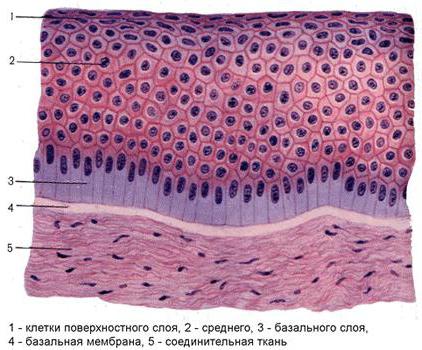
Consists of 3 layers:
- a) basal layer - cylindrical epithelial cells with weakly basophilic cytoplasm, often with a mitotic figure; in small quantities stem cells for regeneration;
- b) spinous (intermediate) layer - consists of a significant number of layers of spinose-shaped cells, the cells are actively dividing.
In the basal and spinous layers in epithelial cells, tonofibrils (bundles of tonofilaments made from keratin protein) are well developed, and between epithelial cells there are desmosomes and other types of contacts.
- c) integumentary cells (flat), aging cells, do not divide, gradually slough off from the surface.
- G Multilayered squamous epithelia have nuclear polymorphism:
- -nuclei of the basal layer are elongated, located perpendicular to the basement membrane,
- -the nuclei of the intermediate (spinous) layer are round,
- -the nuclei of the superficial (granular) layer are elongated and located parallel to the basement membrane.
- 2. Stratified squamous keratinization is the epithelium of the skin. It develops from the ectoderm, performs a protective function - protection from mechanical damage, radiation, bacterial and chemical exposure, demarcates the body from the environment.
- Ш In thick skin (palm surfaces), which is constantly under stress, the epidermis contains 5 layers:
- 1. basal layer- consists of prismatic (cylindrical) keratinocytes, in the cytoplasm of which keratin protein is synthesized, forming tonofilaments. Keratinocyte differon stem cells are also located here. Therefore, the basal layer is called germinal, or rudimentary.
- 2. stratum spinosum- formed by polygonal keratinocytes, which are tightly connected to each other by numerous desmosomes. In place of desmosomes on the surface of cells there are tiny projections - “spines” directed towards each other. In the cytoplasm of spinous keratinocytes, tonofilaments form bundles - tonofibrils, and keratinosomes - granules containing lipids appear. These granules are released into the intercellular space by exocytosis, where they form a lipid-rich substance that cements keratinocytes. In addition to keratinocytes, in the basal and spinous layers there are process-shaped melanocytes with granules of black pigment - melanin, intraepidermal macrophages (Langerhans cells) and Merkel cells, which have small granules and are in contact with afferent nerve fibers.
- 3. granular layer- the cells acquire a rhomboid shape, the tonofibrils disintegrate and the keratohyalin protein is formed inside these cells in the form of grains, this is where the process of keratinization begins.
- 4. shiny layer- a narrow layer, in which the cells become flat, they gradually lose their intracellular structure (not nuclei), and keratohyalin turns into eleidin.
- 5. stratum corneum- contains horny scales that have completely lost their cell structure, are filled with air bubbles, and contain the protein keratin. With mechanical stress and deterioration of blood supply, the process of keratinization intensifies.
- Ш In thin skin that does not experience stress, there is no granular and shiny layer.
- G The basal and spinous layers constitute the germinal layer of the epithelium, since the cells of these layers are capable of division.
- 4. Transitional (urothelium)
There is no nuclear polymorphism; the nuclei of all cells have rounded shapes. Sources of development: the epithelium of the pelvis and ureter - from the mesonephric duct (a derivative of the segmental legs), the epithelium of the bladder - from the endoderm of the allantois and the endoderm of the cloaca. The function is protective.
Lines hollow organs, the wall of which is capable of strong stretching (pelvis, ureters, bladder).
- - basal layer - made of small dark low-prismatic or cubic cells - poorly differentiated and stem cells, provide regeneration;
- - intermediate layer - made of large pear-shaped cells, with a narrow basal part, in contact with the basement membrane (the wall is not stretched, so the epithelium is thickened); when the wall of the organ is stretched, the pyriform cells decrease in height and are located among the basal cells.
- - cover cells - large dome-shaped cells; when the organ wall is stretched, the cells flatten; the cells do not divide and gradually exfoliate.
Thus, the structure of the transitional epithelium changes depending on the state of the organ:
- - when the wall is not stretched, the epithelium is thickened due to the “displacement” of some cells from the basal layer into the intermediate layer;
- - when the wall is stretched, the thickness of the epithelium decreases due to the flattening of the integumentary cells and the transition of some cells from the intermediate layer to the basal layer.
Histogenetic classification (according to sources of development) by N.G. Khlopin:
- 1. Epithelium of the skin type (epidermal type) [cutaneous ectoderm] - protective function
- - multilayered squamous non-keratinizing epithelium;
- - stratified squamous keratinizing epithelium (skin);
- - single-layer multirow ciliated epithelium of the airways;
- - transitional epithelium of the urethra;
- (epithelium of the salivary, sebaceous, mammary and sweat glands; alveolar epithelium of the lungs; epithelium of the thyroid and parathyroid glands, thymus and adenohypophysis).
- 2. Epithelia of the intestinal type (enterodermal type) [intestinal endoderm] - carries out the processes of absorption of substances, performs glandular function
- - single-layer prismatic epithelium of the intestinal tract;
- - epithelium of the liver and pancreas.
- - Renal type epithelium (nephrodermal) [nephrotome] - nephron epithelium; in different parts of the channel:
- - single-layer flat; or - single-layer cubic.
- - Epithelium of coelomic type (coelodermal) [splanchnotome] -
- - single-layer squamous epithelium of the serous integuments (peritoneum, pleura, pericardial sac);
- - epithelium of the gonads; - epithelium of the adrenal cortex.
- 4. Epithelium of neuroglial type / ependymoglial type / [neural plate] -
- - brain cavities;
- - retinal pigment epithelium;
- - olfactory epithelium;
- - glial epithelium of the hearing organ;
- - taste epithelium;
- - epithelium of the anterior chamber of the eye;
- 5. Angiodermal epithelium /endothelium/ (cells lining blood and lymphatic vessels, heart cavities) there is no consensus among histologists: some classify endothelium as single-layer squamous epithelium, others - as connective tissue with special properties. Source of development: mesenchyme.
Tissue is a collection of cells and intercellular substance. It has common structural features and performs the same functions. There are four types of tissues in the body: epithelial, nervous, muscle and connective.
The structure of epithelial and animal tissue is determined, first of all, by its localization. Epithelial tissue is a boundary layer of cells lining the integument of the body, the mucous membranes of internal organs and cavities. Also, many glands in the body are formed by epithelium.
general characteristics
The structure of epithelial tissue has a number of features inherent only to epithelium. The main feature is that the tissue itself looks like a continuous layer of cells that fit tightly together.
The epithelium lining all surfaces in the body has the appearance of a layer, while in the liver, pancreas, thyroid, salivary and other glands it is a cluster of cells. In the first case, it is located on top of the basement membrane, which separates the epithelium from the connective tissue. But there are exceptions when the structure of epithelial and connective tissue is considered in the context of their interaction. In particular, in the lymphatic system there is an alternation of epithelial and connective tissue cells. This type of epithelium is called atypical.
High regenerative capacity is another feature of the epithelium.
The cells of this tissue are polar, which is due to the difference in the basal and apical parts of the cell center.
The structure of epithelial tissue is largely explained by its border position, which, in turn, makes the epithelium an important link in metabolic processes. This tissue is involved in the absorption of nutrients from the intestines into the blood and lymph, in the excretion of urine through the epithelium of the kidneys, etc. We must also not forget about the protective function, which is to protect tissues from damaging influences.
The structure of the substance that forms the basement membrane shows that it contains a large amount of mucopolysaccharides, and also has a network of thin fibrils.
How is epithelial tissue formed?
The structural features of epithelial tissue in animals and humans are largely dictated by the fact that its development is carried out from all three. This feature is inherent only in this type of tissue. The ectoderm gives rise to the epithelium of the skin, oral cavity, a significant part of the esophagus, and the cornea of the eye; endoderm - epithelium of the gastrointestinal tract; and the mesoderm - the epithelium of the genitourinary organs and serous membranes.
In embryonic development, it begins to form at the earliest stages. Since the placenta contains a sufficient amount of epithelial tissue, it is a participant in the metabolism between the mother and the embryo.
Maintaining the integrity of epithelial cells
The interaction of neighboring cells in the layer is possible due to the presence of desmosomes. These are special multiple structures of submicroscopic size that consist of two halves. Each of them, thickening in certain places, occupies adjacent surfaces of neighboring cells. In the slit-like space between the halves of the desmosomes there is a substance of carbohydrate origin.
In cases where the intercellular spaces are wide, desmosomes are located at the ends of cytoplasmic protrusions directed towards each other on the contacting cells. If you examine a pair of these protrusions under a microscope, you will find that they have the appearance of an intercellular bridge.
In the small intestine, the integrity of the layer is maintained due to the fusion of the cell membranes of neighboring cells at the points of contact. Such places are often called end plates.
There are other cases where there are no special structures to ensure integrity. Then the contact of neighboring cells occurs due to the contact of smooth or curved cell surfaces. The edges of the cells may overlap each other in a tiled manner.
The structure of an epithelial tissue cell
Features of epithelial tissue cells include the presence of a plasma membrane on their surface.
In cells involved in the release of metabolic products, folding is observed in the plasma membrane of the basal part of the cell body.
Epithelial cells are the scientific name for cells that form epithelial tissue. The structural features and functions of epithelial cells are closely interrelated. So, according to their shape, they are divided into flat, cubic and columnar. Euchromatin predominates in the nucleus, due to which it has a light color. The nucleus is quite large, its shape coincides with the shape of the cell.
The pronounced polarity determines the location of the nucleus in the basal part, above it there are mitochondria, the Golgi complex and centrioles. In cells performing a secretory function, the endoplasmic reticulum and Golgi complex are especially well developed. The epithelium, which experiences a large mechanical load, has a system of special threads in its cells - tonofibrils, which create a kind of barrier designed to protect the cells from deformation.
Microvilli
Some cells, or rather their cytoplasm, on the surface can form tiny outgrowths directed towards the outside - microvilli. Their largest accumulations are found on the apical surface of the epithelium in the small intestine and the main sections of the convoluted tubules of the kidneys. Due to the parallel arrangement of microvilli in the cuticles of the intestinal epithelium and the brush border of the kidneys, stripes are formed that can be viewed under an optical microscope. In addition, microvilli in these places contain a number of enzymes.
Classification
The structural features of epithelial tissues of different localizations make it possible to classify them according to several criteria.
Depending on the shape of the cells, the epithelium can be cylindrical, cubic and flat, and depending on the location of the cells - single-layer and multilayer.

Glandular epithelium is also isolated, which performs a secretory function in the body.

Single layer epithelium
The name of single-layer epithelium speaks for itself: in it, all cells are located on the basement membrane in one layer. If the shape of all cells is the same (that is, they are isomorphic) and are at the same level, then they speak of single-row epithelium. And if in a single-layer epithelium there is an alternation of cells of various shapes, their nuclei are located at different levels, then this is a multirow or anisomorphic epithelium.

Stratified epithelium
In stratified epithelium, only the bottom layer is in contact with the basement membrane, and the other layers are above it. Cells of different layers differ in shape. The structure of epithelial tissue of this type allows us to distinguish several types of multilayered epithelium depending on the shape and condition: stratified squamous, multilayered keratinized (there are keratinized scales on the surface), multilayered non-keratinized.

There is also the so-called transitional epithelium, lining the organs of the excretory system. Depending on whether or not it is stretched, the fabric takes on a different appearance. Thus, when the bladder is stretched, the epithelium is in a thinned state and forms two layers of cells - basal and integumentary. And when the bladder is in a compressed (shortened) form, the epithelial tissue thickens sharply, the cells of the basal layer become polymorphic and their nuclei are at different levels. The integumentary cells acquire a pear-shaped shape and are layered on top of each other.
Histogenetic classification of epithelia
The structure of epithelial tissue in animals and humans often becomes the subject of scientific and medical research. In these cases, the histogenetic classification developed by Academician N. G. Khlopin is used more often than others. According to it, there are five types of epithelium. The criterion is from which rudiments the tissue developed during embryogenesis.
1. Epidermal type, which originated from the ectoderm and prechordal plate.
2. Enterodermal type, the development of which originated from the intestinal endoderm.
3. Coelonephrodermal type, developed from the coelomic lining and nephrotome.
4. Angiodermal type, the development of which began from a section of mesenchyme that forms the vascular endothelium, which is called angioblast.
5. Ependymoglial type, which originated from the neural tube.
Features of the structure of epithelial tissues forming glands
The glandular epithelium performs a secretory function. This type of tissue is a collection of glandular (secretory) cells called granulocytes. Their function is to carry out synthesis, as well as the release of specific substances - secrets.
It is thanks to secretion that the body is able to perform many important functions. The glands secrete secretions on the surface of the skin and mucous membranes, inside the cavities of a number of internal organs, as well as into the blood and lymph. In the first case, we are talking about exocrine, and in the second, about endocrine secretion.

Exocrine secretion allows the production of milk (in the female body), gastric and intestinal juices, saliva, bile, sweat and sebum. The secretions of the endocrine glands are hormones that perform humoral regulation in the body.
The structure of epithelial tissue of this type can be different due to the fact that granulocytes can take different shapes. It depends on the phase of secretion.
Both types of glands (endocrine and exocrine) can consist of a single cell (unicellular) or many cells (multicellular).
Histology.
Cell: structure, properties. Fabrics: definition, properties. Epithelial, connective, muscle tissue: position, types, structure, significance. Nervous tissue: position, structure, meaning.
The human body is a complex, integral, self-regulating and self-renewing system, which is characterized by a certain organization of its structure. The basis of human structure and development is cell– an elementary structural, functional and genetic unit of a living organism, capable of division and exchange with the environment.
The human body is built of cells and non-cellular structures, united during development into tissues, organs, organ systems and the whole organism. There are a huge number of cells in the human body (10-14), and their size ranges from 5-7 to 200 microns. The largest are the egg and nerve cells (up to 1.5 m together with processes), and the smallest are blood lymphocytes. The science that studies the development, structure and functions of cells is called cytology. The shape of cells, as well as their size, is very diverse: flat, cubic, round, elongated, stellate, spherical, spindle-shaped, which is determined by the function they perform and the conditions of their life.
All cells have a common structural principle. The main parts of a cell are: the nucleus, the cytoplasm with the organelles located in it, and the cytolemma (plasmalemma, or cell membrane).
Cell membrane is a universal biological membrane that ensures the constancy of the internal environment of the cell by regulating the metabolism between the cell and the external environment - it is the transport (transport of necessary substances into and out of the cell) and barrier-receptor system of the cell. With the help of the plasmalemma, special cell surface structures are formed in the form of microvilli, synapses, etc.
Inside the cell is core– the control center of the cell and the regulator of its vital functions. Usually a cell has one nucleus, but there are also multinucleated cells (in the epithelium, vascular endothelium) and non-nucleated cells (erythrocytes and platelets). The nucleus has a nuclear envelope, chromatin, a nucleolus and nuclear sap (nucleoplasm). The nuclear envelope separates the nucleus from the cytoplasm and is actively involved in the exchange of substances between them. Chromatin contains proteins and nucleic acids (chromosomes are formed during cell division). The nucleolus takes part in the synthesis of cellular proteins.
Cytoplasm is the content of the cell and makes up 1-99% of its mass. It contains the nucleus and organelles, products of intracellular metabolism. Cytoplasm unites all cellular structures and ensures their chemical interaction with each other. It consists of proteins (from which cellular structures are built), fats and carbohydrates (a source of energy), water and salts (they determine the physicochemical properties of the cell, create osmotic pressure and its electrical charge) and nucleic acids (participation in protein biosynthesis).
Cytoplasmic organelles. Organelles are microstructures of the cytoplasm that are present in almost all cells and perform vital functions.
Endoplasmic reticulum - a system of tubules, vesicles, the walls of which are formed by cytoplasmic membranes. There are granular and agranular (smooth) endoplasmic reticulum. The agranular endoplasmic reticulum takes part in the synthesis of carbohydrates and lipids, the granular endoplasmic reticulum takes part in the synthesis of protein, because Ribosomes are located on the membranes of the granular endoplasmic reticulum, which can also be located on the nuclear membrane or freely in the cytoplasm. Ribosomes carry out protein synthesis, and in an hour they synthesize more protein than their total mass.
Mitochondria– power energy stations of the cell. In mitochondria, the breakdown of glucose, amino acids, fatty acids and the formation of ATP, the universal cellular fuel, occurs.
Golgi complex– has a mesh structure. Its function is to transport substances, process them chemically and remove waste products from the cell outside the cell.
Lysosomes– contain a large number of hydrolytic enzymes involved in the process of intracellular digestion of nutrients entering the cell, destroyed parts of the cell, and foreign particles that have entered the cell. Therefore, there are especially many lysosomes in cells that take part in phagocytosis: leukocytes, monocytes, liver cells, and small intestine.
Cell center represented by two centrioles located directly at the geometric center of the cell. During mitosis, microtubules of the mitotic spindle diverge from the centrioles, providing orientation and movement of chromosomes, and the zona radiata is formed, and the centrioles form cilia and flagella.
Flagella and cilia are special-purpose organelles - designed to move specialized cells (spermatozoa) or determine the movement of fluid around the cell (epithelial cells of the bronchi, trachea).
Cell properties:
1. Metabolism (metabolism) is a set of chemical reactions that form the basis of the life of a cell.
2. Irritability - the ability of cells to respond to changes in environmental factors (temperature, light, etc.) Cell reaction - movement, increased metabolism, secretion, muscle contraction, etc.
3. Growth – increase in size, development – acquisition of specific functions
4. Reproduction – the ability to reproduce itself. The basis for the preservation and development of cells, the replacement of aging and dead cells, the regeneration (restoration) of tissues and the growth of the body (many cells that perform complex functions have lost the ability to divide, but the appearance of new cells occurs only through the division of cells that are capable of dividing). Physiological regeneration– the process of death of old cells in tissues and the appearance of new ones.
There are two main forms of cell division: mitosis (the most common, ensures an even distribution of hereditary material between daughter cells) and meiosis (reduction division, observed during the development of only germ cells).
The period from one cell division to another is its life cycle.
In addition to cells, the human body also contains non-cellular structures: symplast and intercellular substance. Symplast, unlike cells, contains many nuclei (striated muscle fibers). The intercellular substance is secreted by cells and is located in the spaces between them.
Intercellular (tissue) fluid is replenished by the liquid part of the blood that leaves the bloodstream, the composition of which changes.
Cells and their derivatives are combined into tissues. Textile is a system of cells and non-cellular structures united by a unity of origin, structure and function. Histology- a science that studies the structure of a person at the tissue level.
In the process of evolution, as the needs of the body became more complex, specialized cells appeared that were capable of performing certain functions. The ultrastructure of these cells changed accordingly. The process of tissue formation is long, it begins in the prenatal period and continues throughout a person’s life. The interaction of the organism with the external environment that has developed in the process of evolution and the need to adapt to living conditions led to the emergence of 4 types of tissues with certain functional properties:
1. epithelial,
2. connecting,
3. muscular and
4. nervous.
All types of tissues of the human body develop from three germ layers - mesoderm, ectoderm, endoderm.
In the body, tissues are interconnected morphologically and functionally. The morphological connection is due to the fact that different tissues are part of the same organs. Functional connection is manifested in the fact that the activities of different tissues that make up the organs are coordinated. This consistency is due to the regulatory influence of the nervous and endocrine systems on all organs and tissues - the neurohumoral mechanism of regulation.
Epithelial tissue
Epithelial tissue (epithelium) covers:
1. The entire outer surface of the human and animal body
2. All body cavities, lining the mucous membranes of hollow internal organs (stomach, intestines, urinary tract, pleura, pericardium, peritoneum)
3. Part of the endocrine glands.
Functions:
1. metabolic function - participates in the metabolism between the body and the external environment, absorption (intestinal epithelium) and excretion (renal epithelium, gas exchange (lung epithelium);
2. protective function (skin epithelium) – protection of underlying structures from mechanical, chemical influences and infections;
3. delimiting;
4. secretory – glands.
Features:
1. Located on the border between the external and internal environments of the body
2. Consists of epithelial cells forming continuous layers. Cells are closely connected to each other.
3. Poor development of the intercellular substance is characteristic.
4. there is a basement membrane (a carbohydrate-protein-lipid complex with the finest fibrils that separates the epithelial tissue from the underlying loose connective tissue)
5. cells have polarity (the apical and basal parts differ in structure and function; and in multilayered epithelium there are differences in the structure and function of the layers). Epithelial cells may have organelles for special purposes:
Ø cilia (airway epithelium)
Ø microvilli (intestinal and kidney epithelium)
Ø tonofibrils (skin epithelium)
6. There are no blood vessels in the epithelial layers. Cell nutrition is carried out by the diffusion of nutrients through the basement membrane, which separates the epithelial tissue from the underlying loose connective tissue and serves as a support for the epithelium.
7. Has great regenerative capacity (has a high ability to recover).
Classification of epithelial tissue:
By function differentiate :
1. integumentary;
2. glandular epithelium.
IN integumentary epithelia are divided into single-layer and multilayer epithelium.
1. In single-layer epithelium, all cells are located on the basement membrane in one row,
2. in multilayer – several layers are formed, while the upper layers lose contact with the basement membrane (lining the outer surface of the skin, the mucous membrane of the esophagus, the inner surface of the cheeks, the vagina).
Multilayer epithelium is:
Ø keratinizing(skin epithelium)
Ø non-keratinizing(epithelium of the cornea of the eye) – keratinization is not observed in the surface layer, unlike the keratinizing epithelium.
A special form of stratified epithelium - transition epithelium, which is located in organs that can change their volume (are subject to stretching) - in the bladder, ureters, renal pelvis. The thickness of the epithelial layer changes depending on the functional state of the organ
Single-layer epithelium can be single- or multi-row.
Based on the shape of the cells, they are distinguished:
Ø single-layer squamous epithelium (mesothelium)– consists of one layer of sharply flattened cells of a polygonal shape (polygonal); The base (width) of the cells is greater than the height (thickness). Covers the serous membranes (pleura, peritoneum, pericardium), walls of capillaries and blood vessels, and alveoli of the lungs. Carries out diffusion of various substances and reduces friction of flowing liquids;
Ø single layer cuboidal epithelium - when cut, the cells are as wide as they are high; they line the ducts of many glands, form kidney tubules, small bronchi, and perform a secretory function;
Ø single layer columnar epithelium– in a section, the width of the cells is less than the height; they line the stomach, intestines, gallbladder, kidney tubules, and are part of the thyroid gland.
Depending on the characteristics of the structure and function, they are distinguished:
Ø single-layer prismatic ferrous– present in the stomach, in the cervical canal, specialized for the continuous production of mucus;
Ø single-layer prismatic edged– lines the intestine, on the apical surface of the cells there is a large number of microvilli, specialized for absorption;
Ø single layer ciliated epithelium- often prismatic multirow, the cells of which have outgrowths at the upper, apical end - cilia, which move in a certain direction, creating a flow of mucus. Lines the respiratory tract, fallopian tubes, ventricles of the brain, and spinal canal. Provides transport of various substances. It contains the following types of cells:
1. short and long intercalary cells (poorly differentiated and among them stem cells; provide regeneration);
2. goblet cells – do not perceive dyes well (white in the preparation), produce mucus;
3. ciliated cells - have ciliated cilia on the apical surface; purify and humidify the passing air.
Glandular epithelium makes up the bulk of the glands, the epithelial cells of which are involved in the formation and secretion of substances necessary for the life of the body. Glands are divided into exocrine and endocrine. Exocrine glands secrete secretions into the cavities of internal organs (stomach, intestines, respiratory tract) or onto the surface of the body - sweat, salivary, mammary, etc., endocrine glands do not have ducts and secrete secretions (hormone) into the blood or lymph - pituitary gland, thyroid and parathyroid glands glands, adrenal glands.
By structure, exocrine glands can be tubular, alveolar, or combined - tubular-alveolar.
Epithelial tissue covers the entire outer surface of the human body and lines all body cavities. It lines the mucous membrane of hollow organs, serous membranes, and is part of the glands of the body. Therefore, they distinguish integumentary and glandular epithelium.
Epithelial tissue is located at the border between the external and internal environments of the body. And participates in the metabolism between the body and the external environment. Performs protective role (skin epithelium). Performs functions suction(intestinal epithelium), discharge(epithelium of the renal tubules), gas exchange(epithelium of the lung alveoli). This fabric has high regeneration. glandular epithelium, which forms glands, capable of releasing secrets. This ability to produce and secrete substances necessary for life is called secretion. This epithelium is called secretory.
Distinctive features of epithelial tissue:
-Epithelial tissue is located at the border between the external and internal environment of the body.
- It consists of epithelial cells, these cells form continuous layers.
- In these layers there are no blood vessels.
-Nutrition this tissue occurs by diffusion through the basement membrane, which separates the epithelial tissue from the underlying loose connective tissue and serves as a support for the epithelium.
IN integumentary epithelia secrete single-layered epithelium and multilayered.
IN single-layer epithelia all cells are located on the basement membrane.
IN multilayer epithelia Only the lower layer of cells lies on the basement membrane. The upper layers lose connection with it and form several layers.
Single layer epithelium happens single-row and multi-row.
Epithelial cells – epithelial cells. In epithelial cells they secrete two parts. 1. Basal part is directed towards the underlying tissue. 2. Apical part is facing the free surface. In the basal part lies the nucleus.
The apical part contains organelles, inclusions, microvilli, and cilia. According to the shape of the cells, the epithelium is flat, cubic, cylindrical (prismatic).
Rice. No. 1. Types of epithelium.
Single layer squamous epithelium– mesothelium – covers the serous membranes – pleura, epicardium, peritoneum.
Single layer squamous epithelium – endothelium – lines mucous membrane circulatory and lymphatic vessels.
Single layer cubic epithelium covers renal tubules, excretory ducts of glands And small bronchi.
Single layer prismatic epithelium lines gastric mucosa.
Single layer prismatic edged epithelium lines intestinal mucosa.
Single layer multi-row prismatic ciliated epithelium covers fallopian tubes and respiratory tract.
Stratified squamous epithelium Based on the keratinization of the upper layers of cells, they are divided into keratinizing and non-keratinizing.
Stratified squamous keratinizing epithelium – epidermis. It covers the surface of the skin. The epidermis consists of many dozens of layers of cells. On the surface of the skin, cells die, turning into horny scales. The nucleus and cytoplasm are destroyed in them and keratin accumulates.
Stratified squamous non-keratinizing epithelium lines the cornea of the eye, oral cavity, and esophagus.
There is a transitional form of multilayer epithelium - transition. It covers the urinary tract - renal pelvis, bladder, i.e. organs that can change their volume.
Glandular epithelium makes up the bulk of the body's glands. Glands in the body perform a secretory function. The secretion it secretes is necessary for the processes occurring in the body. Some glands are independent organs, for example the pancreas and major salivary glands. Other glands are part of organs, for example, glands of the intestinal wall and stomach. Most glands are derivatives of epithelium.
There are glands external secretion - exocrine. They have excretory ducts and secrete their secretions into the body cavity or onto the surface of the body. These are mammary glands, sweat glands, salivary glands.
Eat Endocrine glands are endocrine. They do not have excretory ducts and secrete their secretions into the internal environment of the body - blood or lymph. Their secret is hormones.
There are glands of mixed secretion. They have endocrine and exocrine parts, such as the pancreas.
Figure No. 2. Types of glands.
Exocrine glands are very diverse. Highlight unicellular and multicellular glands.
Unicellular glands– goblet cells, located in the epithelium of the intestine; they produce mucus in the respiratory tract.
In multicellular glands there are secretory section and excretory duct. The secretory department consists of cells - glandulocytes, which produce secretions. Depending on whether the excretory duct branches or not, simple and complex glands.
According to the shape of the secretory department, they are distinguished tubular, alveolar and alveolar-tubular glands.
Depending on how the secretion is formed and how it is released from the cells, there are merocrine, apocrine and holocrine glands.
Merocrine glands are the most common. They release their secretion into the duct without destroying the cytoplasm of the secretory cells.
In apocrine glands, partial destruction of the cytoplasm of secretory cells occurs. The apical part of the cell is destroyed and becomes part of the secretion. Then the destroyed cell is restored. These glands include the mammary and sweat glands.
In holocrine glands, secretion is accompanied by cell death. These destroyed cells are the secretion of the gland. These glands include the sebaceous glands.
By the nature of the secret distinguish between mucous, protein and mixed (protein-mucous) glands.


