Foreign bodies in the body of animals. Foreign bodies of the gastrointestinal tract in cats and dogs. How does a foreign body enter the body?
A foreign body of the stomach and intestines is any object, of both food and non-food origin, that is swallowed but cannot leave the body naturally (through vomiting or defecation). Examples of foreign bodies can be objects such as large pieces of bones that cannot be digested, various toys, New Year's decorations (usually tinsel), ropes and threads, clumps of glued hair, stones, various metal objects (usually coins) and some other objects.
Animals are more likely to eat objects as a result of curiosity, which is more common in young animals. Sometimes foreign bodies enter the animal's body when eating food products intended for humans and contained in various types of packaging. Also, eating foreign bodies can develop as a result of pathological appetite against the background of certain diseases.
Clinical signs
The main symptom of foreign bodies in the stomach and intestines is loss of appetite and vomiting. Due to the fact that the body is able to secrete fluid into the cavity of the gastrointestinal tract, vomiting can occur even though the animal does not take food or water. Continued loss of fluid leads to dehydration of the animal, which manifests itself in dry mucous membranes of the oral cavity, decreased skin elasticity and sunken eyeballs. Due to the fact that with vomiting, electrolytes (sodium, potassium, etc.) necessary for normal life are lost, the animal develops muscle weakness and depression. Other possible signs include fever (high temperature), diarrhea and abdominal pain.
Diagnostics
In case of a foreign body of the stomach and intestines, sometimes it can be felt (palpated) in the abdominal cavity, or changes in the shape of the intestine (seals, abnormal location, etc.) characteristic of a foreign body can be detected. In some cases, a foreign body does not cause any changes that can be detected by palpation and therefore, with a simple examination, the presence of a foreign body cannot be completely excluded.
The diagnostic test of choice when a foreign body is suspected is a radiographic (x-ray) examination of the abdominal organs, in which radiopaque objects (eg metal objects, stones) are easily identified or changes in internal organs characteristic of a foreign body are determined. Also, if a foreign body is suspected, a radiographic examination can be performed using contrast, when a substance (usually barium) is injected and its progress through the stomach and intestines is monitored. A gastric foreign body is best identified by endoscopic examination.
Other diagnostic tests used in diagnosing a foreign body may include studies such as ultrasound of internal organs and laboratory tests of blood and urine. Sometimes, a foreign body can only be detected during a diagnostic laparotomy - when the animal's abdominal cavity is opened under anesthesia and the internal organs are examined through inspection and palpation.
Treatment and prognosis
The basis of treatment is the removal of foreign bodies from the stomach and intestines. In most cases, surgical removal of foreign bodies is performed; in rare cases, a foreign body from the stomach can be removed endoscopically. If the animal is severely dehydrated, dehydration is corrected before surgery through intravenous administration of electrolyte solutions.
Prognosis depends on the timeliness of diagnosis and treatment. If you seek help early, the prognosis is often favorable and after surgical treatment the animal makes a full recovery. If a foreign body remains in the gastrointestinal tract for a long time, the likelihood of developing perforation (perforation) of the wall of the stomach and/or intestines and irreversible complications increases.
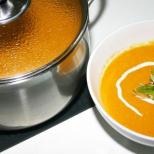
6-year-old cat Stepan was taken to the veterinary clinic on New Year's Eve due to vomiting. When palpating the abdomen, characteristic changes in the intestine were detected and a decision was made to perform a diagnostic laparotomy. During the operation, an accumulation of tinsel was discovered, which was safely removed (upper left corner). Christmas decorations are perhaps the most common cause of foreign body formation in cats.

X-ray of a cat brought to the veterinary clinic due to vomiting; it reveals changes in the intestines characteristic of a linear foreign body. The decision was made to undergo surgery (see below).

View of the intestine removed during surgery, it is strung on a thread like a garland. The operation was carried out in a timely manner; if the operation is refused, intestinal perforation with a thread develops, which leads to irreversible consequences. (see below).

Type of propylene thread extracted from the intestines. After the operation, the cat recovered safely.
Veterinary clinic of Dr. Shubin, Balakovo.
When our pets get sick, we often find ourselves helpless. What was the cause of the disease: unbalanced nutrition, improper placement of the cell, or something else? Is this an acute disease?
This reference book will allow you to quickly assess errors in housing and feeding; it discusses the main symptoms of diseases and provides methods for their treatment.
Experienced veterinarian S. Kaiser describes common diseases of dogs, cats, rabbits, guinea pigs, hamsters and rats, songbirds and budgies, turtles and ornamental fish, modern therapeutic possibilities of allopathy, herbal medicine and homeopathy. Particular attention is paid to home-based treatment.
Veterinarians, pharmacists and pet owners will discover a treasure trove of essential tips and practical guidelines.
This reference book will be a guide that describes the most commonly used treatments through allopathy, homeopathy and herbal medicine.
1100 rub
Neurology of dogs and cats. A reference guide for practicing veterinarians
This is a reference guide to common neurological problems in dogs and cats, providing step-by-step procedures for each disease, including neurological examination, diagnostic tests, important diagnostic, therapeutic and prognostic principles.
The book makes extensive use of intensive care reference tables for treatment with specific drugs and indicates their doses, treatment regimens, as well as tips and warnings that highlight common problems that arise in practice and ways to solve them.
1502 rub

This reference book briefly presents the anatomical, topographical and physiological characteristics of the genital organs of females and males, describes the physiology and diagnosis of pregnancy, diseases of pregnant women, childbirth and delivery operations, complications during childbirth and the postpartum period, postpartum diseases, diseases and care for newborns, breast pathology animal glands. The main attention is paid to the most common animal diseases: mastitis, diseases of the uterus and ovaries; Each disease is defined, its causes, symptoms, diagnosis, treatment and prevention are indicated.
The directory is intended for veterinary specialists (doctors, paramedics, etc.), livestock specialists and artificial insemination technicians.
407 rub

The main diseases, methods of diagnosis, treatment and prevention of domestic and exotic animals, ornamental and songbirds, animals and birds living in living areas are described. Personal protection and safety measures for people caring for these animals and birds are recommended. For a wide range of readers and veterinary specialists.
404 rub

Horse breeding. A practical guide to curing horse diseases and recognizing it by external examination
This book, compiled by veterinarian P.A. Bobarykin, is a reference book on horse breeding and the treatment of horse diseases. The book begins with a brief sketch of the natural history of the horse and the general characteristics of the various breeds. The external and internal structure of the horse is described in detail, and the various temperaments of horses are discussed. Next, the author talks about the horse farm, its structure, caring for horses and breeding horse breeds, keeping mares after giving birth and raising foals. The second half of the book is entirely devoted to horse diseases and methods of treating them. The signs and properties of various diseases are described, and medicines for each disease are given in the form of recipes in Latin and Russian. The publication contains many illustrations.
The book, written about a century and a half ago (the first edition was published in 1869), will still be useful today to specialists in the field of horse breeding and horse breeding, zoologists, livestock specialists, veterinarians, as well as a wide range of interested readers.
779 rub

This passport complies with the requirements of the International Office of Epizootics, based on the principles established by expert panels of the World Health Organization and the World Food and Agriculture Organization of the United Nations.
50 rub

Ferret diseases
Veterinarians have recently begun to see ferrets more and more often at appointments, so there is a need for universal and easily accessible information on the health, diseases and maintenance of these animals.
This book is a useful and comprehensive guide to the care and maintenance of domestic ferrets. Part 1 is devoted to biology and animal husbandry; Part 2 contains material on a systematic approach to the diagnosis and treatment of the most common diseases; Part 3 contains information on anesthesia, general surgery, sampling and administration techniques, fixation and clinical examination; Part 4 contains tables on physiological data and drug doses.
The book contains a wealth of useful information and is intended for veterinarians, animal care professionals and anyone involved in any way with ferrets.
456 rub

Canine obstetrics and gynecology
Written for veterinary surgeons, veterinary students, and breeders, this book examines the various conditions in dogs that affect their ability to reproduce. The book contains complete information on this issue. It outlines the anatomy, physiology and endocrinology of the reproductive system of males and females, as well as methods of treating the most common diseases of this system.
The purpose of this publication is to highlight the most significant aspects of the therapy and surgery of pathological conditions that impair the reproductive function of offspring.
689 rub

Emergency and intensive care for small animals
The book EMERGENCY CARE AND INTENSIVE CARE FOR SMALL DOMESTIC ANIMALS is a reference manual dedicated to providing assistance in the most common critical conditions. The book consists of two sections. First, the basic principles of initial stabilization and maintenance of the vital functions of the entire animal body are described, and then a systematic approach to the most common critical conditions. Each section begins with a clinical approach to a patient presenting with signs of critical illness in each body system, and then describes treatment regimens for specific diseases. This guide also contains information on monitoring and management of critically ill patients.
The material in the book is presented in the form of small abstracts so that you can quickly find the necessary information. All information is numbered and contains numerous references to ensure that the various emergency conditions are described as completely as possible. The book also contains many useful formulas, tables, illustrations and drug doses.
Therapy and surgery for puppies and kittens. Veterinary practice
This book offers a comprehensive overview of canine and feline veterinary neonatology, including detailed descriptions of therapeutic and surgical techniques and techniques related to the problems associated with this particular area of veterinary practice. All common and numerous veterinary problems of puppies and kittens are discussed in great detail. Diseases are considered from the point of view of etiology, pathophysiology, diagnosis and treatment.
Summary tables and detailed figures make this book an even more valuable reference. Descriptions of surgical procedures are given very accurately and are accompanied by supporting illustrations.
This book will become an indispensable guide for all veterinarians and veterinary students.
1363 rub
Foreign bodies in the esophagus in cats and dogs are the most common problem encountered by veterinarians. Often, when pets play, they find small toys, plastic bags, wire, thread, small bones and other items that are not suitable for food. Large and sharp objects can injure the walls of the stomach and cause bleeding. Owners who are not competent in terms of treatment can harm the animal. Such interventions often lead to death.
Symptoms:
- poor appetite or refusal to eat;
- when palpating the abdomen, pain occurs, which is accompanied by distortion of behavior;
- repeated vomiting;
- change in stool frequency or its complete absence;
- lethargy and drowsiness.
If at least 3 of the above symptoms coincide, you should immediately consult a doctor. Under no circumstances should you take any action to save the animal yourself.
Diagnostics
A qualitative examination can be done in a veterinary clinic. To do this, the pet is shown to a doctor and consulted with him. There are several ways to identify an object and its location.
Types of diagnostics
- Ultrasound of the abdominal cavity. This method is not always effective and will only indirectly indicate the presence of a problem.
- X-ray. Diagnostics is carried out in two types - direct and lateral. For a more accurate result, the picture is taken in vertical and horizontal projection. The image shows objects that do not require the injection of a contrast agent (metal, rubber, bones, paper clips).
- To reveal elements that are not visible on x-rays - threads, fabric, plastic - a contrast agent (barium sulfate solution) is introduced.
- Ultrasound examination. Allows you to determine the state of intestinal motility or lumen patency.
- An infection test is required to identify pathologies.
Treatment
Each case is individual. If the object is small and does not have sharp edges, the doctor may not do anything. In most cases, the foreign body comes out naturally naturally. They try to induce vomiting in the animal or use special instruments to remove small things. Large and sharp objects are removed through surgery.
Prevention
- The room in which the animal is located must be kept clean.
- The feed is checked for the presence of foreign objects.
- When walking, pay attention to ensure that your pet does not pick up garbage, bones, or small parts.
- There should be no objects that could harm the pet on the floor or in places accessible to the pet.
- Select toys that are suitable in size, shape and material.
- Brush the animal regularly.
If you go to the hospital quickly, the chance of survival increases significantly if the object that got inside poses a real threat to the animal.
Foreign objects in the body of cats: symptoms and treatment methods
The legendary vitality of cats has given rise to a myth according to which these animals practically do not get sick. These furry pets are credited with 13 lives, and even their owners believe that the animal is capable of healing itself. Cats may be capable of self-healing in the wild, but domestic animals are not endowed with this unique ability. The peculiarity of any disease in cats is that they are able to hide their illness for a long time, but over time, the disease can manifest itself with serious consequences.
Foreign bodies in the animal's body
Often foreign bodies can get stuck in the oral cavity. Fortunately, of all the problems that occur when objects enter a cat's body, getting a foreign body out of the mouth is the easiest.
Symptoms
The animal refuses to eat food and may rub its muzzle on different surfaces or rub its paw on its cheek. Vomiting and difficulty swallowing are also observed.
Treatment methods
You can remove a foreign object using your fingers, of course, while observing safety precautions. After this, using a spray, the oral cavity is sprayed with a solution of potassium permanganate or modern septic tanks. You should not give solid food during the first 24 hours.
Foreign objects in the larynx
Often contact with glass, nails and other sharp objects causes an inflammatory process.
Symptoms
If there is significant damage to soft tissue, bleeding may occur. The animal refuses food, the body temperature rises, asphyxia and a painful cough are observed.
Treatment methods
Only a specialist can make a diagnosis after a thorough examination of the soft tissues, which is carried out by palpation and sometimes fluoroscopy. Removal of the object is carried out under anesthesia. If suffocation occurs, a tracheotomy is performed.
The owner needs to know that in such cases the animal cannot be fed for two days. Therefore, nutritional enemas are used. In addition, the rehabilitation period is carried out using antibiotics, which are administered intravenously.
Foreign objects in the esophagus
A cat can swallow a foreign body that can block the passage in the initial part of the esophagus or pass further. Based on this, the degree of the pathological process will depend, since the object will block the esophagus partially or completely. If the process continues for a long time, it is accompanied by secondary diseases, such as necrosis of the walls.
The animal may retain its appetite, but eating in almost all cases is accompanied by vomiting. The diagnosis can only be established in a veterinary clinic using fluoroscopy and probing. In some situations, veterinarians use emetics, but if sharp objects become stuck, surgery is required. After surgery, liquid food is given only on the third day. Full nutrition is possible only after two weeks.
Don't forget that a cat is a living creature, just like you. Therefore, she may even suffer from a common cold..jpg" alt="4983b1dc86154dff8a421f2bc116c139_i-2230" width="600" height="450" />!}
Foreign bodies of the gastrointestinal tract in cats and dogs
Foreign bodies in the gastrointestinal tract of pets are unfortunately a common and very serious problem. An error in diagnosis has extremely adverse consequences. In such cases, it is extremely important to contact a specialist without delay at a well-equipped veterinary clinic.
The Vetus clinic performs successful operations to remove foreign bodies from the gastrointestinal tract of cats and dogs:
We use the best informative methods of clinical and instrumental diagnostics, allowing us to quickly and accurately identify pathology and begin treatment;
The combination of ultrasound and endoscopic examination methods, as well as plain radiography, makes it possible to accurately differentiate this diagnosis from pathologies with similar symptoms;
During the operation to remove a foreign body from the gastrointestinal tract, the animal receives modern high-quality anesthesia, which allows it to come out of sleep more softly and easily;
After the operation, you will receive advice from leading clinic specialists about postoperative care measures for the animal;
Don't let the situation go! At the first signs of pathology, immediately contact the Vetus clinic!
24/7 telephone:
A typical picture of the disease is:
- vomiting and impaired evacuation of stomach contents into the small intestine;
- refusal to eat for a long time, or prolonged vomiting immediately after eating.
As a result, there is a need for differential diagnosis with a number of conditions, primarily infectious processes, food poisoning, etc.
The set of diagnostic measures includes:
- plain radiography of the abdominal organs of a dog or cat;
- ultrasound examination of the gastrointestinal tract for pendulum movements;
- some objects can be identified by palpation, but this must be done very carefully, as they may have sharp edges (needles, stones, fishhooks, glass) and can easily injure the mucous membrane of the organ.
The task of radiography of the digestive organs is to detect the foreign body itself, as well as to identify signs of the consequences of its presence in the lumen of the gastrointestinal tract, which pose a direct threat to the life and health of the patient.
Survey radiography of the abdominal cavity is correctly performed in the lateral projection - on the right side and in the direct projection - on the back.
The variety of objects that become the contents of the gastrointestinal tract is striking in its diversity. These are Christmas tree decorations, balls, rubber and plastic toys, needles, fishhooks, glass, wool, wires, metal objects and, of course, bones. All of them can be divided into radiopaque and radiopaque or radionegative. The vast majority of such items belong to the first category, i.e. visible on radiographs, however, the mass of soft tissue surrounding the foreign body and the chemical processes to which it is exposed in the stomach under the influence of “food juices” can lead to a significant deterioration in visualization.
Main consequences:
- A diverticulum (pocket) in the wall of the esophagus.
- Traumatic gastritis.
- Intestinal obstruction (partial or complete).
- Necrosis of the intestinal wall, in the place where the foreign body is located.
- Intestinal perforation (rupture).
- Peritonitis.
- Death.

After diagnosis, several options are possible to solve this problem.
The first and less traumatic operation for the animal is endoscopic removal of foreign bodies from the esophagus and stomach, using a fiber-optic gastroscope with additional manipulators. The foreign body is captured and carefully removed from the cavity of the esophagus or stomach, depending on the location.
If objects are in the lumen of the small intestine or they cannot be reached using a gastroscope, abdominal surgery is performed, during which the foreign object is localized and then removed.
The main and most important task for the owner is to never leave small toys, threads, needles and many other items that animals can swallow in places accessible to them. While walking, carefully monitor your animal and do not let it pick up any objects on the street.