Where are nerve cells found in humans? Neurons and nervous tissue. Myelinated nerve fibers
Nerve cell Not to be confused with neutron.
Pyramidal cell neurons in the mouse cerebral cortex
Neuron(nerve cell) is a structural and functional unit of the nervous system. This cell has a complex structure, is highly specialized and in structure contains a nucleus, a cell body and processes. There are more than one hundred billion neurons in the human body.
Review
The complexity and diversity of the nervous system depends on the interactions between neurons, which in turn represent a set of different signals transmitted as part of the interaction of neurons with other neurons or muscles and glands. Signals are emitted and propagated by ions that generate an electrical charge that travels along the neuron.
Structure

Cell body
A neuron consists of a body with a diameter of 3 to 100 μm, containing a nucleus (with a large number of nuclear pores) and other organelles (including a highly developed rough ER with active ribosomes, the Golgi apparatus), and processes. There are two types of processes: dendrites and axons. The neuron has a developed cytoskeleton that penetrates its processes. The cytoskeleton maintains the shape of the cell; its threads serve as “rails” for the transport of organelles and substances packaged in membrane vesicles (for example, neurotransmitters). A developed synthetic apparatus is revealed in the body of the neuron; the granular ER of the neuron is stained basophilically and is known as the “tigroid”. The tigroid penetrates the initial sections of the dendrites, but is located at a noticeable distance from the beginning of the axon, which serves as a histological sign of the axon.
There is a distinction between anterograde (away from the body) and retrograde (toward the body) axon transport.
Dendrites and axon

Neuron structure diagram
Synapse
Synapse- the site of contact between two neurons or between a neuron and the effector cell receiving the signal. It serves to transmit a nerve impulse between two cells, and during synaptic transmission the amplitude and frequency of the signal can be adjusted. Some synapses cause depolarization of the neuron, others cause hyperpolarization; the former are excitatory, the latter are inhibitory. Typically, stimulation from several excitatory synapses is necessary to excite a neuron.
Classification
Structural classification
Based on the number and arrangement of dendrites and axons, neurons are divided into axonless neurons, unipolar neurons, pseudounipolar neurons, bipolar neurons, and multipolar (many dendritic arbors, usually efferent) neurons.
Axonless neurons- small cells, grouped near the spinal cord in the intervertebral ganglia, which do not have anatomical signs of division of processes into dendrites and axons. All processes of the cell are very similar. The functional purpose of axonless neurons is poorly understood.
Unipolar neurons- neurons with a single process, present, for example, in the sensory nucleus of the trigeminal nerve in the midbrain.
Bipolar neurons- neurons having one axon and one dendrite, located in specialized sensory organs - the retina, olfactory epithelium and bulb, auditory and vestibular ganglia;
Multipolar neurons- Neurons with one axon and several dendrites. This type of nerve cells predominates in the central nervous system
Pseudounipolar neurons- are unique in their kind. One tip extends from the body, which immediately divides in a T-shape. This entire single tract is covered with a myelin sheath and is structurally an axon, although along one of the branches the excitation goes not from, but to the body of the neuron. Structurally, dendrites are branches at the end of this (peripheral) process. The trigger zone is the beginning of this branching (i.e., it is located outside the cell body).
Functional classification
Based on their position in the reflex arc, afferent neurons (sensitive neurons), efferent neurons (some of them are called motor neurons, sometimes this not very accurate name applies to the entire group of efferents) and interneurons (interneurons) are distinguished.
Afferent neurons(sensitive, sensory or receptor). Neurons of this type include primary cells of the sensory organs and pseudounipolar cells, whose dendrites have free endings.
Efferent neurons(effector, motor or motor). Neurons of this type include the final neurons - ultimatum and penultimate - non-ultimatum.
Association neurons(intercalary or interneurons) - this group of neurons communicates between efferent and afferent, they are divided into comisural and projection (brain).
Morphological classification
Nerve cells are stellate and spindle-shaped, pyramidal, granular, pear-shaped, etc.
Neuron development and growth
A neuron develops from a small precursor cell, which stops dividing even before it releases its processes. (However, the issue of neuronal division currently remains controversial. (Russian)) As a rule, the axon begins to grow first, and dendrites form later. At the end of the developing process of the nerve cell, an irregularly shaped thickening appears, which, apparently, makes its way through the surrounding tissue. This thickening is called the growth cone of the nerve cell. It consists of a flattened part of the nerve cell process with many thin spines. The microspinuses are 0.1 to 0.2 µm thick and can reach 50 µm in length; the wide and flat region of the growth cone is about 5 µm in width and length, although its shape can vary. The spaces between the microspines of the growth cone are covered with a folded membrane. Microspines are in constant motion - some are retracted into the growth cone, others elongate, deviate in different directions, touch the substrate and can stick to it.
The growth cone is filled with small, sometimes connected to each other, membrane vesicles of irregular shape. Directly below the folded areas of the membrane and in the spines is a dense mass of entangled actin filaments. The growth cone also contains mitochondria, microtubules and neurofilaments found in the body of the neuron.
It is likely that microtubules and neurofilaments elongate mainly due to the addition of newly synthesized subunits at the base of the neuron process. They move at a speed of about a millimeter per day, which corresponds to the speed of slow axonal transport in a mature neuron. Since the average speed of advancement of the growth cone is approximately the same, it is possible that during the growth of the neuron process, neither the assembly nor destruction of microtubules and neurofilaments occurs at its far end. New membrane material is added, apparently, at the end. The growth cone is an area of rapid exocytosis and endocytosis, as evidenced by the many vesicles found here. Small membrane vesicles are transported along the neuron process from the cell body to the growth cone with a stream of fast axonal transport. The membrane material is apparently synthesized in the body of the neuron, transported to the growth cone in the form of vesicles and incorporated here into the plasma membrane by exocytosis, thus lengthening the process of the nerve cell.
The growth of axons and dendrites is usually preceded by a phase of neuronal migration, when immature neurons disperse and find a permanent home.
see also
| Histology: Nervous tissue | |
|---|---|
| Neurons (Gray matter) |
Soma Axon (Axon hillock, Axon terminal, Axoplasm, Axolemma, Neurofilaments) Dendrite (Nissl substance, Dendritic spine, Apical dendrite, Basal dendrite) types: Bipolar neurons · Pseudopolar neurons · Multipolar neurons · Pyramidal cells · Purkinje cells · Granule cells |
| Afferent nerve/ Sensory nerve/ Sensory neuron |
|
The main component of the human or other mammal's brain is the neuron (also known as neuron). It is these cells that form the nervous tissue. The presence of neurons helps to adapt to environmental conditions, feel, and think. With their help, a signal is transmitted to the desired area of the body. Neurotransmitters are used for this purpose. Knowing the structure of a neuron and its features, one can understand the essence of many diseases and processes in brain tissue.
In reflex arcs, it is the neurons that are responsible for reflexes and regulation of body functions. It is difficult to find another type of cell in the body that would be distinguished by such a variety of shapes, sizes, functions, structure, and reactivity. We will find out each difference and compare them. Nervous tissue contains neurons and neuroglia. Let's take a closer look at the structure and functions of a neuron.

Due to its structure, the neuron is a unique cell with high specialization. It not only conducts electrical impulses, but also generates them. During ontogenesis, neurons lost the ability to reproduce. At the same time, there are varieties of neurons in the body, each of which has its own function.
Neurons are covered with an extremely thin and at the same time very sensitive membrane. It is called neurolemma. All nerve fibers, or rather their axons, are covered with myelin. The myelin sheath consists of glial cells. The contact between two neurons is called a synapse.
Structure
Externally, neurons are very unusual. They have processes, the number of which can vary from one to many. Each section performs its own function. The shape of a neuron resembles a star, which is in constant motion. It is formed:
- soma (body);
- dendrites and axons (processes).
An axon and a dendrite are present in the structure of any neuron in an adult organism. They are the ones who conduct bioelectric signals, without which no processes can occur in the human body.
There are different types of neurons. Their difference lies in the shape, size, and number of dendrites. We will consider in detail the structure and types of neurons, divide them into groups, and compare types. Knowing the types of neurons and their functions, it is easy to understand how the brain and central nervous system work.
The anatomy of neurons is complex. Each species has its own structural features and properties. They fill the entire space of the brain and spinal cord. There are several types found in every person's body. They can participate in different processes. Moreover, these cells during the process of evolution lost the ability to divide. Their number and connection are relatively stable.
The neuron is the final point that sends and receives the bioelectrical signal. These cells provide absolutely all processes in the body and are of paramount importance for the body.

The body of the nerve fibers contains neuroplasm and most often one nucleus. The processes are specialized for certain functions. They are divided into two types - dendrites and axons. The name of dendrites is associated with the shape of the processes. They really look like a tree with many branches. The size of the processes ranges from a couple of micrometers to 1-1.5 m. A cell with an axon without dendrites is found only at the stage of embryonic development.
The task of the processes is to perceive incoming irritations and conduct impulses to the body of the neuron itself. The axon of a neuron carries nerve impulses away from its body. A neuron has only one axon, but it can have branches. In this case, several nerve endings (two or more) appear. There can be many dendrites.
Vesicles that contain enzymes, neurosecretions, and glycoproteins constantly run along the axon. They are directed from the center. The movement speed of some of them is 1-3 mm per day. This current is called slow. If the movement speed is 5-10 mm per hour, such a current is classified as fast.
If the axon branches extend from the neuron body, then the dendrite branches. It has many branches, and the terminal ones are the thinnest. On average there are 5-15 dendrites. They significantly increase the surface of nerve fibers. It is thanks to dendrites that neurons easily contact other nerve cells. Cells with many dendrites are called multipolar. There are most of them in the brain.
But bipolar ones are located in the retina and the apparatus of the inner ear. They have only one axon and dendrite.
There are no nerve cells that have no processes at all. In the adult human body there are neurons that have at least one axon and a dendrite. Only embryonic neuroblasts have a single process - the axon. In the future, such cells will be replaced by full-fledged ones.
Neurons, like many other cells, contain organelles. These are permanent components, without which they cannot exist. Organelles are located deep inside cells, in the cytoplasm.
Neurons have a large round nucleus that contains decondensed chromatin. Each nucleus has 1-2 fairly large nucleoli. In most cases, the nuclei contain a diploid set of chromosomes. The task of the nucleus is to regulate the direct synthesis of proteins. Nerve cells synthesize a lot of RNA and proteins.
Neuroplasm contains a developed structure of internal metabolism. There are many mitochondria, ribosomes, and a Golgi complex. There is also Nissl substance, which synthesizes protein in nerve cells. This substance is found around the nucleus, as well as on the periphery of the body, in the dendrites. Without all these components, it will not be possible to transmit or receive a bioelectric signal.

The cytoplasm of nerve fibers contains elements of the musculoskeletal system. They are located in the body and processes. Neuroplasm constantly renews its protein composition. It moves by two mechanisms - slow and fast.
The constant renewal of proteins in neurons can be considered as a modification of intracellular regeneration. Their population does not change, since they do not divide.
Form
Neurons can have different body shapes: stellate, fusiform, spherical, pear-shaped, pyramidal, etc. They make up different parts of the brain and spinal cord:
- stellate are motor neurons of the spinal cord;
- spherical ones create sensitive cells of the spinal ganglia;
- pyramidal ones make up the cerebral cortex;
- pyriforms create cerebellar tissue;
- fusiform are part of the tissue of the cerebral cortex.

There is another classification. It divides neurons according to the structure of their processes and their number:
- unipolar (only one process);
- bipolar (there are a pair of processes);
- multipolar (many processes).
Unipolar structures do not have dendrites, they are not found in adults, but are observed during embryonic development. Adults have pseudounipolar cells, which have a single axon. It branches into two processes at the point of exit from the cell body.
Bipolar neurons have one dendrite and one axon. They can be found in the retina of the eyes. They transmit impulses from photoreceptors to ganglion cells. It is the ganglion cells that form the optic nerve.
Most of the nervous system is made up of neurons with a multipolar structure. They have many dendrites.
Dimensions
Different types of neurons can differ significantly in size (5-120 microns). Some are very short, and some are simply gigantic. The average size is 10-30 microns. The largest of them are motor neurons (they are found in the spinal cord) and Betz's pyramids (these giants can be found in the cerebral hemispheres). The listed types of neurons are classified as motor or efferent. They are so large because they must receive so many axons from other nerve fibers.

Surprisingly, individual motor neurons located in the spinal cord have about 10 thousand synapses. It happens that the length of one shoot reaches 1-1.5 m.
Classification by function
There is also a classification of neurons that takes into account their functions. It contains neurons:
- sensitive;
- insertion;
- motor.
Thanks to the "motor" cells, orders are sent to the muscles and glands. They send impulses from the center to the periphery. But along sensitive cells the signal is sent from the periphery directly to the center.
So, neurons are classified according to:
- form;
- functions;
- number of shoots.
Neurons can be found not only in the brain, but also in the spinal cord. They are also present in the retina of the eyes. These cells perform several functions at once, they provide:
- perception of the external environment;
- irritation of the internal environment.
Neurons are involved in the process of excitation and inhibition of the brain. The received signals are sent to the central nervous system thanks to the work of sensory neurons. Here the impulse is intercepted and transmitted through the fiber to the desired area. It is analyzed by many interneurons in the brain or spinal cord. Further work is performed by the motor neuron.
Neuroglia
Neurons are not able to divide, which is why the statement appeared that nerve cells do not regenerate. That is why they should be protected with special care. Neuroglia cope with the main function of the “nanny”. It is located between the nerve fibers.

These small cells separate neurons from each other and hold them in place. They have a long list of features. Thanks to neuroglia, a constant system of established connections is maintained, the location, nutrition and restoration of neurons is ensured, individual mediators are released, and genetically foreign substances are phagocytosed.
Thus, neuroglia perform a number of functions:
- supporting;
- delimiting;
- regenerative;
- trophic;
- secretory;
- protective, etc.
In the central nervous system, neurons make up the gray matter, and outside the brain they accumulate in special connections, nodes - ganglia. Dendrites and axons create white matter. At the periphery, it is thanks to these processes that the fibers that make up the nerves are built.
"Nerve cells are not restored,” we have become accustomed to hearing and repeating for a long time. And this expression could well be included in the truisms. However, at the first congress on the regeneration of the central nervous system held in the USA in 1970, messages were made that testified : Nerve cells can be regenerated and even to a wider extent than scientists previously thought.
Ten years have passed, and new facts have emerged. Thus, studies conducted at the Maryland Medical Institute have established that nerve cells in the brain and spinal cord, after damage, are regenerated as a result of the massive proliferation of special cells that form a dense plexus at the site of damage. Encouraging results were obtained when parts of peripheral nerve cells were transplanted into damaged areas of the spinal cord, and then parts of the nervous tissue were transplanted into degenerated areas. True, the research is still being conducted on laboratory animals; experiments on humans are considered risky. If you cut the optic nerve of a frog or fish, then, as is known, it often recovers, finding the “right path” for itself. The "guiding factor" is probably a chemical substance discovered by Rita Levi-Montalcini that stimulates nerve cells to grow in the ganglia of the sympathetic nervous system. However, something is also produced by the neurons themselves. Many years ago, neuroscientist Paul Weiss established that matter is constantly moving inside nerve cells, and the speed of its movement varies - from a millimeter to several tens of centimeters per day. Is this related to the process of nerve cell regeneration?
A neuron is a structural and functional unit of the nervous system. These nerve cells have a complex structure; they contain a nucleus, a cell body and processes. There are more than eighty-five billion neurons in the human body.
Nerve cells consist of protoplasm (cytoplasm and nucleus), externally bounded by a membrane of a double layer of lipids (bilipid layer). There are proteins on the membrane: on the surface (in the form of globules), on which growths of polysaccharides can be observed, thanks to which cells perceive external irritation, and integral proteins that penetrate the membrane through, in which ion channels are located. A neuron consists of a body with a diameter of 3 to 130 microns, containing a nucleus and organelles, as well as processes. There are two types of processes: dendrites and axons. The neuron has a developed and complex cytoskeleton that penetrates its processes. The cytoskeleton maintains the shape of the cell.
An axon is usually a long extension of a nerve cell, adapted to conduct excitation and information from the body of a neuron or from a neuron to an executive organ. Dendrites are short and highly branched processes of a neuron, serving as the main site of formation of excitatory and inhibitory synapses influencing the neuron, and which transmit excitation to the body of the nerve cell.
Neuron(from the Greek neuron - nerve) is a structural and functional unit of the nervous system. This cell has a complex structure, is highly specialized and in structure contains a nucleus, a cell body and processes. There are more than 100 billion neurons in the human body.
Functions of neurons Like other cells, neurons must maintain their own structure and function, adapt to changing conditions, and exert a regulatory influence on neighboring cells. However, the main function of neurons is the processing of information: receiving, conducting and transmitting to other cells. Information is received through synapses with sensory organ receptors or other neurons, or directly from the external environment using specialized dendrites. Information is carried through axons and transmitted through synapses.
Neuron structure
Cell body The body of a nerve cell consists of protoplasm (cytoplasm and nucleus), and is externally bounded by a membrane of a double layer of lipids (bilipid layer). Lipids consist of hydrophilic heads and hydrophobic tails, arranged with hydrophobic tails facing each other, forming a hydrophobic layer that allows only fat-soluble substances (eg oxygen and carbon dioxide) to pass through. There are proteins on the membrane: on the surface (in the form of globules), on which growths of polysaccharides (glycocalyx) can be observed, thanks to which the cell perceives external irritation, and integral proteins that penetrate the membrane through, they contain ion channels.
A neuron consists of a body with a diameter of 3 to 100 µm, containing a nucleus (with a large number of nuclear pores) and organelles (including a highly developed rough ER with active ribosomes, the Golgi apparatus), as well as processes. There are two types of processes: dendrites and axons. The neuron has a developed cytoskeleton that penetrates its processes. The cytoskeleton maintains the shape of the cell; its threads serve as “rails” for the transport of organelles and substances packaged in membrane vesicles (for example, neurotransmitters). A developed synthetic apparatus is revealed in the body of the neuron; the granular ER of the neuron is stained basophilically and is known as the “tigroid”. The tigroid penetrates the initial sections of the dendrites, but is located at a noticeable distance from the beginning of the axon, which serves as a histological sign of the axon. There is a distinction between anterograde (away from the body) and retrograde (toward the body) axon transport.
Dendrites and axon
An axon is usually a long process adapted to conduct excitation from the neuron body. Dendrites are, as a rule, short and highly branched processes that serve as the main site of formation of excitatory and inhibitory synapses influencing the neuron (different neurons have different ratios of axon and dendrites lengths). A neuron may have several dendrites and usually only one axon. One neuron can have connections with many (up to 20 thousand) other neurons. Dendrites divide dichotomously, while axons give off collaterals. Mitochondria are usually concentrated at branching nodes. Dendrites do not have a myelin sheath, but axons may have one. The place of generation of excitation in most neurons is the axon hillock - a formation at the point where the axon departs from the body. In all neurons, this zone is called the trigger zone.
Synapse A synapse is a point of contact between two neurons or between a neuron and an effector cell receiving a signal. It serves to transmit a nerve impulse between two cells, and during synaptic transmission the amplitude and frequency of the signal can be adjusted. Some synapses cause depolarization of the neuron, others cause hyperpolarization; the former are excitatory, the latter are inhibitory. Typically, stimulation from several excitatory synapses is necessary to excite a neuron.
Structural classification of neurons
Based on the number and arrangement of dendrites and axons, neurons are divided into axonless neurons, unipolar neurons, pseudounipolar neurons, bipolar neurons, and multipolar (many dendritic arbors, usually efferent) neurons.
- Axonless neurons- small cells, grouped near the spinal cord in the intervertebral ganglia, which do not have anatomical signs of division of processes into dendrites and axons. All processes of the cell are very similar. The functional purpose of axonless neurons is poorly understood.
- Unipolar neurons- neurons with a single process, present, for example, in the sensory nucleus of the trigeminal nerve in the midbrain.
- Bipolar neurons- neurons having one axon and one dendrite, located in specialized sensory organs - the retina, olfactory epithelium and bulb, auditory and vestibular ganglia;
- Multipolar neurons- Neurons with one axon and several dendrites. This type of nerve cells predominates in the central nervous system
- Pseudounipolar neurons- are unique in their kind. One process extends from the body, which immediately divides in a T-shape. This entire single tract is covered with a myelin sheath and is structurally an axon, although along one of the branches the excitation goes not from, but to the body of the neuron. Structurally, dendrites are branches at the end of this (peripheral) process. The trigger zone is the beginning of this branching (i.e., it is located outside the cell body). Such neurons are found in the spinal ganglia.
Functional classification of neurons Based on their position in the reflex arc, afferent neurons (sensitive neurons), efferent neurons (some of them are called motor neurons, sometimes this not very accurate name applies to the entire group of efferents) and interneurons (interneurons) are distinguished.
Afferent neurons(sensitive, sensory or receptor). Neurons of this type include primary cells of the sensory organs and pseudounipolar cells, whose dendrites have free endings.
Efferent neurons(effector, motor or motor). Neurons of this type include the final neurons - ultimatum and penultimate - non-ultimatum.
Association neurons(intercalary or interneurons) - this group of neurons communicates between efferent and afferent, they are divided into commissural and projection (brain).
Morphological classification of neurons The morphological structure of neurons is diverse. In this regard, several principles are used when classifying neurons:
- take into account the size and shape of the neuron body,
- number and nature of branching of processes,
- the length of the neuron and the presence of specialized membranes.
According to the shape of the cell, neurons can be spherical, granular, stellate, pyramidal, pear-shaped, fusiform, irregular, etc. The size of the neuron body varies from 5 μm in small granular cells to 120-150 μm in giant pyramidal neurons. The length of a neuron in humans ranges from 150 μm to 120 cm. Based on the number of processes, the following morphological types of neurons are distinguished: - unipolar (with one process) neurocytes, present, for example, in the sensory nucleus of the trigeminal nerve in the midbrain; - pseudounipolar cells grouped near the spinal cord in the intervertebral ganglia; - bipolar neurons (have one axon and one dendrite), located in specialized sensory organs - the retina, olfactory epithelium and bulb, auditory and vestibular ganglia; - multipolar neurons (have one axon and several dendrites), predominant in the central nervous system.
Neuron development and growth A neuron develops from a small precursor cell, which stops dividing even before it releases its processes. (However, the issue of neuronal division currently remains controversial.) Typically, the axon begins to grow first, and dendrites form later. At the end of the developing process of the nerve cell, an irregularly shaped thickening appears, which, apparently, makes its way through the surrounding tissue. This thickening is called the growth cone of the nerve cell. It consists of a flattened part of the nerve cell process with many thin spines. The microspinuses are 0.1 to 0.2 µm thick and can reach 50 µm in length; the wide and flat region of the growth cone is about 5 µm in width and length, although its shape can vary. The spaces between the microspines of the growth cone are covered with a folded membrane. Microspines are in constant motion - some are retracted into the growth cone, others elongate, deviate in different directions, touch the substrate and can stick to it. The growth cone is filled with small, sometimes connected to each other, membrane vesicles of irregular shape. Directly below the folded areas of the membrane and in the spines is a dense mass of entangled actin filaments. The growth cone also contains mitochondria, microtubules and neurofilaments found in the body of the neuron. It is likely that microtubules and neurofilaments elongate mainly due to the addition of newly synthesized subunits at the base of the neuron process. They move at a speed of about a millimeter per day, which corresponds to the speed of slow axonal transport in a mature neuron.
Since the average speed of advancement of the growth cone is approximately the same, it is possible that during the growth of the neuron process, neither the assembly nor destruction of microtubules and neurofilaments occurs at its far end. New membrane material is added, apparently, at the end. The growth cone is an area of rapid exocytosis and endocytosis, as evidenced by the many vesicles present there. Small membrane vesicles are transported along the neuron process from the cell body to the growth cone with a stream of fast axonal transport. The membrane material is apparently synthesized in the body of the neuron, transported to the growth cone in the form of vesicles and incorporated here into the plasma membrane by exocytosis, thus lengthening the process of the nerve cell. The growth of axons and dendrites is usually preceded by a phase of neuronal migration, when immature neurons disperse and find a permanent home.
The human body is made up of trillions of cells, and the brain alone contains approximately 100 billion neurons, in a variety of shapes and sizes. The question arises, how is a nerve cell structured, and how does it differ from other cells in the body?
The structure of a human nerve cell
Like most other cells in the human body, nerve cells have nuclei. But compared to the others, they are unique because they have long, thread-like branches through which nerve impulses are transmitted.
The cells of the nervous system are similar to others because they are also surrounded by a cell membrane, have nuclei containing genes, cytoplasm, mitochondria and other organelles. They are involved in fundamental cellular processes such as protein synthesis and energy production.

Neurons and nerve impulses
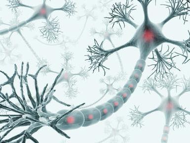
Consists of a bundle of nerve cells. A nerve cell that transmits certain information is called a neuron. The data that neurons carry is called nerve impulses. Like electrical impulses, they carry information at incredible speeds. Fast signal transmission is ensured by neuron axons covered with a special myelin sheath.
This sheath covers the axon, similar to the plastic coating on electrical wires, and allows nerve impulses to travel faster. What is a neuron? It has a special shape that allows it to transmit a signal from one cell to another. A neuron consists of three main parts: a cell body, many dendrites, and one axon.

Types of neurons
Neurons are usually classified based on the role they play in the body. There are two main types of neurons - sensory and motor. Sensory neurons carry nerve impulses from the senses and internal organs to Motor neurons, on the contrary, carry nerve impulses from the central nervous system to organs, glands and muscles.

The cells of the nervous system are designed in such a way that both types of neurons work together. Sensory neurons carry information about the internal and external environment. This data is used to send signals through motor neurons to tell the body how it should respond to the information received.

Synapse
The place where the axon of one neuron meets the dendrites of another is called a synapse. Neurons communicate with each other through an electrochemical process. When this happens, chemicals called neurotransmitters react.

Cell body
The structure of a nerve cell presupposes the presence of a nucleus and other organelles in the cell body. Dendrites and axons connected to the cell body resemble rays emanating from the sun. Dendrites receive impulses from other nerve cells. Axons transmit nerve impulses to other cells.
A single neuron can have thousands of dendrites, so it can communicate with thousands of other cells. The axon is covered with a myelin sheath, a fatty layer that insulates it and allows signal transmission much faster.
Mitochondria
When answering the question of how a nerve cell is structured, it is important to note the element responsible for the supply of metabolic energy, which can then be easily utilized. Mitochondria play a primary role in this process. These organelles have their own outer and inner membrane.
The main source of energy for the nervous system is glucose. Mitochondria contain the enzymes needed to convert glucose into high-energy compounds, mainly adenosine triphosphate (ATP) molecules, which can then be transported to other areas of the body that need their energy.

Core
The complex process of protein synthesis begins in the cell nucleus. The nucleus of a neuron contains genetic information, which is stored as encoded strings of deoxyribonucleic acid (DNA). Each contains for all the cells in the body.
It is in the nucleus that the process of constructing protein molecules begins, by writing the corresponding part of the DNA code on complementary ribonucleic acid (RNA) molecules. Released from the nucleus into the intercellular fluid, they trigger the process of protein synthesis, in which the so-called nucleoli also take part. This is a separate structure within the nucleus that is responsible for building molecular complexes called ribosomes, which are involved in protein synthesis.

Do you know how a nerve cell works?
Neurons are the most tenacious and longest cells in the body! Some of them remain in the human body throughout life. Other cells die and are replaced by new ones, but many neurons cannot be replaced. With age there are fewer and fewer of them. This is where the expression comes from that nerve cells do not regenerate. However, research data from the late 20th century proves the opposite. In one area of the brain, the hippocampus, new neurons can grow even in adults.

Neurons can be quite large and several meters long (corticospinal and afferent). In 1898, the famous nervous system specialist Camillo Golgi announced his discovery of a ribbon-shaped apparatus specializing in neurons in the cerebellum. This device now bears the name of its creator and is known as the “Golgi apparatus.”

From the way a nerve cell is structured, it is defined as the main structural and functional element of the nervous system, the study of simple principles of which can serve as the key to solving many problems. This mainly concerns the autonomic nervous system, which includes hundreds of millions of interconnected cells.


