Foci of endometriosis in the abdominal cavity. Endometriosis of the abdominal cavity: what underlies the pathology. Approximate duration of disability for endometriosis
Main symptoms:
Endometriosis is a gynecological non-tumor disease, accompanied by the growth of the inner lining of the uterus (endometrium) outside its cavity. Simply put, tissue that is found in the uterus of healthy women with endometriosis grows in other organs. Endometriosis, the symptoms of which women experience, develops for unknown reasons, although there is some evidence for the identification of immune, hormonal, hereditary and some other factors.
general description
So, in order to better understand what we are talking about when considering this disease, it is necessary to dwell on what the endometrium actually is, and also delve a little into the features of the organs of the woman’s reproductive system.
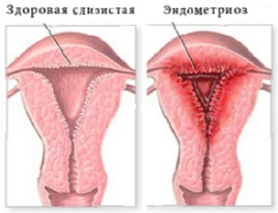
In women, the pelvis contains the uterus, a muscular organ connected on both sides to fallopian tubes that open to the abdominal cavity. The uterus is covered by three main layers, this is the inner layer endometrium, middle (muscle) layer myometrium, And perimetry- the outer serous thin shell of the organ, acting as a continuation for the layers of peritoneum from the bladder.
The layer we are interested in, the endometrium, includes two more layers, this is the functional layer and the basal layer. The functional layer includes a layer of cells that, in their own structure, resemble cylinders, which, in fact, determines their name - this is cylindrical epithelium. Between such cells there are glandular cells - due to them, the required mucus is produced, and there are also large numbers of terminal branches belonging to small spiral arteries.
Throughout the entire menstrual cycle, the functional layer is subject to constant changes due to the effects of female sex hormones. When menstruation occurs, it is rejected and finally released. Further, where the functional layer in the uterus has been rejected, the process of cell division of the basal layer begins. As a result, new cells are formed, replacing the rejected layer and forming a new layer.
Experts note that in terms of prevalence, endometriosis ranks third among gynecological diseases, following uterine fibroids and inflammatory processes affecting the genital organs of women. Most often, endometriosis is diagnosed in women of reproductive age, mainly between the ages of 40 and 44 years. According to various data, the average incidence of endometriosis in this category is about 12%. In infertile women, endometriosis is diagnosed more often - they account for approximately 30-40%, while multiparous women encounter this disease somewhat less frequently - about 27%.
Interestingly, teenage girls can also develop this disease. For example, it is known that about 50% of patients in this group who underwent surgery due to pain arising in the pelvic area were diagnosed with endometriosis. The premenopausal period also does not exclude women from developing this disease - here its frequency on average is about 2-5%. We would like to add that after menopause, women in similar age groups can also develop endometriosis, which, however, occurs somewhat less frequently.
At the same time, it is impossible to determine the true incidence of the disease in question, due to the difficulties associated with its diagnosis, as well as the fact that in some cases endometriosis occurs without symptoms at all. On average, about 70% of cases of patients seeking medical help due to pain in the pelvic area result in a diagnosis of endometriosis.
These data, if readers approach them properly, are a strong argument in favor of regular preventive visits to a doctor such as a gynecologist. This especially applies to those women who experience certain embarrassment associated with seeing this specialist, as well as those women who completely ignore such recommendations and do not visit a gynecologist at all.
Endometriosis: causes
The disease we are considering is polyetiological, which, in turn, indicates the presence of many different probable causes that determine it. However, as has already been highlighted, the true cause of endometriosis has still not been determined. Let's look at some of the options that are currently considered as the main ones.
- Retrograde menstruation. Or, as it is called, “reverse” menstruation. This phenomenon is determined by the following process: a certain amount of menstrual blood released during menstruation is directed into the abdominal cavity through the fallopian tubes. Menstruation according to a similar “scenario” is not uncommon; moreover, it often occurs in healthy women. The only difference from patients with endometriosis is that in healthy women the immune system restrains the endometrium, preventing it from growing in the area in which it ends up, that is, in the abdominal cavity.
- Heredity. This factor is relevant for many diseases that a person has to face, and endometriosis can also be considered as a disease associated with this factor. Accordingly, it is believed that the risk of developing the disease in question increases if it is present in close relatives.
- Immune system disorders. This reason is also considered as a putative factor leading to the development of endometriosis. If the immune system is weakened, then finding itself in the abdominal cavity during the already considered option of “reverse” menstruation, endometrial cells not only are not destroyed, but also attach to the tissues and organs located here, thereby forming foci of endometriosis.
- Surgical intervention in gynecology. Any kind of surgical intervention, such as curettage (curettage), abortion, cesarean section, cauterization of erosion, etc. - all this is usually considered as significant predisposing factors to the development of endometriosis.
- Hormonal changes. This factor is also believed to contribute to the development of endometriosis. The fact is that the endometrium is quite sensitive to the effects of female sex hormones, and foci of endometriosis react to them in a similar way. The growth of such lesions, for example, is promoted by female sex hormones and estrogens.
- Endometrial metaplasia. This factor implies a transformation in which one tissue is transformed into another. There is a theory according to which the endometrium, once outside the uterus, can transform into another tissue in a similar way. Meanwhile, the causes of metaplasia are currently unclear; moreover, any assumptions about it give rise to a lot of controversy among researchers.

In addition to the factors listed, there are some other factors, and they also cannot be excluded when considering the connection with endometriosis. In particular these include:
- iron deficiency in the body;
- environmental impact;
- urinary tract infections, as well as STDs;
- dysfunction of the liver organ;
- obesity;
- use of an intrauterine device, etc.
Endometriosis: forms and types
Classification of endometriosis is made in gynecology in accordance with the area of localization of its foci. In particular, they highlight genital And extragenital endometriosis. Genital endometriosis can be internal (this is adenomyosis) or external, extragenital, in turn, can be extraperitoneal or peritoneal.
Internal genital endometriosis refers to the growth of endometrial foci in the area of the muscular uterine layer, namely in the cervix and in the uterine canal.
As for extragenital endometriosis, it mainly develops in the environment of the kidneys, bladder, intestines, lungs, and in the area of some postoperative scars.
Extragenital peritoneal endometriosis primarily affects the fallopian tubes, ovaries and pelvic peritoneum.
The localization of extraperitoneal endometriosis is concentrated on the side of the external genitalia. The main forms of this variant of the disease are endometriosis of the vaginal part of the cervix, endometriosis of the vagina, retrocervical endometriosis, endometriosis of the rectovaginal septum.
Endometriosis can occur in so-called “minor” forms or in severe forms. In the latter variant, the localization of lesions may correspond to a mixed form, which is why endometriosis is sometimes not subject to a clear classification at all. In addition, severe forms of endometriosis, according to the observations of specialists, develop as a result of ignoring therapeutic and preventive measures at the stages necessary for this.
Based on the depth of the lesions, the corresponding stages of endometriosis are distinguished. In particular, these are the minimal stage, the mild stage, the moderate stage and the severe stage. The severe stage, as can easily be assumed, is the most painful for patients, and also the most difficult in terms of implementing measures aimed at treating endometriosis. In internal endometriosis, the lesion according to specific stages is as follows:
- Stage 1 - the mucous membrane is affected up to the myometrial layer (up to the middle, muscular layer, as stated earlier);
- Stage 2 - the myometrial layer is affected to the middle;
- Stage 3 - the lesion reaches the serous (peritoneal) lining of the uterus;
- Stage 4 - here the parietal peritoneum is affected.
Thus, we can distinguish a group of abdominal and pelvic organs (namely, they are most often affected by lesions), which will determine the types of endometriosis:
- Endometriosis of the uterine body (aka adenomyosis);
- Ovarian endometriosis;
- Peritoneal endometriosis (also known as peritoneal endometriosis);
- Vaginal endometriosis;
- Cervical endometriosis;
- Rectovaginal endometriosis;
- Bladder endometriosis;
- Endometriosis affecting other organs (at this point the disease is much less common): diaphragm, pleura of the lungs, the lungs themselves, intestines, eyes, stomach, skin, etc.

Endometriosis of the uterine body: symptoms
Endometriosis of the uterine body, or, as we previously designated, adenomyosis, is one of the main forms of endometriosis, in which the myometrium is affected by foci of endometrioid tissue. The symptoms of this form of the disease are as follows:
- Painful menstruation. This symptom also has its own medical definition - aldismenorrhea. The intensity of pain does not correspond to the severity of pain, in general. The appearance of pain is caused by the fact that fluid begins to accumulate in the tissues, which occurs due to the actual adhesive process affecting the uterine cavity, the accumulation of menstrual blood in the foci, and the inflammatory process.
- Cycle disorders. This symptom is quite characteristic of adenomyosis, although, however, not only for it - many gynecological diseases and disturbances in the functioning of the body are accompanied, as is known, by such “failures”. With adenomyosis, cycle disorders are mainly reduced to bleeding. A fairly important symptom for this case is the appearance of brownish or bloody discharge; they appear 1-2 days before the start of menstruation and last the same, 1-2 days after it. An important signal is also a change in the nature of menstrual flow. So, if before menstruation proceeded normally, then with adenomyosis they can become, for example, excessively abundant. This is also often accompanied by severe exhaustion of the patient.
- Dark color of menstrual discharge. A characteristic feature of the manifestation of endometriosis during menstruation is also the presence of blood clots.
- Prolonged menstrual flow. Often, menstruation with endometriosis lasts longer, exceeding the average duration.
- Infertility. Infertility is caused by two main reasons, namely the fact that there is no possibility of implantation of the fertilized egg and its further gestation due to the prevalence of the process, and also the fact that the adhesive process is developed in a pronounced form, which is accompanied by damage to the uterine cavity. In both cases, the result is the same - all this leads to infertility. At the same time, this is not the final verdict on the disease, because in at least 20% of cases in practice, pregnancy is recorded among patients even with a severe form of the disease in question.
- miscarriage, that is, in this case we are talking about spontaneous abortion/miscarriage. The reasons for this outcome are related to the general picture of changes against which infertility develops.
- Endocrine disorders. This symptom is mainly relevant for extragenital endometriosis, although it may also be present during adenomyosis. It manifests itself in particular in hypothalamic-pituitary insufficiency of the ovarian system. Due to hormonal imbalances, spotting may appear in women between menstruation, which occurs quite often with endometriosis.
In most cases, the disease progresses. Without treatment for six months, approximately 47% of patients experience a worsening of their condition, while spontaneous improvement occurs in approximately 30%. What is noteworthy is that during pregnancy, patients experience some regression of the disease, and even a significant improvement in their general condition. The fact is that pregnancy is a condition in which a decidual reaction begins to develop in the formed lesions, as a result of which they begin to decrease.
Decidualization consists of changes in the endometrium during pregnancy in which a special type of cellular layer of the endometrium is formed - decidual tissue. During pregnancy, decidual changes occur quite intensively: cells accumulate fat and glycogen, and the size of these cells increases. At the same time, the growth of blood vessels in the endometrium is enhanced.
As for the role of this decidual tissue, its role has not been fully determined. Meanwhile, it is generally accepted that due to this tissue, control is exercised over the introduction of the fertilized egg into the wall of the uterus, where it acts as a kind of layer, first between the trophoblast, and then the wall of the uterus and placenta. We also add that the decidual reaction acts as an integral stage of implantation.
Ovarian endometriosis: symptoms
The ovaries with endometriosis can be affected due to the introduction of endometrial cells into them through the lumen of the fallopian tube, which occurs with the flow of lymph and blood. The causes of ovarian endometriosis are also not completely clear at the moment; foci of endometriosis can be located both externally on the ovary and directly in it. Symptoms of ovarian endometriosis may manifest differently in each case, depending on the size of the lesions and the specific area of their localization. Let's highlight the general symptoms:
- Lower abdominal pain. Such pain is not necessarily associated with a specific period of the menstrual cycle, that is, it can appear at any time. Constant pain in the lower abdomen can be caused by inflammation of the peritoneum due to irritation due to the formation of endometriotic lesions.
- Pain in the lower abdomen during physical activity and sexual intercourse.
- Increased pain in the period before menstruation, especially severe pain on the first day.
- Spread of pain to the groin or lumbar region, to the rectum.
Peritoneal endometriosis: symptoms
Peritoneal endometriosis (peritoneal endometriosis) is characterized by the fact that in its development a significant role is played by the interaction of endometrial elements with peritoneal mesothelial cells. The “reverse” reflux of menstrual blood, which we have already discussed earlier, can contribute to the development of this form of the disease, which is caused by certain disturbances in the functioning of the immune and endocrine systems.
Peritoneal endometriosis can be of two types. Thus, the first type is characterized by a limited extent of damage - only the peritoneum is affected. The second type, accordingly, is characterized by the fact that damage to endometrioid foci occurs not only within the peritoneum, but also behind them, that is, the uterus, ovaries and fallopian tubes are affected.
With minor forms of endometriosis, there may be no clinical symptoms for a long time - the disease occurs in a latent form. At the same time, infertility with such a course of the disease, even in a small form, often exceeds 90%. If foci of endometriosis have spread beyond the peritoneum and “taken root” in the rectum and its muscular layer, also affecting the perirectal tissue, then a similar course is accompanied by the appearance of pelvic pain, painful sexual intercourse (which is more pronounced before the onset of menstruation, as well as after it) .
Endometriosis of the vagina and perineum: symptoms
Basically, the perineum and vagina are affected by endometriosis as a result of germination from the side of the retrocervical lesion, somewhat less often this occurs due to the appearance of endometrial lesions in the area of the area affected during childbirth.
The leading complaint for this form of the disease is pain that occurs both in the vagina itself and in the depths of the pelvis, and the severity of pain in this case varies from moderate to quite pronounced, often painful and exhausting. Increased pain is observed during sexual intercourse, as well as a week before and after menstruation. Severe pain appears particularly if the anterior perineum, as well as the external sphincter of the rectum, is involved in the process.
There are also certain kinds of difficulties in the act of defecation, which is accompanied by excruciating pain during those periods during which endometriosis worsens. The nature of the pain is pulsating and burning (analogy with an abscess). When menstruation occurs, patients detect swelling, nodes or a cystic type of formation when palpated.
After the end of menstruation, the detected formations either decrease in size or completely disappear, after which scars remain in their place, they are painful, and have areas of brownish pigmentation. If in this case the diagnosis is made erroneously and unreasonably (sphincteritis, rectitis) on the basis of damage to the external sphincter of the rectum, and thermal procedures are prescribed (including warm sitz baths), then the pain only intensifies.
Pain in the vagina can also be combined with local itching. Some patients experience brownish and bloody discharge coming from the vagina, both spontaneously and during sexual intercourse. Such discharge appears during the almost standard period for endometriosis for this symptom - a few days before menstruation and a few days after it.
Cervical endometriosis: symptoms
This form of the disease is also quite common, and the reason for this is the location of the affected area - the cervix most often “comes under attack” during various manipulations in gynecology (abortion, curettage, etc.).
Some symptoms of cervical endometriosis, in general, can be called common with other areas affected by this disease. These include:
- spotting brownish discharge that appears before menstruation;
- pain and discomfort during sexual intercourse;
- the appearance of brownish discharge during sexual intercourse (this symptom mainly occurs in the second half of the cycle).
As for other pain sensations (in the lower abdomen, for example), in this form of the disease they are not so characteristic of the overall clinical picture.
Rectovaginal endometriosis: symptoms
Rectovaginal endometriosis can be deep (or internal), which is accompanied by the development of pathological foci in the uterus characteristic of endometriosis, as well as external, which is accompanied by damage to the fallopian tubes, wide uterine and intestinal ligaments, ovaries, pouch of Douglas and peritoneum.
The symptoms of this form of the disease are similar to other forms: there are also pain sensations that occur during sexual intercourse, as well as pain in the lower abdomen before and after menstruation.
Bladder endometriosis: symptoms
Some time ago, endometriosis in this form was considered to be a rare disease; any information about this disease in the medical literature flashed in a rather meager volume. Meanwhile, now cases of this disease are diagnosed more often, and this is most likely due to the past insufficient familiarization of urologists and gynecologists with it, rather than the rarity of its occurrence. A role in this is also played by the fact that often specialists, in attempts to establish such a diagnosis as endometriosis of the bladder, adhere to the direction of another pathology - cyclic hematuria, which in any case is incorrect, moreover, the last specified diagnosis is rarely relevant for patients for whom it was delivered.
The bladder with endometriosis can be affected in different ways. So, for example, it is possible that the contents that are in the endometriotic ovarian cysts get on its surface, as well as the ingress of menstrual blood (according to the “scenario” of retrograde reflux), which includes viable endometrial particles, or the growth of the endometrium from the isthmus and the anterior uterine wall to the bladder. An important role is also played by the isthmus left during supravaginal amputation of the uterus, which is affected by endometriosis, as well as the gentle surgical effect on the uterus during certain manipulations. Caesarean section plays some role. An acceptable option is the hematogenous entry of endometrial elements into the wall of the bladder organ.
The features of the clinical picture of endometriosis in this case are determined by the features of its genesis. Thus, foci of endometriosis formed during the implantation of endometrial particles on the surface of the bladder organ may not manifest themselves for a long time, in other words, there are no symptoms. Detection of lesions occurs accidentally, for example, during the process of abdominal dissections for actual diseases of certain pelvic organs, as well as in the area of the lower abdominal cavity. Naturally, the detection of pathology is allowed by those specialists who are familiar with it.
When endometriosis spreads to the posterior wall of the bladder from the uterine stump or from the isthmus, it leads to the occurrence of quite severe dysuric phenomena in patients. If we are talking about a pathology such as congenital endometriosis of the bladder, in which the location of the lesions is concentrated on the side of the orifices of the ureter, then the picture of the disease can also be quite severe.
Most often, symptoms of endometriosis of the bladder include complaints of a feeling of heaviness that occurs in the depths of the pelvis and lower abdomen. It intensifies before menstruation, as well as after it. At the same time, urination in patients becomes more frequent, in some cases it is accompanied by pain. The severity of pain can vary; accordingly, it can be either moderate or quite severe, up to the loss of normal ability to work during such a period. During urological examinations and repeated urine tests, no reasons are found to explain the patients’ suffering, which is why the diagnosis of “cystalgia” is established. The therapy used to address the manifestations of symptoms determines the lack of sufficient effectiveness. During thermal procedures, the pain intensifies. At the same time, experts do not attach due importance to the relationship determined between the menstrual cycle and complaints.
Gradually, painful urination is complemented during menstruation by hematuria (blood in the urine), the severity of its manifestation may vary. At this stage of disease progression, a diagnosis such as recurrent hemorrhagic cystitis can be established. Therapy to address the manifestations of actual symptoms is still ineffective.
Soon the disease becomes chronic. According to some data, about 3-5 years pass from the onset of such a symptom as painful urination until the onset of hematuria. What is noteworthy is that many patients experience some relief from pain when urinating from the time the hematuria began to appear. In most cases, the listed symptoms lead to fear in patients that they may have a tumor in the bladder.
Let us add that blood in the urine with endometriosis of the bladder, according to some observations, is a symptom that occurs in 25% of patients with this disease. If we are talking about extensive endometriosis, in which the bladder neck is captured by the lesion, then a symptom such as a problem with urinary retention (incontinence) may also make itself felt.
Endometriosis and pregnancy
If we consider this disease in combination with infertility, then we cannot unequivocally assert equality between them. In other words, pregnancy is not impossible with endometriosis. Another thing is that pregnancy with endometriosis significantly reduces the chances of pregnancy. In practice, there are cases of conception with this disease, but it is important to understand that the success rate of conception in this case is lower, and, of course, that with endometriosis there is a certain risk to the fetus, which consists of spontaneous miscarriage. If you still succeed in conceiving a child, then it is imperative to see a doctor, strictly following his recommendations.
As for the cause-and-effect mechanism in the “endometriosis - infertility” scheme, there is no clear clarity here yet. Meanwhile, there are certain assumptions regarding the factors that provoke infertility in endometriosis:
- Immunological and endocrine disorders that are relevant in parallel with endometriosis. These factors negatively affect ovulation, fertilization and subsequent implantation of the egg into the uterus.
- Mechanical disorders causing obstruction of the fallopian tubes; pathology of ovarian anatomy; adhesions that make it difficult for the egg to be released.
- Processes accompanying local inflammation.
- Luteinized follicle syndrome.
- Frequent miscarriages in the early stages.
- Pathology of the transport function of the fallopian tubes, caused by an increase in prostaglandins against the background of endometriosis.
Interesting in its own way and, at the same time, important is such a factor as unfavorable conditions for the future fetus in the body (womb). More precisely, the essence of this is as follows: the body independently decides whether a woman can now bear (and then give birth to) a healthy baby.
At the same time, new studies show that the majority of women with endometriosis, regardless of the nature of the menstrual cycle (even normal and regular), there is no true ovulation as such, that is, we are talking about anovulation. Let us add that without ovulation, pregnancy is in principle impossible.
Data from some sources indicate that after treatment and organ-preserving surgical interventions, pregnancy occurs on average in 15-56% of cases - such a significant gap in rates is determined by the characteristics of the pathological process and the severity of the disease. Basically, gynecologists note that after treatment carried out in the right direction, pregnancy occurs within six months to a year. Accordingly, the wait for pregnancy can last from 6 to 14 months.
At the same time (albeit rarely), in practice, such cases are not excluded in which successful treatment of endometriosis does not result in the onset of a long-awaited pregnancy after six months or more. In this case, you will need to undergo additional examination, which will allow you to identify other factors involved in the problem of infertility.
Complications of endometriosis
If treatment of endometriosis is ignored as a necessity or it is initially implemented illiterately, then you may subsequently encounter a number of complications:
- infertility;
- development of adhesive processes in the abdominal cavity and small pelvis;
- development of posthemorrhagic anemia in patients against the background of heavy chronic blood loss accompanying menstruation;
- neurological disorders caused by compression of nerve trunks;
- formation of endometrioid ovarian cysts;
- transformation of endometrioid tissue into a malignant tumor formation.
Diagnosis
In order to establish a diagnosis of endomketriosis, it is necessary to obtain the results of certain studies, these include in particular:
- Ultrasound of the pelvic organs using a special vaginal sensor;
- hysterosalpingography is a method in which a contrast agent is used, due to which one can assess how widespread the process of formation of lesions turned out to be, as well as understand how much the patency of the fallopian tubes has been affected by this, which is especially important if the patient is experiencing infertility;
- hysteroscopy - this method makes it possible to examine the features of the surface of the uterus, endometrioid ducts and the mouths of the fallopian tubes (on the pale pink mucous membrane they look like dark red dots);
- laparoscopy is a microsurgical procedure that makes it possible to diagnose any form of the disease, and with the simultaneous possibility of treatment during it;
- blood test to detect endometriosis markers;
In general, the need for one or another version of the study is determined by the attending physician; depending on the characteristics of the pathological process, the diagnostic scheme may vary.

Treatment
Treatment of endometriosis is carried out in two main directions, and this is surgical elimination of foci in areas affected by endometriosis (or removal of organs with them completely), as well as drug treatment, aimed at providing hormonal correction of activity characteristic of the endometrium.
Surgical treatment often has no alternatives due to the fact that the patient’s condition often rapidly deteriorates and there is a threat to subsequent infertility. In many cases, the pain that accompanies endometriosis becomes almost unbearable; in addition, the pain is accompanied by the rapid growth of lesions, which causes an unfavorable prognosis.
Surgical intervention can be performed in different ways, this is determined based on the location of the lesions, the possibility of access to them through one or another technique of the required effect. If we are talking about endometriosis of the vagina, cervix or perineum, then the preferred option is endoscopy (excision of lesions and cauterization is carried out either through the vaginal cavity or from the outside). If the lesions are located in the uterine cavity, then an option such as removal of the uterus (the question of whether or not to remove the appendages) or hysteroscopic surgery, which provides access through the vagina to the affected organ of the uterus, can be considered.
If endometriotic lesions are located in the area of the fallopian tubes, ovaries or peritoneum, then laparoscopy can be performed - several small holes are made in the abdomen in certain areas for subsequent access to the affected areas.
As for drug treatment, it is aimed at suppressing the growth/reproduction of endometrial cells. The following groups of drugs are mainly used (only the attending physician can decide on their prescription!):
- combined action oral contraceptives (Marvelon, Femoden, Diane-35, etc.);
- drugs representing the group of antigonadotropins (gestrinone, danazol, etc.);
- drugs representing the group of progestins (Depostat, Duphaston, etc.);
- drugs of the agonist group (decapeptyl-depot, Zoladex, etc.);
- antiestrogens (tamoxifen, etc.).
Information on these groups of drugs is provided for general information only! Self-medication with them is unacceptable; use is possible only on the basis of the testimony of the treating gynecologist!
Endometriosis: prevention measures
Prevention of endometriosis is an equally pressing issue both for those women who suffered from this disease and were cured of it, and for those women who encountered this disease only through certain information sources. We highlight the following recommendations for prevention:
- regular visits to the treating gynecologist, at least once every 6 months;
- sexual abstinence during menstruation;
- timely treatment of gynecological diseases;
- fighting excess weight (doing exercises, dieting, etc.);
- avoidance of stressful situations as such, as far as possible;
- exclusion of abortions, selection of optimal solutions for contraception.
Endometriosis: some facts about this disease
Some women ignore this disease, believing that it will go away on its own, some believe that it is only “their problem,” and some even believe that it will not affect them at all. Is it so? Let's look at some established facts related to endometriosis.
- Endometriosis is a disease of active and business women
A number of studies in this area and their results, in particular, indicate that endometriosis is more often diagnosed in women in large cities, as well as in women whose professional activities are associated with increased mental stress. Basically, we are talking about socially successful business women, as well as women whose activities are supplemented by systematic changes in climatic conditions due to business trips, etc. Here we are talking about the desire for self-realization while postponing motherhood for “later.” Those women whose sex life is “excessively active” are also at increased risk, especially if it is combined with frequent changes of sexual partners (which, as we know, is not very rare). It is quite possible that in addition to treating endometriosis, we will have to take care of some restructuring of consciousness, which is also an important part in this matter.
- Endometriosis negatively affects the sexual partners of women with this disease
Relatively recently it became known that the problem of endometriosis is not only a purely female problem, but also a problem of a woman’s sexual partner. There are two substantiated statements on this matter:
Seminal fluid contains certain molecules (such as estradiol, prostaglandins, etc.), due to which a stimulating effect is exerted on the endometrium. During sexual intercourse, seminal fluid ends up in the uterine cavity or in the abdominal cavity through the posterior and anterior vaginal fornix. Prostaglandins enhance cell proliferation and also lead to the suppression of apoptosis (that is, a protective mechanism that acts as part of the development of endometriosis), while inducing the production of certain enzymes, due to which endometriosis develops with greater intensity. Accordingly, it can be summarized that when sperm enters the body of a patient with endometriosis, it acts as a factor supporting the development of this disease.
Endometriosis negatively affects the quality characteristics of sperm. It has also been proven that hypoxia acts as one of the trigger mechanisms for endometriosis. In other words, women with endometriosis are always in a state of oxidative stress, in which cells are damaged as a result of oxidation. At the same time, there is an accumulation of oxygen free radicals in their body, which, as it has become known, are characterized by a toxic effect on sperm - in particular, cell membranes are affected, incl. and DNA, apoptosis (cell death) is also triggered. That is, those processes that “control” the disease in women negatively affect the general condition of sperm. Accordingly, in case of infertility, it is extremely important to begin treatment, thereby eliminating the negative impact for both partners that leads to such a result.
- Endometriosis and IVF
In particular, on this point, patients are interested in whether the IVF procedure for endometriosis will be effective. There are also certain facts about this:
Common forms of the disease negatively affect the ovarian reserve, reducing the number of eggs obtained during stimulation; why this happens is still unclear;
Endometriosis itself acts as a basis for including patients in the appropriate risk group regarding the possibility of developing an ectopic pregnancy after the embryo transfer has been performed;
Internal endometriosis (i.e. adenomyosis) helps to reduce the possibility of successful embryo implantation within the framework of RVT programs in the same way as with the usual (natural) method of conception.
Let us summarize that the only correct solution in the treatment of endometriosis is laparoscopy (surgical intervention) with the addition of certain drugs of conservative therapy.
Joseph Addison
With the help of exercise and abstinence, most people can do without medicine.
Which doctor should I contact?
If you suspect a disease such as Endometriosis, you should consult a doctor:

Peritoneal endometriosis is a common pathology characterized by the penetration and infiltrative growth of endometrial cells into the peritoneum. Benign growth of the uterine mucosa beyond its boundaries can lead to serious consequences.
Reasons for the development of pathology
The causes of endometriosis in various organs have not been fully studied by doctors. Many versions of the appearance of this pathology have been put forward, but preference is given to the implantation theory (retrograde menstruation). This phenomenon is understandable and occurs in some healthy women.
According to him, part of the menstrual blood, together with particles of the endometrial layer (heterotopias), which should come out, rush into the abdominal cavity. For various reasons, they are consolidated in this body. Further functioning of the particles occurs cyclically. If pregnancy does not occur, heterotopias are rejected and minor hemorrhages occur in the peritoneum.
This pathology can develop in 2 variants:
- endometrial cells cover only the anterior part of the peritoneum;
- The pathological process involves the abdominal area, fallopian tubes, ovaries, uterus, etc.
The following factors can contribute to the formation of pathological foci in the peritoneum:

Other theories of the development of endometriosis have shown their inconsistency.
Symptoms of the disease
Endometriosis, having spread to the peritoneum, does not manifest itself in any way at the initial stage of its development. During this period, gynecologists can detect the disease only during a routine examination.
As the pathology progresses, the following symptoms begin to manifest:

The listed symptoms of endometriosis are similar to those of other female diseases, so it is important to conduct a thorough examination to prescribe effective treatment.
Diagnostic measures
Diagnosis of the patient begins with a conversation in order to establish a hereditary predisposition to the disease, and by listening to complaints. Then a visual inspection and palpation of the abdomen is performed. To make an accurate diagnosis, a complete examination is necessary, including:

When carrying out diagnostic measures to identify endometriosis, it is distinguished from other diseases of the genitourinary system with similar symptoms.
Features of the treatment of the disease
An integrated approach is used to treat endometriosis. It is important not only to get rid of the symptomatic manifestations of the pathology, but also to eradicate the cause and prevent serious consequences.
The main directions are:
- conservative therapy;
- surgical intervention;
- mixed treatment.
The initial stage of the disease, asymptomatic course, small areas of peritoneal damage are considered direct indications for therapeutic treatment. It includes:

Properly selected drugs help stop the growth of endometriosis and reduce the production of estrogen. To accomplish these tasks, several hormonal drugs may be prescribed:
- progestogens;
- estrogen-gestagens;
- GnRH agonists;
- antigestagens.
To obtain a lasting effect, drugs of this action should be taken for a long time. Treatment with them is carried out strictly under the supervision of a specialist due to numerous side effects.
Depending on the manifestations of the disease, anti-inflammatory and painkillers may be prescribed.
For anemia, the doctor prescribes iron supplements.
If the therapeutic treatment has not brought the desired result, then surgical intervention is proposed, during which the areas of endometriosis and the consequences - adhesions, endometrioid cysts, etc. are removed. Depending on the severity of the pathological process, a suitable surgical intervention is selected using a gentle laparoscopic or radical method.
Laparoscopy may include:

The listed minimally invasive types are characterized by speed and painlessness, the absence of postoperative complications, and a short recovery period.
After any surgical intervention, you should take hormonal medications for more than six months to prevent relapse of the disease.
An indicator of complete elimination of endometriosis is the absence of relapses within 5 years.
What could be the consequences?
The unfavorable consequences of endometriosis result from the lack of timely diagnosis of the disease and proper treatment. The severity of complications is directly dependent on the degree of involvement of the peritoneum and other internal organs of the small pelvis in the pathological process.
In complicated conditions, the following may occur:

Many of these consequences often occur simultaneously and complicate each other.
Prevention

During the absence of menstruation (during pregnancy or menopause), the pathological process subsides.
Only careful attention to your health will help you avoid endometriosis or identify it at an initial, easily treatable stage.
Peritoneal endometriosis - what is it?
Peritoneal endometriosis is considered one of the most common and at the same time unclear pathologies in gynecology. This diagnosis is made quite often. However, as a rule, it is difficult for women to understand exactly what they have been diagnosed with, why it is necessary to treat peritoneal endometriosis and how dangerous this condition can be.
In order to understand, firstly, you should have an idea about menstruation and the endometrium.
The inside of the uterine cavity is lined with mucous membrane. This membrane is called the endometrium. It consists of two layers. Functional (if pregnancy has not occurred) is rejected during menstruation every month. Every month a new functional layer grows from the basal layer.
Menstrual discharge is a mixture of endometrial fragments and blood. In almost every woman they not only come out (through the vagina). A certain part of the secretions enters the abdominal cavity through pipes. There they are normally destroyed with the help of special protective cells.
However, cleansing of menstrual fluid does not always occur in the abdominal cavity. Fragments of rejected mucosa are able to attach to tissues, implant and take root in them.
In other words, endometriosis is a disease in which the endometrium of the uterus is located outside its cavity in the form of separate foci. The mucous membrane can take root in various parts of the body. However, endometriosis of the peritoneum is most often detected.
After the fragments of the mucous membrane have taken root, they begin to develop according to the same principle as they existed in the uterine cavity. Under the influence of the ovarian hormone, the lesions (explants) begin to increase in size. Then some of them are rejected. Thus, peritoneal endometriosis provokes many small menstruation along with the main one.
Due to the development of miniature rejections in the peritoneum, which is quite well innervated, the processes are accompanied by painful sensations. This symptom is the main one accompanying the disease “endometriosis”.
This theory of the development of pathology is called “implantation”. It is considered one of the oldest and most likely. There are, of course, other theories about the occurrence of endometriosis. Thus, it is assumed that pathological foci can form as a result of the transformation of peritoneal cells into endometrial cells, genetic predisposition, immunological disorders, or as a result of hormonal effects.
Anything that provokes more frequent entry of secretions into the peritoneal cavity can contribute to the development of pathology.
Foci of endometriosis are found not only in the peritoneum, but also in completely different tissues and organs. It should be noted that such phenomena are rarely detected. Presumably, fragments of mucosal tissue can spread throughout the body through the circulatory or lymphatic system, and also penetrate into wounds during surgery.
Taking into account the localization of foci, determine
External endometriosis of the pelvic peritoneum, ovarian tubes, broad ligaments of the uterus, retrouterine space;
Internal endometriosis affecting the body of the uterus;
Extragenital with damage to the intestines, bladder, scar on the perineum after dissection during labor, lungs, and other organs.
The surviving fragments can be of different sizes, colors or shapes. As a rule, the lesions are represented by small red, black, yellow, brown and other compactions scattered throughout the peritoneum. In some cases, fusion of foci and tissue infiltration occurs. As a rule, this phenomenon is characteristic of the retrouterine region and the area of the uterine ligaments.
Endometriosis of the pelvic peritoneum: how to identify the pathological process
All systems of the female body are very susceptible to the influence of hormones, and the slightest changes in a woman’s hormonal levels immediately affect the functioning of almost all systems and organs of the female body. It's no secret that women are much more emotional than men because of their susceptibility to hormonal influences. It is as a result of hormonal imbalances, as well as under the influence of some other factors, that a gynecological pathology such as endometriosis can develop in the female body. Estet-portal.com talks in detail about one of the forms of this disease – endometriosis of the pelvic peritoneum.
Endometriosis of the pelvic peritoneum: symptoms and methods of diagnosing pathology
Endometriosis of the pelvic peritoneum occurs as a result of pathological benign growth of uterine tissue with its subsequent spread to the organs and structures of the pelvis. This pathology significantly affects the functioning of the organs of the female reproductive system and affects the general condition of the body. The clinical picture of endometriotic lesions differs depending on the form of endometriosis, and often the disease can go undetected for a long time. However, timely diagnosis of endometriosis of the pelvic peritoneum will help protect a woman from complications of this condition.
Endometriosis of the pelvic peritoneum:
- the main forms of endometriosis are the pelvic peritoneum;
- what symptoms indicate endometrioid lesions of the peritoneum;
- main methods for diagnosing endometriosis of the pelvic peritoneum.
Main forms of endometriosis pelvic peritoneum
The pathogenetic mechanism of development of endometriosis of the pelvic peritoneum is based on the interaction of peritoneal mesotheliocytes and elements of the endometrium of the uterus. The process is triggered as a result of retrograde reflux of menstrual blood from the uterine cavity into the pelvic cavity, under the influence of endocrine disorders in the woman’s body, as well as with a general weakening of the body’s defense mechanisms. Depending on the prevalence of the pathological process, there are two main forms of endometriosis of the pelvic peritoneum:
- in the first form, the pathological process is limited only to the pelvic peritoneum;
- in the second form, in addition to damage to the peritoneum, the ovaries, fallopian tubes and the uterus itself may be involved in the pathological process.
What symptoms indicate endometriosis-induced damage to the peritoneum?
The clinical picture of endometriosis of the pelvic peritoneum is not specific. In many cases, especially with small forms of endometriotic lesions, the pathological process can be asymptomatic for a long time. Only if the pathological process spreads from the pelvic peritoneum to the muscular layer of the rectum and pararectal tissue can the first symptoms of the disease occur. In the clinical picture, pain syndrome comes to the fore: the patient is bothered by intense pain in the pelvic area, which intensifies on the eve of menstruation and after it. In addition, pain may occur during sexual intercourse. In almost 90% of cases, even with minor forms of endometriosis of the pelvic peritoneum, patients experience infertility.
Basic methods for diagnosing endometriosis of the pelvic peritoneum
The diagnostic process of endometriosis of the pelvic peritoneum begins with a careful collection of anamnestic data. The patient's characteristic complaints of constant pain in the pelvis, pain during sexual intercourse and the inability to conceive a child may lead the doctor to think about endometriosis. Laparoscopy, which is the main method for diagnosing endometriosis of the pelvic peritoneum, most effectively helps to clarify the localization of the pathological process in this form of pathology. The following are the main manifestations of endometriosis foci on the peritoneum:
- atypical vesicles;
- hemorrhagic vesicles;
- pigmented spots and tubercles of yellow-brown color;
- typical superficial and deep lesions are blue, purple or black.
Peritoneal endometriosis: modern women's problems with ancient roots
Medical interest in endometriosis has grown over the past two decades. And there was something to show it off from! Gynecologists began to diagnose the disease en masse. Any compaction immediately raises suspicions and concerns - what is its nature, whether there are malignant cells in it. After a detailed study, endometriosis nodules no longer raise such questions, but no one has been able to fully understand the mystery of the female illness. There are theories and concepts, but they are all controversial.
 Endometriosis
However, the disease is not as young as it seems. Descriptions of its symptoms were found in Egyptian manuscripts dating back to 1855 BC. Later, Hippocrates dedicated his research to her. Several millennia have passed, and there is still no clarity, despite the fact that modern science has technical capabilities at the highest level.
Endometriosis itself
Ancient scientists, of course, did not have at their disposal the equipment and instruments that are available to modern doctors. As a result of long-term research and observation, it was found that the nodules consist of pieces of endometrial tissue. Their source is located in the uterine cavity, the inner lining of which is covered with a layer of endometrium. During menstruation, it is periodically rejected.
The process of the menstrual cycle is controlled by the pituitary gland, which gives the female body the necessary commands. First, it stimulates the maturation of the egg. Then it promotes its movement to the uterus. If fertilization has occurred, then the next stage is the attachment of the fertilized egg in the uterine cavity to the nutrient soil of the endometrium. If not, then it is removed from it. Menstruation is precisely the endometrial tissue that, along with blood, exits through the vagina.
All this happens with the participation of several hormones produced in the female body. The action of each of them is aimed at achieving a specific goal. Their level at different periods of the cycle either increases or decreases, reacting to changes occurring in the woman.
Why is the endometrium outside the uterus?
If medicine could answer this question, then perhaps the topic of endometriosis would be closed. Proponents of the hormonal and metaplastic theories put forward various assumptions regarding the occurrence of foci of endometrial tissue outside the uterine cavity. Until now, they still remain only hypotheses.
 Abdominal endometriosis - foci of inflammation outside the uterine cavity
Whatever the reasons for the development of pathology, scientists have no disagreement about their prerequisites. They are unanimous in their opinion that endometriosis is provoked and aggravated by:
- Disruptions in the hormonal system, weakened immunity;
- Retrograde menstruation, in which menstrual blood moves in an unusual reverse direction and enters the peritoneum;
- Abortions, cesarean sections, diagnostic uterine curettages;
- Other interventions in the uterus - operations, cauterization of erosions, installation of intrauterine devices;
- Sexual intercourse during menstruation;
- Hereditary predisposition;
- Inflammatory processes in the genitourinary system;
- Anomalies in the structure of the genital organs.
What forms does endometriosis exist in?
There are three groups of forms of endometriosis:
- Genital. Foci of endometriosis with it develop inside the genital organs;
- Extragenital, in which endometrial tissue is found outside the genital organs;
- Combined, combining the two previous ones.
In the extragenital form, endometriosis spreads far beyond the uterus. The intestines, lungs, and urinary system are attacked. If there are postoperative scars, then they also serve as an object for consolidation for endometriosis. In some cases, the organs of vision are affected, and then such a characteristic phenomenon as bleeding through the eyes is observed.
The genital form is more common. When leaving the uterine cavity, endometrial tissue settles on its outer wall, moves towards the fallopian tubes and reaches the ovaries. Damage to the peritoneal walls by endometriosis is a common occurrence. The external genitalia, cervix and cervical canal are also affected.
The longer endometriosis goes untreated, the more lesions it forms. Gradually, from the 1st and 2nd stages, when relatively small areas are affected, endometriosis develops into a chronic disease with 3-4 degrees of development.
 To prevent the disease from progressing, it is necessary to cure it as early as possible
Endometriosis of the pelvic peritoneum
Endometrial nodules are capable of increasing in size (and quite quickly) and affecting one organ after another. The process resembles metastasis, which is characteristic of cancer. But endometriosis forms benign formations, and its degeneration into a malignant form occurs relatively rarely.
The internal organs located in the pelvic cavity are in close proximity to the walls of the peritoneum. Over time, foci of endometriosis form on them as well. In such cases, endometriosis of the peritoneum is diagnosed, which in medicine is called peritoneal.
The similarity of the symptoms of this type of disease with signs of the inflammatory process is so great that an incorrect diagnosis is often made. Treatment in this case is with antibiotics and does not bring a positive result. A new round of examination begins, which takes some time. But you can’t hesitate, because endometrial tissue grows deeper and deeper into the affected organs, not excluding the peritoneum.
Symptoms indicating endometriosis
The first thing to suspect endometriosis is changes in vaginal discharge. In inflammatory processes, they are purulent and have an unpleasant odor. In the case of endometriosis, they bother a woman between periods. Their color is brown, turning to reddish. Menstrual bleeding at the same time becomes too heavy and also painful.
Pain not only accompanies menstruation, but also appears on other days of the cycle. They radiate down the abdomen, and their strength is so great that the woman is forced to take painkillers. Together with bleeding, this leads to significant blood loss. Anemia develops. A woman complains of weakness and dizziness. Her appearance is unhealthy, her skin becomes pale.
Pain syndrome is also observed during sexual intercourse, but to a lesser extent. If endometriosis affects the rectum, blood is released during bowel movements, and the process itself causes pain. With endometriosis of the abdominal cavity, a woman experiences discomfort when examined by a gynecologist. Sometimes during this procedure it is possible to palpate lumps indicating the presence of a disease.
 With endometriosis, the patient experiences severe pain
How is endometriosis determined?
A woman cannot determine that she has endometriosis based on her symptoms. The gynecologist will not do this until the examination results are in front of him. To do this, use one of the methods: ultrasound, hysterosalpingography (HSG) or laparoscopy. Typically, examination of organs in the pelvic area is performed. If foci of endometriosis cannot be identified during the procedure, then the area of research is expanded.
Due to its prevalence, ultrasound is available in most medical institutions. The report form is a graphic image obtained after a transvaginal examination. The presence of endometriosis is reflected in the form of circles and ellipses.
The set of equipment for HSG includes a fluoroscope, an X-ray tube and a monitor on which the image is received. After administration of the contrast agent, an x-ray is taken and its description is made.
The most accurate data is obtained during laparoscopy. The technique is reliable and gives comprehensive results. It is used for both diagnosis and treatment of endometriosis. Moreover, the operation is often performed without removing the patient from anesthesia after suspicions of endometriosis are confirmed.
How is the treatment carried out?
It takes a long time to get rid of endometriosis, even if this involves surgical intervention. After removing foci of the disease using a laser or electrocoagulation, it is necessary to take hormonal medications for six months or more. They are designed to restore hormonal balance and thus stop the formation of new endometrial lesions.
When peritoneal endometriosis is diagnosed, surgical treatment can eliminate adhesions. They form in the area of the fallopian tubes and ovaries, thereby preventing natural conception.
Drug therapy in some cases is possible without surgery, but it is effective only in the early stages. It is based on taking hormonal drugs that change the ratio of hormones in the body and at the same time have a contraceptive effect. Therefore, it is impossible to get pregnant during the treatment period, but as soon as it ends, doctors strongly recommend conceiving a child. In this way, two problems are solved at once: it is possible to avoid a relapse of the disease and give birth to a healthy baby.
 Treatment requires hormonal medications
What are the advantages of laparoscopy
By giving consent to the operation, a woman can expect that all her genitals will be preserved. This opportunity is provided by laparoscopy, a minimally invasive and gentle method used to treat endometriosis. You won’t be able to do without anesthesia, but it is quite possible to reduce the time you spend under it to half an hour and get rid of huge stitches and postoperative scars. There are only three small puncture wounds left on the body, which heal quickly and are almost invisible over time.
The recovery process is much faster. The fact that on the day of the operation or, in extreme cases, the next day the woman leaves the hospital speaks for itself. It is also important that during laparoscopy the chances of damage to other organs located in close proximity to the peritoneum are minimized. It is not always possible to avoid this during open abdominal surgery.
Since endometrioid nodules can degenerate into oncological tumors, it is advisable to send the removed areas for histological examination. With laparoscopy this is possible, except in cases where thermal effects are used.
Traditional medicine experience
Since endometriosis was known to ancient doctors, then there must be some methods that have come down to us from those times. Unfortunately, for a long time the disease was considered to be the possession of the devil in a woman, hysteria. The reason was the woman’s behavior during attacks. She was forced to writhe in unbearable pain, which bewildered those around her.
From the heritage of doctors and healers, which has survived to this day, treatment with leeches, acupuncture, and medicinal herbs is successfully used.
Herbal medicine has a greater analgesic and hemostatic effect and does not lead to the disappearance of endometrial lesions. But hirudotherapy is considered a fairly effective technique and has become widespread. It has been approved by the Ministry of Health of the Russian Federation and has been experiencing another surge in its use since the end of the last century. Leeches bite at biologically active points and inject their saliva. The enzymes it contains help thin the blood, restore hormonal balance, and improve blood circulation.
 One of the methods of traditional medicine is treatment with leeches.
Despite all the positive properties of folk remedies, it is not recommended to use them without consulting a gynecologist.
The connection between endometriosis and pregnancy
Endometriosis and pregnancy can be at opposite poles, or they can maintain a friendly relationship. The hostility between them arises due to infertility, which is caused by endometriosis. In exceptional cases, a woman with endometriosis is able to conceive a child. If she does not know that she is sick, then the diagnosis is made after childbirth or during curettage required after a frozen or ectopic pregnancy. Often such a pregnancy ends in miscarriage.
At the same time, pregnancy that occurs after treatment of endometriosis contributes to final recovery. This happens because menstruation stops and hormonal levels change. Foci of endometriosis destroy themselves and after at least 10-12 months the disease does not recur. After this period, which covers the period of gestation and feeding the child, there may be no relapse. Of course, for this it is necessary to take care to exclude the causes that led to the development of endometriosis.
Peritoneal endometriosis
Endometriosis is a disease that manifests itself as hormone-dependent pathological growths of the endometrium outside the uterus, namely in the ovaries, fallopian tubes, in the thickness of the uterus, in the rectum, on the peritoneum and in some other organs.
Heterotopias - fragments of the endometrium - when growing in other organs, undergo the same changes during the menstrual cycle as the endometrium in the uterus. In this case, a pain syndrome occurs, the affected organ increases in size, and bloody discharge appears from heterotopias every month.
In addition, menstrual function is disrupted, discharge from the mammary glands is likely, and the risk of infertility increases. Other clinical symptoms are determined by the location of endometriosis.
Classification
Pathology is classified according to the location of heterotopic lesions. There are genital and extragenital endometriosis. In turn, the genital form of the disease is divided into the following types:
- Peritoneal: the ovaries, fallopian tubes, and pelvic peritoneum are affected.
- Extraperitoneal: lesions are localized in the lower parts of the reproductive system (external genitalia, vagina, vaginal portion of the cervix, rectovaginal septum).
- Internal, or adenomyosis, develops in the muscular wall of the uterus. The organ takes on a spherical shape, the size of the uterus increases (corresponds to 5-6 weeks of pregnancy).
Sometimes the localization of endometriosis is mixed, this is possible in the case of an advanced form of the pathology.
Heterotopic foci in the case of the extragenital form are localized in the intestines, kidneys, navel, postoperative scars, and lungs.
The depth and prevalence of heterotopia foci make it possible to determine 4 degrees of pathology:
- The first is single, superficial lesions.
- The second is that the lesions are more numerous and deep.
- The third - the lesions are deep and multiple, on one or both ovaries - endometrioid cysts, individual adhesions are located on the peritoneum.
- Fourth - the lesions are deep, multiple, on the ovaries there are large bilateral endometrioid cysts, dense adhesions. The endometrium grows into the walls of the vagina and rectum. Difficult to undergo therapeutic correction.
Heterotopic lesions differ in shape and size: there are both round-shaped formations several millimeters in size and shapeless growths reaching several centimeters in diameter.
Most often, foci of endometriosis are dark cherry in color. They are separated from the surrounding tissue by whitish scar tissue. The lesions are more noticeable on the eve of menstrual bleeding due to cyclic maturation. Heterotopias can be located superficially or grow deep into the tissue.
Endometriosis is a common cause of pelvic adhesions, which limit the mobility of the ovaries, fallopian tubes and uterus and provoke menstrual irregularities, and also significantly increase the risk of infertility.
Causes
Endometriosis is a polyetiological disease, that is, several provoking factors can lead to its progression. Their list includes:
- Retrograde menstruation. In this phenomenon, a small amount of menstrual fluid enters the abdominal cavity through the fallopian tubes. In the presence of other predisposing factors (deterioration of the immune system), the endometrium that has thus reached the peritoneum begins to grow.
- Hereditary predisposition.
- Impaired functionality of the immune system.
- Gynecological surgical interventions - abortion, curettage, caesarean section and others.
- Hormonal imbalance. Excessive concentrations of estrogen can
- stimulate the proliferation of endometrioid lesions.
Other predisposing factors include:
- unfavorable environmental conditions;
- the use of an intrauterine device as a means of contraception;
- infections of the genitourinary system, including sexually transmitted diseases;
- excess body weight;
- violation of the functional state of the liver;
- iron deficiency in a woman's body.
Symptoms
The pathogenesis of the peritoneal form of pathology lies in the interaction of endometrial cells with peritoneal mesotheliocytes. This type of disease can progress due to the reflux of menstrual blood in combination with impaired functionality of the immune system and endocrine system.
In the peritoneal form of the pathology, either only the peritoneum can be affected, or the process may involve both the peritoneum and the uterine appendages - the ovaries and fallopian tubes, as well as the uterus itself.
Heterotopic lesions on the peritoneum can be represented by the following formations:
- hemorrhagic vesicles;
- pigmented tubercles and yellow-brown spots;
- typical superficial or deep infiltrated lesions of blue, purple, black color;
- atypical non-pigmented or whitish vesicles.
Symptoms may be absent in cases of low prevalence of the disease, that is, endometriosis has a latent form. With this form, the risk of infertility is 90%.
When lesions spread beyond the peritoneum and progress in the muscular layer of the rectum, the perirectal tissue is also affected. In this case, pelvic pain appears, sexual intercourse becomes painful (dyspaurenia), which is more pronounced a few days before the expected start of menstruation.
About half of patients with endometriosis experience painful menstruation, which may be due to bleeding into the cystic cavity and an increase in pressure in it, irritation of the peritoneum by blood discharge from their heterotopias, and spasm of the uterine vessels. The duration of menstrual bleeding often increases and its volume increases - which is referred to as menorrhagia.
Also, with this pathology, discomfort and pain during urination and bowel movements are possible.
Due to significant chronic blood loss during menstruation, posthemorrhagic anemia progresses with endometriosis, which is expressed in weakness, pallor or yellowness of the skin and mucous membranes, and dizziness.
For many patients, the most worrying consequence of endometriosis is infertility. Due to changes in the ovaries and fallopian tubes, impaired functionality of the immune system, and ovulation disorders, the likelihood of pregnancy is significantly reduced.
Even with successful conception, you need to understand that bearing a child in the presence of such a pathology is not at all easy: there is a high risk of spontaneous miscarriage. Therefore, expectant mothers who have been diagnosed with pathology should be under constant medical supervision. After treatment for endometriosis, the chance of conception is 15-56% in the first 6-14 months.
Complications
Due to hemorrhages and scar changes in this disease, adhesive processes are triggered in the pelvis. Another common complication is the formation of endometrioid ovarian cysts, which fill with old menstrual blood. These complications can cause infertility.
As mentioned earlier, due to blood loss, posthemorrhagic anemia develops, which manifests itself in weakness, irritability, dizziness, changes in skin tone and mucous membranes.
Neurological disorders may occur due to compression of nerve structures. In some cases, a complication may be malignant degeneration of endometriotic lesions.
Diagnostics
During the diagnostic search, other pathologies that may occur with similar clinical manifestations should be excluded. If there is a suspicion of endometriosis, the doctor collects anamnesis and the patient’s complaints. Next, the following manipulations are carried out:
- Examination on a gynecological chair. It is most informative on the eve of menstruation.
- Colposcopy and hysterosalpingoscopy. They are carried out to determine the location, shape of lesions, and obtain biopsy samples.
- Ultrasound examination of the pelvic organs. It is necessary both for primary diagnosis and for monitoring the dynamics during treatment of the disease.
- Spiral computed tomography or magnetic resonance imaging. Allows you to clarify the nature and location of heterotopias.
- Hysterosalpingography (x-ray of the uterus and fallopian tubes) and hysteroscopy (endoscopic examination of the uterine cavity). Necessary for the diagnosis of adenomyosis.
- Blood test for tumor markers CA-125, CA-19-9, CEA, PO test. In endometriosis, the concentration of these analytes is increased several times.
Treatment
When treating endometriosis, the doctor takes into account the woman’s age, the number of pregnancies and births, the location and nature of the distribution of heterotopias, the intensity of clinical symptoms, the presence of concomitant diseases, and the patient’s desire to have children. There are medicinal, surgical and combined methods for correcting this disease.
The goal of treatment is to eliminate the active manifestations of the pathology and get rid of its consequences. Conservative tactics are used in asymptomatic cases, in the case of a young woman, if it is necessary to restore or preserve reproductive function, in the premenopausal period. In this case, hormone therapy with the following groups of medications is rational:
- Combined estrogen-progestin drugs. Effective at the initial stage of pathology.
- Gestagens. They can be prescribed at any stage of the pathology. Use continuously for 6-8 months.
- Antigonadotropic drugs. Prescribed for 6-8 months, not used in case of hyperandrogenism.
- Gonadotropin releasing hormone agonists. The advantage of this group of medications is that they can be taken once a month, as well as the absence of severe side effects. The use of these drugs suppresses the ovulation process, reduces estrogen levels, which leads to suppression of the spread of heterotopias.
In addition to hormonal therapy, immunostimulating drugs and symptomatic drugs: analgesic, antispasmodic and anti-inflammatory drugs may be prescribed as part of drug treatment.
For moderate and severe endometriosis, organ-preserving surgical treatment with removal of heterotopic foci is indicated. In this case, lesions in different organs are removed, endometrioid cysts are also removed, and adhesions are dissected.
Such treatment is necessary if there is no effect from conservative measures, as well as if there are contraindications to the use of certain medications. Surgical treatment is also indicated for heterotopias with a diameter greater than 3 centimeters, and for impaired functionality of the ureters, bladder, kidneys and intestines. Often surgical treatment is combined with medication.
Radical surgical treatment such as hysterectomy and adnexectomy is carried out if patients are over 40 years old, active progression of the pathology is observed, and if conservative and organ-preserving surgical methods of treatment are ineffective.
Endometriosis tends to recur, and repeated surgery may often be required. It is extremely important to regularly see a doctor to determine timely measures in case of recurrence of the pathological process.
Recovery criteria are satisfactory health, no complaints, no relapse for 5 years after the course of treatment. In reproductive age, the criterion for recovery is also the restoration or preservation of reproductive function.
Prevention
To minimize the risk of developing endometriosis, you must follow these recommendations:
- Be examined if you have complaints of dysmenorrhea to exclude pathology.
- Regularly see a doctor after undergoing gynecological surgical interventions.
- Complete and timely treatment when acute and chronic pathologies of the reproductive system are detected.
Regular visits to the doctor for a preventive examination and compliance with all recommendations will reduce the risk of developing such a dangerous pathology as endometriosis, or identify it at an early stage and take the most effective measures to correct it.
Endometriosis of the abdominal cavity: what underlies the pathology

Endometriosis of the abdominal cavity is a pathological proliferation of endometrial cells outside the uterus, their growth into the soft tissue of neighboring internal organs located in the pelvis. The process of cell germination in the abdominal cavity is benign, but without timely treatment, in the presence of concomitant diseases, the possibility of degeneration of endometriosis foci into a malignant tumor cannot be ruled out.
Causes of proliferation of uterine epithelium
Scientists still have not been able to figure out why endometrial cells grow into the abdominal cavity. However, there are a number of factors that can significantly increase the risk of endometriosis in the peritoneum:
- depressed immune system – weak immunity is observed in women who have suffered severe infectious or viral diseases, which is why the body cannot cope with the growing endometrial cells;
- failure of the menstrual cycle, leading to incorrect and untimely maturation and increase in the thickness of the epithelial uterine layer;
- injuries to the cervix and the organ itself - cases of medical termination of pregnancy, undergoing a curettage procedure for therapeutic or diagnostic purposes, other types of surgical interventions;

- chronic inflammation of the pelvic organs;
- lack of hemoglobin - this pathological condition is observed in diseases of the circulatory system, anemia, and poor nutrition;
- overweight, obesity;
- liver diseases;
- poor environmental conditions in the region of residence;
- Hormonal imbalance is a common cause of the disease;
- heredity.
If a woman has had cases of peritoneal endometriosis among her blood relatives, the risk of developing the disease increases significantly, especially in the presence of provoking factors. The likelihood of pathological proliferation of epithelial cells increases in girls with an early onset of menstruation. This refers to situations where a teenager’s periods began before the age of 14-15.

The etiology of the disease is due to the fact that during menstruation, for unknown reasons, the cells of the uterine layer of the endometrium do not penetrate the cervical canal, but leak into the peritoneum through the passages of the fallopian tubes. In normal health, the immune system itself copes with pathogenic cells by producing antibodies to them - macrophages. But with weak immunity, this process occurs too slowly or is absent altogether. Then peritoneal endometriosis begins to develop. Endometriosis of the pelvic peritoneum is more often diagnosed in women aged 35 to 40 years.
Symptoms
Endometriosis of the pelvic peritoneum in the early stages of development may not have a pronounced symptomatic picture. Signs of the disease appear gradually. Their nature and intensity depend on the tissue of which organ the endometrial cells grow.
Common signs of pathology include:
- severe pain in the lower abdomen;
- increased pain during menstruation, heavy periods;
- constant feeling of discomfort in the abdomen, bloating;
- discomfort in the groin area during sexual intercourse;
- bleeding not associated with menstruation.

If there is damage to the tissues of the bladder, cervical canal, endometriosis of the anterior abdominal wall (see photo) or rectum, the woman experiences stool disturbances, frequent constipation is replaced by prolonged diarrhea, and there are blood clots in the stool and urine. Over time, endometrial cells affect an increasing amount of tissue, which leads to blockage of the fallopian tubes and the inability to conceive a child.
The disease has a negative impact on the emotional background. Knowing that every sexual intercourse will cause abdominal pain, a woman deliberately refuses sex. Each arrival of menstruation causes a feeling of panic; heavy discharge, which occurs even in the middle of the cycle, depresses a woman and prevents her from leading an active life.

Diagnostics
Damage to soft tissues by endometriosis is a serious disease that, without timely treatment, can lead to serious consequences. The specificity of endometriosis is that it very quickly becomes chronic, and even surgical treatment does not guarantee a complete recovery.
A woman should immediately consult a doctor as soon as she discovers that, in addition to menstruation, she has discharge, and bleeding during menstruation has become heavy, and her stomach has begun to hurt frequently. The diagnosis of abdominal endometriosis is made after the doctor conducts a gynecological examination of the woman in a chair, takes a thorough medical history, and analyzes complaints.
To clarify the primary diagnosis, determine the degree of development of the disease and the presence of complications, a medical examination is carried out, including the following methods:
- Ultrasound of the pelvic organs– carried out transvaginally. The insertion of a special sensor into the vagina makes it possible to obtain an accurate image of the condition of the cervix and uterine cavity.
- Hysteroscopy– carried out to analyze the condition of the uterine fundus, the degree of patency of the fallopian tubes is analyzed.
- Laparoscopy– minimally invasive surgical intervention is performed for both therapeutic and diagnostic purposes. This examination method is used if complications from the pelvic organs are suspected.
To assess the general health of a woman, a general and detailed blood test is performed. If blood clots are present in the urine or stool, they are tested in a laboratory. Based on the data obtained during the medical examination, the doctor selects a treatment method.

Treatment of abdominal endometriosis
In the early stages of the development of the pathological process, when pathogenic cells have not yet completely grown into the soft tissues of the abdominal organs, conservative treatment is prescribed. It consists of taking medications aimed at normalizing the hormonal levels of the reproductive system, relieving signs of endometriosis and stopping the further spread of the pathological process.
At a severe stage, when the disease has become chronic and provoked a number of complications, there is no point in drug therapy. A positive result can only be achieved through surgical intervention.
Drug treatment
To stop the process of pathological proliferation of endometrial cells, hormonal drugs are prescribed. As a rule, women are prescribed oral contraceptives, long-term use of which helps normalize hormonal levels and inhibit the production of large amounts of the female hormone estrogen.

What does this give? When estrogen concentration decreases, there is no ovulation. The organs of the reproductive system cease to prepare monthly for the possible fertilization of the egg, and the endometrium stops growing. Hormonal therapy includes the use of certain drug groups to alleviate the general condition of a woman:
- Antigestagens– medications quickly reduce the pain symptoms of the disease. Drugs in this group trigger the process of atrophy of the uterine layer of the endometrium, preventing further growth of pathogenic cells. Despite the effectiveness of this therapy, it is used only if a woman has contraindications to surgery and the disease is rapidly developing. The disadvantage of this treatment is that hormonal drugs from the antigestagen group often cause side symptoms - weight gain, hot flashes, acne.
- Agonists– drugs in this group slow down the production of hormones in the female reproductive system. These medications need to be taken in a short course, since their long-term use creates an artificial menopause. It is not recommended for patients planning pregnancy.
- Preparations of the estrogen-progestogen group– are prescribed for an intense symptomatic picture, when endometrial cells have grown deep into the tissue of the pelvic organs, and the woman has severe pain in the pelvic region.
- Progestogens– cause endometrial atrophy. Effective for symptomatic therapy. Medicines relieve severe pain in a short time. The disadvantage of treatment is the high risk of complications such as rapid weight gain and extensive soft tissue swelling. Hormonal drugs from the progestogen group are taken over a long course of 6 to 12 months.

In addition to hormonal treatment, symptomatic therapy is carried out. To relieve attacks of abdominal pain, painkillers and non-steroidal anti-inflammatory drugs are prescribed.
To restore the protective functions of the immune system, immunomodulators are prescribed. It is allowed to use traditional medicine methods - taking decoctions of medicinal herbs (chamomile, St. John's wort, celandine). Decoctions are also used for douching. The traditional method of treatment helps to normalize the functioning of the reproductive system and reduce the intensity of pain.
Doctors allot 6 months for conservative treatment; ultrasound is periodically performed to monitor the condition of the endometrium. If after six months there is no positive dynamics, surgery is prescribed.

Surgery
Surgery to treat endometriosis is carried out in two ways - laparoscopy and classical surgery. Depending on the severity of the clinical case, during the operation the uterus and appendages are preserved or partially or completely removed.
Preference is given laparoscopy. This method of operation is characterized by minimal trauma, and the rehabilitation period is short. Complications after laparoscopy are quite rare. Due to the absence of an abdominal incision, the woman does not need to stay in the hospital for a long time. Discharge from the hospital is carried out the next day.
During laparoscopy, the doctor makes several punctures in the abdominal cavity, through which surgical instruments and a device - an endoscope - are inserted, through which the doctor receives an image from the peritoneum on the screen.
The foci of endometriosis themselves are removed by cauterization. The following methods are used for this:
- cryodestruction – destruction of pathogenic cells with liquid nitrogen;
- electrocoagulation – exposure to high-frequency current;
- laser vaporization - a method of evaporating pathogenic tissues with a laser;
- radiocoagulation - exposure of lesions to radio waves.
During laparoscopy, the removed tissue from endometriosis lesions is sent to the laboratory for histological examination, which determines the nature of the formations. Such an examination is carried out for the reason that there is a risk of the lesions degenerating into malignant neoplasms.
After surgery, a woman needs to undergo a course of hormonal therapy to restore the functioning of the reproductive system, normalize the menstrual cycle, and prevent the re-growth of endometrial cells.

Possible complications
Ascites is dropsy in the abdominal cavity that occurs due to inflammation of tissues damaged by endometriosis cells.
Ascites is manifested by an increase in abdominal volume, severe pain, and signs of intoxication of the body. The disease can only be treated through surgery. If the operation is not performed in a timely manner, the fluid-filled dropsy will burst, leading to peritonitis.
Despite the fact that during surgical treatment of abdominal endometriosis the foci of the disease are removed, there is still a risk of relapse of the disease. Recurrence of lesions occurs if the cause of endometriosis has not been treated.

Another complication after surgery is endometriosis of the postoperative scar. The pathology develops gradually and may manifest itself after a few years. With the development of this disease, endometrial cells begin to rapidly grow outside the uterine cavity and grow into the postoperative scar. Symptoms and treatment are identical to the general clinic of endometriosis.
A woman experiences severe abdominal pain and the cyclicity of menstruation is disrupted. Therapy is conservative if the disease was diagnosed early, or surgery is prescribed when medications do not help, the pathology is advanced and there are complications.
Pregnancy and pathology
It is possible to become pregnant with abdominal endometriosis in the early stages of the development of the pathological process, when there are still no extensive adhesions blocking the passages of the fallopian tubes. If laparoscopy was performed, it is recommended to become pregnant immediately after completion of restorative hormonal therapy. Under the influence of natural changes in hormonal levels, there is a possibility that the pathological process will stop.
Endometriosis (endometrioid disease) is a disease characterized by the growth of tissue similar to the endometrium outside the normally located mucous membrane of the uterine body.
Endometriosis is capable of infiltrative growth with penetration into surrounding tissues and their destruction, can grow into any tissue or organ: the wall of the intestine, bladder, ureter, peritoneum, skin, and can metastasize by lymphogenous or hematogenous route. Foci of endometriosis are found in the lymph nodes, subcutaneous tissue of the anterior abdominal wall or in the area of a postoperative scar, as well as in distant areas of the body, such as the navel and conjunctiva of the eye. Endometriosis differs from a true tumor in the absence of pronounced cellular atypia and the dependence of clinical manifestations on menstrual function.
Classification of endometriosis. Depending on the location, genital and extragenital endometriosis are distinguished. Genital endometriosis is divided into internal (uterine body, isthmus, interstitial parts of the fallopian tubes) and external (external genitalia, vagina and vaginal part of the cervix, retrocervical region; ovaries, fallopian tubes, peritoneum lining the pelvic organs). With extragenital endometriosis, endometrioid implants are detected in other organs and tissues of the woman’s body (lungs, intestines, navel, postoperative wounds, etc.).
Epidemiology. Endometriosis is one of the most common diseases of the reproductive system in women 20-40 years old, the frequency of its detection sharply decreases in postmenopause. Endometriosis is diagnosed in 6-8% of patients in gynecological clinics, and in patients with infertility its detection increases to 35-44%. The main part is genital endometriosis (92-94%), extragenital endometriosis is much less common (6-8%).
Causes of endometriosis
The translocation theory (implantation) considers the possibility of the development of endometrioid heterotopias from endometrial elements transferred retrogradely with menstrual flow into the abdominal cavity and spreading to various organs and tissues. Implantation of endometrial cells and its further development can only be carried out under additional conditions: when endometrial cells have an increased ability for adhesion and implantation and when there is a violation of the hormonal and immune systems.
The theory of endometrial origin considers the possibility of the development of endometrioid heterotopias from endometrial elements displaced into the thickness of the uterine wall. It has been proven that intrauterine medical manipulations (abortions, diagnostic curettage of the uterine mucosa, manual examination of its cavity after childbirth, cesarean section, enucleation of myomatous nodes, etc.) contribute to the direct germination of the endometrium into the wall of the uterus, leading to the development of internal endometriosis of the uterine body. During gynecological operations, endometrial elements can also spread to other organs and tissues through the blood and lymph flow. Lymphogenous and hematogenous routes of spread lead to the development of endometriosis of the lungs, skin, and muscles.
Embryonic and dysontogenetic theories consider the development of endometriosis from displaced areas of embryonic material, from which female genital organs and, in particular, the endometrium are formed during embryogenesis. The detection of clinically active endometriosis at a young age and its frequent combination with anomalies of the genital organs, urinary system and gastrointestinal tract confirm the validity of the embryonic or dysontogenetic concept of the origin of endometriosis.
Metaplastic concept. According to this hypothesis, endometriosis develops as a result of metaplasia of the embryonic peritoneum or coelomic epithelium. The possibility of transformation of the endothelium of lymphatic vessels, mesothelium of the peritoneum and pleura, epithelium of renal tubules and other tissues into endometrium-like tissue is allowed.
Among the numerous factors contributing to the development and spread of endometriosis, hormonal disorders and dysfunction of the immune system should be highlighted.
Hormonal disorders are not the direct cause of the formation of endometrioid lesions, but are only predisposing conditions for the occurrence of a pathological process. In patients with endometriosis, the presence of unsystematic peaks of FSH and LH, as well as a decrease in the basal level of progesterone secretion, was noted; the development of follicle luteinization syndrome is noted. However, 40% of patients with endometriosis maintain a normal two-phase menstrual cycle. In these patients, the mechanism of cytoplasmic binding of progesterone is disrupted, which leads to a distortion of the biological action of hormones.
Estrogens stimulate the growth of the endometrium; their excess leads to the growth of endometriosis.
In patients with endometriosis, hyperprolactinemia and impaired androgenic function of the adrenal cortex are often detected.
Autoimmune reactions play an important role in the pathogenesis of endometriosis. In case of hormonal imbalances, dysfunction of the immune system is expressed in the suppression of the activity of natural killer cells, as well as in an increase in the concentration of vascular endothelial growth factor, which causes excessive angiogenesis.
Cervical endometriosis
The prevalence of endometriosis in this localization is associated with damage to the cervix during gynecological manipulations and diathermoconization of the cervix. Trauma during childbirth, abortion and various manipulations can contribute to the implantation of the endometrium into damaged cervical tissue. Cervical endometriosis may arise from elements of the Müllerian tubercle of the primary vaginal plate. In addition, lymphogenous and hematogenous spread of endometriosis to the cervix from other foci cannot be excluded.
Depending on the depth of the lesion, ectocervical and endocervical endometriosis of the vaginal part of the cervix are distinguished; less often, endometrioid heterotopias affect the cervical canal.
Diagnosis of cervical endometriosis
With cervical endometriosis, there may be complaints of spotting on the eve of menstruation or during sexual intercourse. Pain is observed with atresia of the cervical canal or endometriosis of the uterine isthmus. Sometimes cervical endometriosis does not manifest itself clinically and is diagnosed as red or dark purple lesions only upon examination of the cervix. Endometrioid heterotopias increase significantly on the eve of or during menstruation. During this period, individual lesions may open and empty. At the end of menstruation, endometrioid heterotopias decrease in size and turn pale. During colposcopy, differential diagnosis of cysts of the nabothian glands, ectopia, erythroplakia, polyps of the mucous membrane of the cervical canal, ectropion and endometriosis of the cervix is carried out. Data from a cytological examination of prints from the mucous membrane of the cervix are not very informative for the diagnosis of endometriosis, but they allow one to judge the state of the stratified squamous epithelium of the cervix and identify cellular atypia.
Endometriosis of the vagina and perineum
The vagina and perineum are more often affected by endometriosis secondary to germination from a retrocervical lesion, less often as a result of implantation of endometrial particles into the damaged area during childbirth.
The leading complaint with endometriosis of this localization is pain in the vagina - from moderate to very strong and painful. Pain appears cyclically, on the eve of and during menstruation, and intensifies during sexual intercourse. Severe pain is observed when the perineum and external rectal sphincter are involved in the process. Defecation during periods of exacerbation is accompanied by severe pain.
Diagnosis is based on complaints related to the menstrual cycle and data from a gynecological examination, which includes examination of the cervix and vagina using speculum, bimanual vaginal-abdominal and rectovaginal examination. In the thickness of the vaginal wall or in the rectovaginal cavity, dense painful scars, nodes or thickenings are palpated. On examination, brown or dark blue lesions are detected on the vaginal mucosa. On the eve and during menstruation, endometrioid heterotopias increase in size and may bleed.
To determine the extent of the process, additional research methods are used: sigmoidoscopy, ultrasound of the pelvic organs, laparoscopy, tissue biopsy and histological examination of the biopsy.
Retrocervical endometriosis
With retrocervical endometriosis, the pathological process is localized in the projection of the posterior surface of the cervix and its isthmus at the level of the uterosacral ligaments. The lesions are capable of infiltrative growth, usually in the direction of the rectum, posterior vaginal fornix and rectovaginal recess.
Clinical picture of retrocervical endometriosis
Complaints with retrocervical endometriosis are caused by the proximity of the rectum and the pelvic nerve plexus. Patients complain of aching pain in the depths of the pelvis, lower abdomen and lumbosacral region. On the eve and during menstruation, the pain intensifies, becomes throbbing or jerking, and can radiate to the rectum and vagina. Less commonly, pain radiates to the side wall of the pelvis, to the leg. Patients may complain of constipation, and sometimes the discharge of mucus and blood from the rectum during menstruation. Severe endometriosis in 83% of cases causes periodic disability and in a significant number of cases imitates diseases of other organs.
Diagnosis of retrocervical endometriosis
Patients' complaints and gynecological examination data are taken into account. With retrocervical endometriosis, a dense formation is palpated in the rectovaginal tissue behind the cervix. Ultrasound data is quite informative; a formation behind the cervix, a smoothness of the isthmus, and an unclear contour of the rectum are determined. To clarify the extent of the process, sigmoidoscopy, colonoscopy, excretory urography, cystoscopy, and MRI are necessary.
Ovarian endometriosis
Most often, ovarian endometriosis is localized in the ovarian cortex; widespread endometriosis also affects the medulla. Endometrioid heterotopias are pseudocysts with a diameter of up to 5-10 mm, filled with a brown mass. The walls of heterotopia consist of layers of connective tissue.
There are several histological types of ovarian endometriosis: glandular, cystic, glandular-cystic and stromal. When endometriosis foci merge, endometrioid or “chocolate” cysts are formed, the walls of which are lined with columnar or cuboidal epithelium.
Endometriotic glands are often found in the cytogenic stroma and tissue of the affected ovary. This form of ovarian endometriosis corresponds to a true epithelial tumor - ovarian endometrioma. Glandular and glandular-cystic endometriosis has the greatest capacity for proliferative growth and malignancy.
Clinical picture. Ovarian endometriosis may not manifest itself until a certain time. During menstruation, microperforations may occur in endometrioid heterotopias or endometrioid cysts. When endometriotic contents enter the abdominal cavity, the parietal and visceral peritoneum is involved in the pathological process, further spread of endometriosis foci and the formation of adhesions occur. There are complaints of dull aching pain in the lower abdomen, which intensifies during menstruation. The adhesive process and the spread of endometriosis foci throughout the peritoneum increase pain during physical activity and sexual intercourse. Algomenorrhea and dyspareunia are observed in 70% of patients with ovarian endometriosis.
Diagnosis of ovarian endometriosis
External endometriosis involving the ovaries in the early stages of the disease is indicated by chronic pain syndrome. Small cystic heterotopias of endometriosis do not lead to noticeable enlargement of the ovaries and are practically not diagnosed during a gynecological examination. With the formation of an adhesive process, the mobility of the uterus may be limited; often the ovaries are palpated in a single conglomerate with the uterus. Data from a gynecological examination and additional research methods are more informative when endometrioid transudate accumulates and endometrioid cysts form. The volume of endometriotic formations varies depending on the phase of the menstrual cycle: their size before menstruation is smaller than after it.
With small endometrioid heterotopias of the ovaries, the cystic cavity is not formed and, therefore, their ultrasound visualization is difficult. When an endometrioid formation forms, the information content of ultrasound increases to 87-93%. Endometrioid formations of the ovaries have a round shape with a pronounced echo-positive capsule, contain a fine echo-positive suspension against the background of liquid contents, are often bilateral, and are localized posterior to the uterus. The internal relief of the walls may be uneven due to wall sediment. The size of endometrioid cysts can reach 15 cm in diameter. Highly resistant blood flow is recorded in the endometrioma wall during CDC. For the differential diagnosis of endometriosis and malignant tumors, the determination of the following oncoantigens is important: CA 19-9, CEA and CA 125, which are analyzed by ELISA. It has been established that in patients with endometriosis the concentration of CA 19-9 averages 13.3-29.5 U/ml, the oncoantigen CA 125 averages 27.2 U/ml and in 95% of cases does not exceed 35 U/ml. The content of carcinoembryonic antigen (CEA) is 4.3 ng/ml. For more complete and accurate screening, as well as to control the treatment of endometriosis, it is advisable to use testing with three tumor markers.
The greatest diagnostic value for ovarian endometriosis is laparoscopy, which detects inclusions in the ovarian stroma of small sizes (2-10 mm) of a bluish or dark brown color, sometimes with leakage of dark blood. Endometriotic formations have a whitish capsule with a pronounced vascular pattern and a smooth surface. The capsule of endometriotic formations is often intimately fused to the posterior surface of the uterus, fallopian tubes, parietal peritoneum, and serous cover of the rectum. The contents are tar-like, thick, and colored chocolate brown.
Fallopian tube endometriosis
Its frequency ranges from 7 to 10%. Endometrioid lesions affect the mesosalpinx and can be located on the surface of the fallopian tubes. The accompanying adhesive process often contributes to the disruption of the functional usefulness of the pipe.
The main method for diagnosing fallopian tube endometriosis is laparoscopy.
Endometriosis of the pelvic peritoneum
There are two main types of peritoneal endometriosis. In the first case, endometrioid lesions are limited to the pelvic peritoneum; in the second variant, endometriosis affects the ovaries, uterus, fallopian tubes and pelvic peritoneum in the form of foci.
Minor forms of endometriosis do not manifest themselves clinically for a long time. However, the incidence of infertility in isolated minor forms of endometriosis can reach 91%.
With the spread and invasion of foci of endometriosis into the muscular layer of the rectum and pararectal tissue, pelvic pain and dyspareunia appear, which are more pronounced before and after menstruation.
Endometriosis diagnosis
The main diagnostic method is laparoscopy, which allows to identify pathological changes. More than 20 types of superficial endometrioid lesions on the pelvic peritoneum have been described. There are red and flame-like lesions, hemorrhagic vesicles, vascularized polypoid or papular lesions, wrinkled black inclusions, pigmented scar tissue or white lesions, as well as other types of heterotopias that can be confirmed histologically. Red lesions, according to their morphological and biochemical properties, represent the most active stage of the development of endometriosis. Petechial and blistering lesions are more often diagnosed in adolescents and may spontaneously disappear during reproductive age. In premenopause, red lesions are replaced by pigmented and fibrous heterotopias, and in postmenopause, black and white scar lesions predominate.
The modern approach to the treatment of patients with endometriosis involves the following combinations:
A surgical method aimed at maximizing the removal of endometriotic lesions;
Hormone modulating therapy;
Immunocorrection for common forms of the disease.
In the combination therapy of genital endometriosis, the leading role belongs to surgical treatment. The choice of method and access for surgical intervention depends on the location and extent of the process.
Treatment of cervical endometriosis involves the use of electro-, radiocoagulation or laser vaporization, as well as cryodestruction of ectocervical endometriosis.
To prevent relapse of endometriosis of the cervix and vagina, it is advisable to prescribe low-dose estrogen-progestin drugs for 3-6 months.
In case of the retrocervical form of the disease, surgical intervention is performed with excision of foci of endometriosis. In severe cases of the disease, surgery involving extirpation of the uterus and appendages is indicated. If necessary, plastic surgery is performed on the rectum, vagina, and organs of the urinary system. As preoperative preparation to reduce the area of destruction and blood loss, hormone therapy with GnRH agonists (GnRH a) is used for 3-6 months.
For common forms of external endometriosis with the formation of adhesions and infiltration of other organs, hormonal drugs are used in the preoperative period to limit the pathological process and facilitate surgical intervention (a-GnRH and antigestagens). In patients with minor forms of endometriosis and external endometriosis, prescribing hormonal therapy before surgery is not advisable, as this may complicate the complete removal of endometrioid heterotopias due to atrophic changes and partial regression. Foci of endometriosis during laparoscopy are subjected to electrocoagulation, cryo- or laser treatment, removal with an argon beam coagulator or an ultrasonic scalpel after a preliminary biopsy.
The basic principle of hormonal therapy is the suppression of ovulation processes, as well as the induction of hypoestrogenism and amenorrhea. In this regard, two main strategies for hormonal treatment of endometriosis can be distinguished. The first is to create an acyclic environment with low estrogen content, since low estrogen levels contribute to endometrial atrophy. The effectiveness of hormonal therapy is determined by the degree and duration of inhibition of the hormone-secreting function of the ovary. The second strategy is to transform the hormonal status into a highly androgenic one, in which estrogen levels decrease and atrophic processes occur in the endometrium and endometrioid implants. For hormonal therapy of endometriosis, various groups of drugs are used: progestogens, estrogen-progestogen agents, GnRH a, antigestagens, etc.
Progestogens (synthetic analogues of progesterone) cause hypoestrogenism and hyperprogestenemia, which ultimately leads to endometrial atrophy. For the treatment of patients with endometriosis, the following are used: medroxyprogesterone, dydrogesterone, etc. Treatment is carried out from the 5th to the 26th day of the menstrual cycle or from the 16th to 25th day for 3-12 months. Progestogens are effective against pain. Therapy with progestogens is well tolerated, but is associated with side effects: weight gain, edema, breast tension, irregular uterine bleeding.
Estrogen-gestagen drugs (Femoden, Marvelon, Rigevidon, Zhanin, Lindinet 30, Regulon, Novinet, etc.) are effective as first-line therapy for women suffering from pelvic pain and menorrhagia. Treatment is carried out for at least 6-9 months. The administration of low-dose estrogen-gestagen drugs in a continuous mode has justified itself. Side effects are determined by the estrogen component; the most important of these is the increased risk of thrombosis.
GnRH agonists. The following drugs are currently used: goserelin, triptorelin (decapeptyl-depot, diferelin), buserelin. There are several forms of administration - intranasal, subcutaneous and intramuscular. Depot forms are most convenient for long-term use. It is advisable to carry out treatment for a long time, for 6 months.
The effect of these drugs is to temporarily block the gonadotropic function of the pituitary gland, which leads to reversible amenorrhea. While taking GnRH agonists, 60% of patients experience regression of endometriosis, and 85% experience clinical improvement. Relapses of the disease within a year are observed in 15-20% of patients. The widespread use of this group of drugs is limited by the development of estrogen deficiency symptoms in young women (hot flashes, depression, mineral metabolism disorders, etc.).
Antigestagens. Currently, two drugs from this group are used: gestrinone (non-mestran) and mifepristone.
Gestrinone (ethynylnortestosterone derivative) has antiestrogenic, antiprogesterone and weak androgenic effects. The drug suppresses the secretion of LH and FSH and leads to a decrease in the levels of estradiol and progesterone by 50-70%; this causes the development of endometrial atrophy and leads to amenorrhea. The antiprogesterone effect is also associated with the binding of progesterone receptors. The androgenic effect is caused by a decrease in the amount of sex hormone binding protein and an increase in the concentration of free testosterone. The use of gestrinone for 4 months leads to a decrease in clinical manifestations in 75-95% of patients with endometriosis. However, side effects were observed in 30-45% of patients; they are expressed in weight gain, acne, seborrhea, hirsutism, deepening of voice, depression, hot flashes, decreased levels of high-density lipoproteins and increased levels of low-density lipoproteins.
Mifepristone is a synthetic steroid drug classified as a progesterone inhibitor; has a strong antiprogestagenic and antiglucocorticoid effect. In addition, mifepristone has a direct antiangiogenic effect, which leads to a sharp decrease in the content of vascular endothelial growth factor (VEGF) and the expression of the VEGF RNA molecule in the endometrium. The use of high doses of mifepristone for a long time can lead to the development of endometrial hyperplasia, since the antiproliferative effect of progesterone is absent. The use of mifepristone in patients with endometriosis is at the stage of testing doses and duration of treatment. The modern approach to the complex treatment of endometriosis involves the use of drugs that act on its pathogenetic component - aromatase inhibitors, prostaglandin synthesis inhibitors and angiogenesis inducers. This type of therapy has not yet received widespread use in the treatment of endometriosis, but is considered a promising direction in its complex treatment.
Symptomatic therapy. Along with pathogenetic therapy, which significantly reduces pain in endometriosis, it is advisable to carry out symptomatic treatment. To relieve pain and as anti-inflammatory therapy, NSAIDs (indomethacin, ketoprofen, naproxen, celecoxib, etc.) are used. It is possible to use antispasmodics and analgesics.
For posthemorrhagic anemia, administration of iron supplements is required.
The effectiveness of treatment depends on correctly selected hormonal therapy and timely surgical treatment.
Prevention of endometriosis. When surgically treating endometriosis, one should remember about the implantation spread of endometrioid heterotopias and avoid contact of the endometrium and tissues affected by endometriosis with the peritoneum and surgical wound. With laparoscopic access, removal of endometriotic formations of the ovaries and uterus affected by adenomyosis through the operating canal should be carried out in a container. When carrying out organ-preserving treatment for endometriosis in order to prevent relapse, it is advisable to prescribe hormonal therapy in the postoperative period. The use of hormonal contraception in young women prevents the occurrence and development of endometriosis.
Prognosis of endometriosis. Endometriosis is a recurrent disease, the relapse rate within up to 5 years is 40%, and after 5 years it reaches 74%. The prognosis is more favorable after hormonal treatment of endometriosis in premenopausal women, since the onset of physiological postmenopause prevents relapse of the disease. In patients who have undergone radical surgery for endometriosis, the process does not resume.
For more detailed information please follow the link
Consultation on treatment using traditional oriental medicine methods (acupressure, manual therapy, acupuncture, herbal medicine, Taoist psychotherapy and other non-drug treatment methods) is carried out at the address: St. Petersburg, st. Lomonosova 14, K.1 (7-10 minutes walk from the Vladimirskaya/Dostoevskaya metro station), with 9.00 to 21.00, no lunches and weekends.
It has long been known that the best effect in the treatment of diseases is achieved with the combined use of “Western” and “Eastern” approaches. Treatment time is significantly reduced, the likelihood of disease relapse is reduced. Since the “eastern” approach, in addition to techniques aimed at treating the underlying disease, pays great attention to the “cleansing” of blood, lymph, blood vessels, digestive tracts, thoughts, etc. - often this is even a necessary condition.
The consultation is free and does not oblige you to anything. on her All data from your laboratory and instrumental research methods are highly desirable over the last 3-5 years. By spending just 30-40 minutes of your time you will learn about alternative treatment methods, learn How can you increase the effectiveness of already prescribed therapy?, and, most importantly, about how you can fight the disease yourself. You may be surprised how logically everything will be structured, and understanding the essence and reasons - the first step to successfully solving the problem!
The most modern methods of treating extragenital endometriosis in the largest gynecology clinic in Moscow at affordable prices. Call!
Extragenital endometriosis is a benign gynecological disease in which cells from the internal cavity of the uterus enter distant parts of the body that are in no way connected with the reproductive organs. This is a rare problem (6-8%), but extremely unfavorable from the point of view of identifying the disease and maintaining human health.
Lesions can enter any part of the female body, however, the most common forms of extragenital endometrioid disease are::
- retrocervical, which, due to its proximity to the reproductive organs, can be classified as genital endometriosis;
- peritoneal, caused by damage to the inner surface of the abdominal cavity;
- endometriosis of the postoperative scar associated with both gynecological and any other surgical interventions;
- endometriosis of internal organs (bladder and intestines).
1. Retrocervical endometriosis
This variant of endometrioid disease can be called combined, because when endometriosis affects the retrouterine tissue, the reproductive organs will certainly suffer. The severity of the disease depends on the stage of the process:
- at stage 1, endometrioid heterotopias are found only in the rectovaginal tissue between the vagina and rectum;
- at stage 2, cystic lesions appear in the vaginal wall and cervix;
- at stage 3, endometriotic changes involve the ligamentous apparatus (sacrouterine ligaments) and the outer surface of the rectum;
- at stage 4, foci of endometriosis penetrate the wall of the rectum and the peritoneum of the small pelvis.
The main causes of retrocervical endometriosis include:
- violations of the immune defense, allowing endometrial particles to take root in those places in the woman’s body where they should not be;
- any pelvic injuries;
- surgical interventions on the perineum;
- chronic inflammatory processes of the pelvic organs.
Symptoms of the disease
The severity of manifestations depends on the stage. The main symptom is pain associated with the onset of menstruation. The pain is most often aching or pressing, temporarily decreasing in intensity after menstruation and increasing as the next critical days approach. There will definitely be irradiation to the rectum or tailbone. The pain intensifies during sexual intercourse or during bowel movements. At stage 4, there will be blood in the stool, but only on perimenstrual days.
Retrocervical endometriosis is difficult to diagnose. It is optimal to use transvaginal ultrasound and magnetic resonance imaging. Any x-ray examinations are not very informative.
When endometriosis affects rectovaginal tissue, treatment is combined. Conservative methods must be used - both basic therapy and in preparation for future surgery. If there are foci of endometriosis in the wall of the rectum, then proctological surgery is required.

2. Peritoneal endometriosis
This variant of damage to the inner surface of the abdominal cavity can be partially attributed to external genital endometriosis, especially if the heterotopias are located on the surface of the pelvic peritoneum and on the reproductive organs. The main factors that determine the severity and severity of the disease are:
- the depth of damage to the peritoneum, which can be superficial (up to 1 cm) and deep (more than 3 cm);
- area of endometriosis foci, from minimal to extensive;
- the presence and severity of adhesive disease, from single adhesions to complete obliteration of the retrouterine space.
The most common cause of the formation of peritoneal endometriosis is a combination of reduced immunity and any kind of trauma to the inner surface of the abdominal cavity. This may be mechanical trauma, surgery or an acute inflammatory process. Genetic predisposition is of great importance.
Symptoms of the disease
1. Pain
There will definitely be pain. Most often it is a debilitating nagging or aching pain in the lower abdominal cavity. The connection with the arrival of critical days is obvious - the closer to menstruation, the more pronounced the pain.
Due to the adhesive process, the egg released from the ovary cannot enter the fallopian tube. Often due to adhesions there is no normal ovulation.
3. Abdominal organ dysfunction
Changes in internal organs that occur against the background of endometriosis contribute to typical manifestations. Impaired urination and defecation, sharp backward bending of the uterus due to adhesive disease.
Quite often, the detection of the peritoneal form of endometriosis occurs during diagnostic or surgical laparoscopy. During a visual examination of the abdominal cavity, the doctor detects foci of endometriosis. The optimal treatment method is laser coagulation of lesions. In the future, conservative treatment methods should be used to prevent relapse.
3. Endometriosis of postoperative scar
Entry of endometrioid tissue into any postoperative wound with subsequent engraftment of endometrial particles can lead to the development of the disease. The most common locations are:
- incision after caesarean section;
- scar after episiotomy or perineotomy;
- stitched perineal tears;
- postoperative scars after interventions on the rectum or bladder.
The reasons for the penetration of endometrioid tissue into the wound area are:
- non-compliance with the surgical technique;
- performing surgery during menstruation;
- congenital disorders of autoimmune defense.
The main symptoms of endometriosis in a postoperative scar:
- detection of a cystic formation in the wound area, which gradually increases in size as menstruation approaches, and disappears after the end;
- red or bluish color of the tumor formation;
- constant aching pain in the wound area, intensifying during critical periods.
Typically, endometriosis in a postoperative scar is discovered by surgeons, to whom a woman approaches with complaints of pain and visible changes at the site of the surgical incision.
The only effective treatment method is surgical removal of the endometriosis lesion within healthy tissue. This method does not always lead to a guaranteed cure: at the site of the next scar, a lesion that gradually grows in size can again form. Conservative treatment methods are ineffective.
4. Endometriosis of the bladder
The entry of endometrial particles into the urinary system occurs from the outside in the peritoneal form of endometrioid disease. Germination of the bladder wall or penetration of disease foci through the ureter leads to the formation of an extremely unpleasant condition - endometriosis of the bladder.
In addition to extragenital forms of endometriosis, possible causes of the disease can be:
- bladder injuries;
- operations or diagnostic interventions on the urinary organs.
The most common symptoms of bladder endometriosis are the following:
- pain in the lower abdomen, gradually increasing as menstruation approaches;
- the appearance of blood in the urine before, during and after menstruation;
- pain when urinating.
The examination should be carried out by a urologist. In a general urine test, the laboratory technician will find blood elements. The optimal diagnostic method is cystoscopy. It is best to conduct a diagnostic test before menstruation. A visual examination of the inner surface of the bladder will help to detect foci of endometriosis without much difficulty. To identify problems in the ureters and kidneys, intravenous urography must be done. Perhaps the doctor will suggest undergoing MRI diagnostics. With diagnostic laparoscopy, the doctor will be able to identify endometrioid heterotopias located in the bladder area.
Combination treatment will be required. It is imperative to use conservative methods to reduce the intensity of pain and reduce the size of endometrioid lesions. The main method of therapy is surgical removal, performed by cystoscopy and laparoscopy. It is optimal to use the method of laser coagulation of lesions, which allows for effective and minimally invasive treatment of endometriosis of the bladder. Further observation is with a urologist and gynecologist.
5. Intestinal endometriosis
The intestinal wall is affected by endometrioid disease with the penetration of endometrial cellular elements in the retrocervical or peritoneal form of the disease. The most commonly affected areas are the rectum, descending colon, and sigmoid colon. In addition to extragenital forms of the disease, intestinal endometriosis can occur:
- after any injuries to the abdominal and pelvic organs;
- after surgical interventions on the intestines;
- after invasive diagnostic studies.
Typical symptoms of intestinal endometriosis:
- the appearance of blood in the stool, which is most often associated with the arrival of menstrual periods;
- pain during bowel movements;
- pain during menstruation, which is localized in the sacrum and rectum.
All diagnostic studies should be carried out by a proctologist.
In addition to the usual examination, the following examinations will be required:
- sigmoidoscopy;
- irrigography;
- irrigoscopy;
- colonoscopy;
- CT scan of the lumbosacral spine;
- MRI diagnostics.
The extent of the operation depends on the degree of damage to the intestinal wall by endometriosis. In each specific case, the surgeon determines the need for partial removal of lesions or resection of part of the intestine. Most often, conservative treatment methods are used before surgery to reduce the size of the lesions and reduce the intensity of pain.
Basic principles of surgical treatment of extragenital endometriosis:
- surgical treatment should be performed strictly after menstruation;
- the optimal method of surgical access should be laparoscopic;
- It is imperative to remove foci of endometriosis within healthy tissue;
- During the operation, it is necessary to cut all adhesions;
- to reduce the possibility of relapse of endometrioid disease, it is necessary to treat the bed of distant foci of endometriosis using laser coagulation;
- in young women, it is desirable to preserve the reproductive organs and use conservative treatment methods;
- If treatment is ineffective or it is impossible to preserve the internal genital organs, a radical operation is performed - extirpation of the uterus and appendages.
Extragenital endometriosis has a negative impact on a woman’s normal life, disrupting not only the ability to bear children, but also changing the daily functioning of the body. As a rule, doctors of several specialties - a surgeon, urologist, proctologist, gynecologist, endocrinologist - will examine and treat extragenital forms of endometrioid disease. The only effective treatment is surgical removal of endometriosis.
Other related articles
Treatment of endometriosis in women of reproductive age should not interfere with the possibility of subsequent conception. Therefore, a conservative method of treatment is preferable for them....
A very common factor that provokes long-term absence of pregnancy is endometriosis. For the possibility of conception, the main thing is the location of the foci....
Endometriosis is a disease of the female reproductive system in which endometrial cells spread in atypical places. The pathology affects both the genitals and other organs of a woman....
The discovery of endometrial cells outside the uterus, but on the reproductive organs, is called external genital endometriosis. This localization of foci of the disease is most common during examination....
Treating
doctors
Our center employs the most experienced and qualified personnel in the region
Attentive
and experienced staff
Zhumanova Ekaterina Nikolaevna
Head of the Center for Gynecology, Reproductive and Aesthetic Medicine, Candidate of Medical Sciences, doctor of the highest category, Associate Professor of the Department of Regenerative Medicine and Biomedical Technologies of Moscow State Medical University named after A.I. Evdokimova, board member of the Association of Aesthetic Gynecologists ASEG.
- Graduated from the Moscow Medical Academy named after I.M. Sechenova, has a diploma with honors, completed clinical residency at the Clinic of Obstetrics and Gynecology named after. V.F. Snegirev MMA named after. THEM. Sechenov.
- Until 2009, she worked at the Clinic of Obstetrics and Gynecology as an assistant at the Department of Obstetrics and Gynecology No. 1 of the MMA named after. THEM. Sechenov.
- From 2009 to 2017 she worked at the Federal State Institution “Treatment and Rehabilitation Center” of the Ministry of Health of the Russian Federation
- Since 2017, he has been working at the Center for Gynecology, Reproductive and Aesthetic Medicine of Medsi Group of Companies JSC.
- She defended her dissertation for the degree of Candidate of Medical Sciences on the topic: “Opportunistic bacterial infections and pregnancy”
Myshenkova Svetlana Aleksandrovna
Obstetrician-gynecologist, candidate of medical sciences, doctor of the highest category
- In 2001 she graduated from the Moscow State Medical and Dental University (MGMSU)
- In 2003, she completed a course of study in the specialty “obstetrics and gynecology” at the Scientific Center for Obstetrics, Gynecology and Perinatology of the Russian Academy of Medical Sciences
- He has a certificate in endoscopic surgery, a certificate in ultrasound diagnostics of pathologies of pregnancy, fetus, newborn, in ultrasound diagnostics in gynecology, a certificate of a specialist in the field of laser medicine. He successfully applies all the knowledge acquired during theoretical classes in his daily practice.
- She has published more than 40 works on the treatment of uterine fibroids, including in the journals “Medical Bulletin” and “Problems of Reproduction”. He is a co-author of methodological recommendations for students and doctors.
Kolgaeva Dagmara Isaevna
Head of pelvic floor surgery. Member of the scientific committee of the association for aesthetic gynecology.
- Graduated from the First Moscow State Medical University named after. THEM. Sechenov, has a diploma with honors
- She completed clinical residency in the specialty “obstetrics and gynecology” at the Department of Obstetrics and Gynecology No. 1 of the First Moscow State Medical University. THEM. Sechenov
- Has certificates: obstetrician-gynecologist, specialist in laser medicine, specialist in intimate contouring
- The dissertation is devoted to the surgical treatment of genital prolapse complicated by enterocele
- The sphere of practical interests of Dagmara Isaevna Kolgaeva includes:
conservative and surgical methods of treating prolapse of the walls of the vagina, uterus, urinary incontinence, including the use of high-tech modern laser equipment
Maksimov Artem Igorevich
Obstetrician-gynecologist of the highest category
- Graduated from Ryazan State Medical University named after academician I.P. Pavlova with a degree in general medicine
- Completed clinical residency in the specialty “obstetrics and gynecology” at the Department of Obstetrics and Gynecology Clinic named after. V.F. Snegirev MMA named after. THEM. Sechenov
- Proficient in a full range of surgical interventions for gynecological diseases, including laparoscopic, open and vaginal access
- The scope of practical interests includes: laparoscopic minimally invasive surgical interventions, including single-puncture access; laparoscopic operations for uterine fibroids (myomectomy, hysterectomy), adenomyosis, widespread infiltrative endometriosis
Pritula Irina Alexandrovna
Obstetrician-gynecologist
- Graduated from the First Moscow State Medical University named after. THEM. Sechenov.
- She completed clinical residency in the specialty “obstetrics and gynecology” at the Department of Obstetrics and Gynecology No. 1 of the First Moscow State Medical University. THEM. Sechenov.
- She is certified as an obstetrician-gynecologist.
- Possesses the skills of surgical treatment of gynecological diseases on an outpatient basis.
- He is a regular participant in scientific and practical conferences on obstetrics and gynecology.
- The scope of practical skills includes minimally invasive surgery (hysteroscopy, laser polypectomy, hysteroresectoscopy) - Diagnosis and treatment of intrauterine pathology, cervical pathology
Muravlev Alexey Ivanovich
Obstetrician-gynecologist, gynecological oncologist
- In 2013 he graduated from the First Moscow State Medical University named after. THEM. Sechenov.
- From 2013 to 2015, he completed clinical residency in the specialty “Obstetrics and Gynecology” at the Department of Obstetrics and Gynecology No. 1 of the First Moscow State Medical University named after. THEM. Sechenov.
- In 2016, he underwent professional retraining at the State Budgetary Institution of Healthcare of the Moscow Region MONIKI named after. M.F. Vladimirsky, specializing in Oncology.
- From 2015 to 2017, he worked at the Federal State Institution “Treatment and Rehabilitation Center” of the Ministry of Health of the Russian Federation.
- Since 2017, he has been working at the Center for Gynecology, Reproductive and Aesthetic Medicine of Medsi Group of Companies JSC.
Mishukova Elena Igorevna
Obstetrician-gynecologist
- Doctor Mishukova Elena Igorevna graduated with honors from the Chita State Medical Academy with a degree in general medicine. She completed clinical internship and residency in the specialty “obstetrics and gynecology” at the Department of Obstetrics and Gynecology No. 1 of the First Moscow State Medical University. THEM. Sechenov.
- Mishukova Elena Igorevna has a full range of surgical interventions for gynecological diseases, including laparoscopic, open and vaginal access. He is a specialist in providing emergency gynecological care for diseases such as ectopic pregnancy, ovarian apoplexy, necrosis of myomatous nodes, acute salpingoophoritis, etc.
- Mishukova Elena Igorevna is an annual participant in Russian and international congresses and scientific and practical conferences on obstetrics and gynecology.
Rumyantseva Yana Sergeevna
Obstetrician-gynecologist of the first qualification category.
- Graduated from the Moscow Medical Academy named after. THEM. Sechenov with a degree in general medicine. She completed clinical residency in the specialty “obstetrics and gynecology” at the Department of Obstetrics and Gynecology No. 1 of the First Moscow State Medical University named after. THEM. Sechenov.
- The dissertation is devoted to the topic of organ-preserving treatment of adenomyosis using FUS ablation. He has a certificate as an obstetrician-gynecologist and a certificate in ultrasound diagnostics. Proficient in the full range of surgical interventions in gynecology: laparoscopic, open and vaginal approaches. He is a specialist in providing emergency gynecological care for diseases such as ectopic pregnancy, ovarian apoplexy, necrosis of myomatous nodes, acute salpingoophoritis, etc.
- Author of a number of published works, co-author of a methodological guide for doctors on organ-preserving treatment of adenomyosis using FUS ablation. Participant of scientific and practical conferences on obstetrics and gynecology.
Gushchina Marina Yurievna
Gynecologist-endocrinologist, head of outpatient care. Obstetrician-gynecologist, reproductive specialist. Ultrasound diagnostics doctor.
- Gushchina Marina Yurievna graduated from Saratov State Medical University. V.I. Razumovsky, has a diploma with honors. She was awarded a diploma from the Saratov Regional Duma for excellent achievements in studies and scientific activities, recognized as the best graduate of Saratov State Medical University named after. V. I. Razumovsky.
- She completed a clinical internship in the specialty “obstetrics and gynecology” at the Department of Obstetrics and Gynecology No. 1 of the First Moscow State Medical University. THEM. Sechenov.
- He is certified as an obstetrician-gynecologist; ultrasound diagnostics doctor, specialist in laser medicine, colposcopy, endocrinological gynecology. She has repeatedly completed advanced training courses in “Reproductive Medicine and Surgery” and “Ultrasonic Diagnostics in Obstetrics and Gynecology.”
- The dissertation work is devoted to new approaches to differential diagnosis and management tactics for patients with chronic cervicitis and early stages of HPV-associated diseases.
- Proficient in the full range of minor surgical interventions in gynecology, performed both on an outpatient basis (radiocoagulation and laser coagulation of erosions, hysterosalpingography) and in a hospital setting (hysteroscopy, cervical biopsy, cervical conization, etc.)
- Gushchina Marina Yuryevna has more than 20 scientific published works, is a regular participant in scientific and practical conferences, congresses and conventions on obstetrics and gynecology.
Malysheva Yana Romanovna
Obstetrician-gynecologist, gynecologist for children and adolescents
- Graduated from the Russian National Research Medical University. N.I. Pirogov, has a diploma with honors. She completed clinical residency in the specialty “obstetrics and gynecology” at the Department of Obstetrics and Gynecology No. 1 of the Faculty of Medicine of the First Moscow State Medical University. THEM. Sechenov. Graduated from the Moscow Medical Academy named after. THEM. Sechenov with a degree in general medicine
- She completed clinical residency in the specialty “Ultrasound Diagnostics” at the Research Institute of Emergency Medicine named after. N.V. Sklifosovsky
- Has a Certificate from the FMF Fetal Medicine Foundation confirming compliance with international requirements for 1st trimester screening, 2018. (FMF)
- Abdominal organs
- Kidney, retroperitoneum
- Bladder
- Thyroid gland
- Mammary glands
- Soft tissues and lymph nodes
- Pelvic organs in women
- Pelvic organs in men
- Vessels of the upper and lower extremities
- Vessels of the brachiocephalic trunk
- In the 1st, 2nd, 3rd trimester of pregnancy with Doppler ultrasound, including 3D and 4D ultrasound
Proficient in ultrasound examination techniques:
Kruglova Victoria Petrovna
Obstetrician-gynecologist, gynecologist for children and adolescents.
- Victoria Petrovna Kruglova graduated from the Federal State Autonomous Educational Institution of Higher Education "Russian Peoples' Friendship University" (RUDN).
- She completed clinical residency in the specialty “Obstetrics and Gynecology” on the basis of the department of the Federal State Budgetary Educational Institution of Additional Professional Education “Institute for Advanced Training of the Federal Medical and Biological Agency.”
- He has certificates: obstetrician-gynecologist, specialist in the field of colposcopy, non-operative and operative gynecology of children and adolescents.
Baranovskaya Yulia Petrovna
Ultrasound diagnostics doctor, obstetrician-gynecologist, candidate of medical sciences
- Graduated from the Ivanovo State Medical Academy with a degree in general medicine.
- She completed an internship at the Ivanovo State Medical Academy, a clinical residency at the Ivanovo Research Institute named after. V.N. Gorodkova.
- In 2013, she defended her thesis on the topic “Clinical and immunological factors in the formation of placental insufficiency” and was awarded the academic degree “Candidate of Medical Sciences.”
- Author of 8 articles
- Has certificates: ultrasound diagnostics doctor, obstetrician-gynecologist.
Nosaeva Inna Vladimirovna
Obstetrician-gynecologist
- Graduated from Saratov State Medical University named after V.I. Razumovsky
- Completed an internship at the Tambov Regional Clinical Hospital, specializing in obstetrics and gynecology.
- He is certified as an obstetrician-gynecologist; ultrasound diagnostics doctor; specialist in the field of colposcopy and treatment of cervical pathology, endocrinological gynecology.
- Repeatedly took advanced training courses in the specialty “obstetrics and gynecology”, “Ultrasonic diagnostics in obstetrics and gynecology”, “Fundamentals of endoscopy in gynecology”
- Proficient in the full range of surgical interventions on the pelvic organs, performed by laparotomy, laparoscopic and vaginal approaches.