Biomicroscopy: an informative diagnostic method. Methods of eye biomicroscopy Biomicroscopy using a slit lamp
26-07-2012, 20:39
Description
Research methodology
It does not present any difficulties, since the main parts of the conjunctiva are easily accessible to examination through a slit lamp. In some cases, when examining the conjunctiva of the eyelids, an assistant is needed whose role is to evert and hold the eyelids.
During biomicroscopy of the conjunctiva of the eyelids, it is often necessary to examine especially carefully transitional folds. When the upper eyelid is everted, the upper transitional fold does not protrude forward enough and, as a result, cannot be examined with due care. To facilitate the study of transitional folds, V.P. Filatov in 1923 proposed injecting 2 ml of a 0.5% novocaine solution under the conjunctiva. The transitional fold protrudes forward. Novocain straightens the folds of the conjunctiva, which makes the tissue more accessible to inspection. On the stretched conjunctiva, follicles, papillae and scars that develop with trachoma are better visible.
When biomicroscopy of the conjunctiva can be used almost all lighting options. A general view of the conjunctiva is usually performed under diffuse illumination under low microscope magnifications. Examination in an optical section with a narrow slit is advisable in the presence of edematous conjunctiva, follicular formations, and conjunctival cysts.
Silhouettes of the meibomian glands and cicatricial changes in the conjunctiva can be identified by examining the method of indirect illumination.
- Diaphanoscopic illumination helps in the differential diagnosis between translucent follicles and opaque papillary formations.
- The conjunctiva of the eyeball is a transparent, translucent tissue, so it can be examined in transmitted light. Rays of light penetrating freely through the conjunctiva fall on the underlying sclera. A strongly illuminated screen is formed, against which numerous conjunctival vessels and cystic formations are clearly visible.
- To study the state of the conjunctival vasculature, you can use a study in red-free light (green filter). In this case, the vessels appear more clearly in the form of dark branches on a blue or even green background.
- Examination with a sliding beam makes it possible to identify various types of irregularities on the surface of the conjunctival tissue.
- When using the mirror field method, the prominent formations of the conjunctiva give a clearly visible, peculiar intense brilliant reflex.
The conjunctiva is normal
Unchanged conjunctiva When examined with a slit lamp, the eyelid has a smooth, shiny surface without folds or thickenings and appears translucent, which allows one to see its adenoid layer. The rich vascularization of the conjunctiva is noteworthy. The vessels of the conjunctiva of the eyelids are distinguished by the correct vertical arrangement of the main large branches. In diffuse light, perforating branches of blood vessels are visible, arising from arterial arches located in the thickness of the upper and lower eyelids, and posterior conjunctival vessels. On the upper eyelid, three zones of these vessels can be distinguished (Fig. 14).
Rice. 14. Zones of the posterior conjunctival vessels of the upper eyelid. 1-first zone; 2-second zone; 3-third zone.
First zone is represented by 8-10 rather short vascular trunks arising from the marginal arterial arch of the upper eyelid and appearing on the conjunctiva 2 mm from the free edge of the eyelid. Second vascular zone consists of a smaller number of longer perforating branches originating from the peripheral arterial arch of the upper eyelid and appearing on the conjunctiva corresponding to the upper edge of the cartilage. Both vascular zones in the lower third of the conjunctiva of the upper eyelid anastomose with each other, forming in this place the third zone of connecting and intertwining vascular branches.
On the lower eyelid peripheral arterial arch is often absent, and upon examination of the conjunctiva, only one zone of the posterior conjunctival vessels is visible, originating from the marginal arterial arch of the eyelid. Many small branches arise from the main arterial trunks, forming a more superficial choroid plexus.
The conjunctiva of the sclera is transparent and is recognized mainly by the existing vessels. With biomicroscopy it is possible to distinguish two vascular systems(Fig. 15).

Rice. 15. Vessels of the conjunctiva of the eyeball (optical section).
One of them, more superficial, subepithelial, consists of posterior conjunctival vessels, passing from the conjunctiva of the eyelid and anastomosing in the circumference of the limbus with the anterior conjunctival vessels. These vessels lie in the superficial parts of the optical section of the conjunctiva. Another vascular system is located deeper and belongs to the episcleral category. These vascular systems differ not only in the depth of their location, but also in the color of the vascular trunks, their caliber, and the possibility of displacement along with the conjunctiva of the eyeball during blinking movements of the eyelids.
Superficial vessels of the conjunctiva They are bright red in color, quite thin and branched, and easily move along with the conjunctiva as it slides along the surface of the eyeball.
In most of these vessels it is usually visible granular blood flow- a physiological phenomenon. It is not always possible to distinguish an artery from a vein by the direction of blood flow, since it changes from time to time. In some cases, especially with vasodilation, oscillatory movements of the blood column in one direction or another and a complete stop of blood flow are observed - a phenomenon of stasis. When vasoconstrictors, in particular adrenaline, are instilled, the granular cryo-current is restored.
Deeper located vessels are more saturated in color and larger in caliber. When the conjunctiva is displaced, they do not change their location. Distinguish episcleral artery from vein often quite difficult, since the difference in their color is subtle, and the direction of blood flow is almost impossible to determine due to the significant width of the vessels.
In the limbus area the conjunctiva imperceptibly passes into the transparent tissue of the cornea. For many, during biomicroscopy, especially in the area of the upper and lower limbus, one can see this transition in the form of peculiar radial stripes of a whitish color: between these stripes - strands of the conjunctiva - transparent areas of tissue of the cornea are clearly visible. Alternating transparent and opaque zones will give the limb a characteristic striation. This is the so-called palisade zone (Fig. 16).

Rice. 16. Palisade area.
Sometimes pigment is deposited on this zone, making the radial striations appear more clearly.
In the area of the limbus, biomicroscopy reveals a very rich network of vessels with a unique architecture, which are mainly branches of the anterior conjunctival arteries and veins. Here you can also highlight three vascular zones(Fig. 17).

Rice. 17. Limbal vessels.
- The first, most peripherally located zone of the palisades is characterized by the presence of parallel, non-anastomosing vascular branches located about the corresponding radial recesses of the limbus. Its length is 1 mm.
- Further towards the cornea there is a second, middle zone, characterized by a large number of anastomosing vessels. Its length is 0.5 mm.
- The third - the zone of terminal capillaries - occupies a space equal to 0.2 mm.
Normally, no matter how wide the limbus is, the terminal capillaries do not penetrate into the transparent tissue of the cornea. One of them does not end freely. At the top of each vascular loop (terminal capillary), the direction of blood flow changes, becomes reversed, and the vessel itself expands. This is the beginning of the venous knee of the capillary.
Biomicroscopy of limbal vessels plays an important role in the early diagnosis of trachoma.
When examining the limbal and perilimbal area, you can see vessels containing very light (liquefied) blood, and sometimes colorless liquid. This water veins, described in 1912 by Ascher. Histologically it was found that they arise from the scleral wall of Schlemm's canal, pierce the sclera in an oblique direction and appear on its outer surface in the circumference of the limbus.
Water veins visible in every third or fourth patient, mainly in the area of the palpebral fissure, slightly above or below the horizontal meridian of the eye. The number of visible veins varies individually. If it is not always possible to immediately notice a vein, then the conjunctival or episcleral vessel that perceives it is usually clearly visible. In some of these vessels it is possible to see two fractions of liquid, different in color (blood and transparent aqueous humor). In these cases the vessel appears two-layer, and sometimes three-layer (Fig. 18).

Rice. 18. Water vein.
When the endothelial septa disappear between these layers, the fluids stick together into one common current and the vessel (vein) takes on a light pink and then its normal red color. If you trace the course of such a vessel to the limbus, you can see the water vein.
During long-term observation of the place where the water vein flows into the receiving vessel, Z. A. Kaminskaya (1950) saw a phenomenon she called piston phenomenon. From time to time, often synchronously with the pulse, a small column of blood flows into the aqueous vein and then flows back. This phenomenon resembles a pump that seems to pump out the intraocular fluid contained in the aqueous vein. According to Z.L. Kaminskaya, the piston phenomenon plays a certain role in the mechanism of drainage of intraocular fluid along the anterior outflow tract.
When biomicroscopy of the conjunctiva, especially with glaucoma, attention should be paid anterior ciliary vessels associated with scleral emissaries. They are visible at some distance from the limb. Arteries enter the eye, veins exit from it.
It is difficult to distinguish an artery from a vein even with a slit lamp. The artery is usually more tortuous than the vein and has fewer lateral branches. To more accurately differentiate an artery from a vein, you need to thread the vessel (after instilling anesthetics) with the edge of a glass rod. If the central segment of the vessel expands and fills with blood, then it is a vein; if the peripheral segment, then the vessel is an artery.
With age the conjunctiva undergoes changes. In older people, there is a thinning of the optical section of the conjunctiva, a decrease in the transparency of the tissue, which acquires a yellowish tint. In the area of the palpebral fissure in the conjunctiva of the eyeball, fatty and hyaline deposits are often observed. The conjunctival and episcleral vessels become denser and become tortuous. When examined in an optical section, it is clear that they lift the conjunctival tissue above themselves, protruding above its surface. Varicose veins often occur with the formation of petechiae.
Pathological changes in the conjunctiva
Diseases of the conjunctiva occupy one of the leading places among other types of eye pathology, accounting, according to various authors, from 30 to 47% of the total number of eye diseases.
Inflammatory diseases
The conjunctiva is in wide contact with the external environment and is therefore most susceptible to inflammatory diseases associated with the introduction of exogenous infection.
Trachoma
Trachoma- chronic infectious proliferative inflammation of the conjunctiva, characterized by tissue hypertrophy with the development of follicles and papillae in it and ending with scarring.
Trachoma belongs to the group of diseases for which biomicroscopy is leading research method throughout the clinical course of the process. Microscopy is necessary for the early diagnosis of trachoma, determining its stage, monitoring the dynamics of the disease under the influence of tone or other therapy, which makes it possible to determine when it is necessary to strengthen, weaken or change the treatment. Biomicroscopy plays a big role in determining whether a patient is cured. Dynamic observation of patients with trachoma shows that in most cases only biomicroscopic examination makes it possible to establish the true recovery of the patient, the complete elimination of the trachomatous process.
Clinical manifestations of trachoma varied - from pronounced to subtle changes in the area of the conjunctiva and limbus. There are erased and mild forms of trachoma. In the latter case, biomicroscopic examination is extremely important for epidemiological reasons.
When examining a patient with the naked eye in the early, initial stage of trachoma, the conjunctiva in some cases may appear almost unchanged. The researcher's attention is attracted only red dots on conjunctival cartilage.
- When biomicroscopy, these points appear as expanded, newly formed capillaries extending from the main vascular trunks of the conjunctiva and their branches in a direction perpendicular to the conjunctival surface. As the process develops, each of these vessels begins to branch, forming capillary arches (vascular bouquets), located parallel to the surface of the connective membrane.
- When examined in an optical section, it is clear that the vessels lie under the epithelium in the adenoid tissue of the conjunctiva. A conjunctival papilla is formed in the circumference of each vascular trunk. Group accumulations of hypertrophied papillae are more often visible on the conjunctiva of the cartilage of the upper eyelid, mainly in the area of the corners of the eyelids. where in connection with this a peculiar mosaic picture arises.
However, early biomicroscopic diagnosis of trachoma, based only on the observation of an increase in the number and hypertrophy of the papillary formations of the conjunctiva, may be erroneous. Papillary hypertrophy It is also observed in a number of commonplace chronic conjunctivitis with a benign course and a favorable outcome.
Dynamic observation of patients with trachoma soon after the detection of hypertrophy and an increase in the number of papillae, and sometimes in parallel with them, allows us to identify the presence follicles. They appear on the conjunctiva of the transitional fold, and then on the cartilage, located in the diffusely infiltrated tissue between the papillae, as if pushing them apart and pressing them to the sides (Fig. 19).

Rice. 19. Stage I trachoma. Changes in the conjunctiva of the eyelid.
Follicles, unlike papillae, develop not only in the conjunctiva of the eyelids, but also on the lacrimal caruncle and the semilunar fold.
Initial follicles have the appearance of gray, slightly protruding above the surface of the conjunctiva, vaguely contoured formations, located mainly in places of bifurcation of blood vessels; they do not yet have their own vessels. As each follicle grows and matures, newly formed vessels are directed to it from the surrounding tissue, which weave around it like a mesh, at the same time giving branches that penetrate into the depths of its tissue.
In some cases distinguishing papillae from follicles is not easy. An inexperienced researcher may mistake the papillae for follicles and vice versa. In order to correctly recognize them during examination with a slit lamp and correct interpretation of the process, it is recommended to dry the surface of the conjunctiva before the study using a damp sterile swab, removing the existing mucus deposits and tears. When differentiating a papilla from a follicle, the appearance of the formation, its size, shape, color, degree of transparency, and nature of vascularization are taken into account.
The conjunctival papilla is smaller in size compared to the follicle, has a polygonal shape, and a more saturated red color. Its fabric is only relatively transparent. The pattern of vascularization of the papilla is typical. The feeding vessel is located inside it (in the center or slightly eccentric, Fig. 20),

Rice. 20. Stage I trachoma. Papillae of the conjunctiva of the eyelid (optical section).
The appearance of the vessel usually precedes the formation of the papilla.
Trachomatous follicle 4-6 times larger than the papilla, has a spherical shape, gray-yellow color. Its tissue is more transparent than the tissue of the papilla. The follicle has a sharply different type of vascularization from the papilla. The vessels are located mainly on the surface of the follicle (Fig. 21)

Rice. 21. Stage I trachoma. Follicles of the conjunctiva of the eyelid (optical section).
and develop later than the follicle itself.
In the first stage of trachoma, in addition to follicles and papillae, biomicroscopic examination reveals changes in the epithelium and diffuse cellular infiltration of adenoid subepithelial tissue. The layers of the epithelium are thickened and less transparent than normal. Adenoid tissue is swollen, loose, granular, which makes the optical section of the conjunctiva much thicker and less transparent. In the vessels of the conjunctiva there is stagnation of blood with the presence of small hemorrhages in the circumference. The correct course of the vessels is disrupted, and numerous anastomoses appear between them.
In stage II of trachoma many papillae undergo reverse development. Only in the papillary form of trachoma is a well-developed mosaic of papillae visible throughout the cartilage conjunctiva. There is an increase in the number of follicles, but at the same time, some of them undergo liquefaction necrosis in the center. Such follicles acquire a dull gray color, unclear boundaries, and often open. The scarring process begins.
Helps identify initial, even hidden, deep scars maximum narrowing of the lighting gap and maximum brightness of the light beam during biomicroscopy. The scars that appear at the site of the follicles look like very delicate white lines located between the papillae. They must be distinguished from between the papillary fissures, which are usually filled with mucus and leukocytes.
In the circumference of the scars there is a significant number of newly formed vessels (Fig. 22).

Rice. 22. Scars of the conjunctiva of the eyelid with trachoma.
In stage III trachoma is characterized by progressive scarring, leading to connective tissue replacement of the affected conjunctiva. With biomicroscopy, islands of infiltration and hypertrophied papillae are visible among smooth, shiny, well-defined scars.
For trachoma stage IV cicatricial cords of a silvery appearance are found, located mainly in areas of richer vascularization of the conjunctiva. Severe scarring of the conjunctival tissue is noted in the area of the sulcus subtarsalis, i.e. there. where the main trunks of the posterior conjunctival vessels arise, as well as in the area of anastomoses between individual vascular branches. Scars are usually located along the vessels or cross them at an angle. In the latter case, the scars stand out more prominently against the background of the vascular trunks.
In the end untreated or poorly treated trachoma Scar tissue completely interferes with all follicles and papillae and leads to obliteration of blood vessels. In these cases, it is impossible to obtain an optical section of the conjunctiva in the area of the formed scar.
In the end successfully treated trachoma Scars also develop, but they are gentle, translucent, do not tighten the conjunctival tissue, and do not lead to the closure of the excretory ducts of the conjunctival glands. Against the background of such scars, the conjunctival section appears almost normal. Scars can be noticed only by delicate silvery layers of denser tissue, visible at different depths of the optical section.
Biomicroscopic studies have proven that with trachoma, in parallel with changes in the conjunctiva, and sometimes even preceding them, changes in limbo. Superficial, diffuse vascular keratitis, or pannus, occurs.
It allowed us to come to a correct understanding of the trachomatous process of the cornea and regard it not as a complication, but as one of the components of the sometimes early clinical manifestation of trachoma. It has been proven that in some cases the cornea may be the site of primary localization of the trachomatous virus.
According to the observation of a number of authors (L. S. Slutskin, 1940; N. N. Nurmamedov, 1960), damage to the cornea during the trachomatous process during the study slit lamp observed in 95-100% of patients. When examined using conventional methods with the naked eye, pannus is detected in only 7-10% of patients (V.V. Chirkovsky, 1953).
When examined using a slit lamp, it is clear that in the early stages of trachoma the transparency of the upper limb decreases, and its characteristic radial striation disappears. The limbus acquires a grayish tint and protrudes slightly above. surface of the cornea, its border becomes uneven. The vessels of the limbus are usually filled with blood and are visible down to the smallest branches.
Soon, when examining in transmitted light in the region of the upper limb, one can notice very mild cloud-like opacification of the cornea, consisting of a mass of gray dots and thin threads. In an optical section, the corneal tissue in this zone appears opalescent, opacities are located in the subepithelial zone. The number of limbal vessels increases, from which capillaries extend, penetrating into the corneal tissue along grayish infiltrating threads. These vessels, like the infiltrates, lie very superficially (Fig. 23).
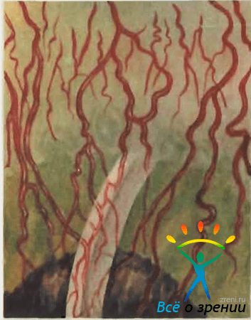
Fig. 23. Trachomatous pannus (optical section)
Pannus vessels consist mainly of tortuous, densely branching veins; the artery has a more linear course and is located deeper.
Somewhat later, in the limbus zone, you can see small, round, grayish gelatinous islands - follicles. They, just like the follicles of the conjunctiva, go through the entire cycle of their development; in some cases they have abortive development. Follicles often merged together form a zone of pronounced infiltration, visible to the naked eye. As a result, small scar impressions covered with epithelium remain in place of the follicles. These impressions are facets, also known as “eyes”, arise as a result of degeneration and disintegration of trachomatous follicles.
With a malignant course of pannus, its ulceration, infiltration and vessels penetrate into the deeper layers of the cornea. In such cases, they are visible in the middle and deep parts of the optical section. In this case, changes of a secondary nature may also develop - deposition of calcium and lipoids in the affected cornea. As a result of trachomatous pannus, a more or less intense clouding of the cornea with a tendon tint remains. Permeated with vessels.
When diagnosing trachoma, one must take into account the possibility of mixing this disease with other inflammatory lesions of the conjunctiva.
When differential diagnosis of trachoma with follicular conjunctivitis, it should be taken into account that with follicular conjunctivitis there is no diffuse infiltration of subepithelial tissue and hypertrophied papillae. When examined with a slit lamp, the follicles appear small, transparent, and do not have a developed network of capillaries (Fig. 24).

Rice. 24. Follicular conjunctivitis.
With follicular conjunctivitis, as with acute conjunctivitis of other origins, thickening of the limbus, dilation of the terminal capillaries and a slight lengthening of individual capillary loops can be observed. However, these changes soon disappear. Biomicroscopic examination does not detect either newly formed vessels or infiltration in the corneal tissue. There is no pannus with follicular conjunctivitis; There is no scarring of the conjunctiva as a result of the disease. After the process is eliminated, the conjunctival tissue appears completely unchanged.
In the differential diagnosis of trachoma with spring catarrh the pale, sometimes milky-white color of the conjunctiva, characteristic of the latter, and the predominant localization of changes on the conjunctiva of the cartilage of the upper eyelid (tarsal form) are taken into account. A lethal examination of these changes by bioncroscopy can be carried out only after removing a thick coating of viscous secretion from their surface.
Macroscopically revealed uneven surface of the conjunctiva. This is explained by the presence of dense, pale, shiny, as if polished, formations. These growths are known to be based on hyaline degeneration of subepithelial tissue.
Hyaline growths on the conjunctiva during spring catarrh they are fused, degeneratively degenerated papillae. When examined with a slit lamp, they look like multiple flat, polygonal and oval formations adjacent to each other with a smooth surface. Vascular bundles are visible, extending from large arterial trunks and running perpendicular to the surface of the conjunctiva. From these bundles a more superficial network of vessels arises (Fig. 25, a).

Rice. 25. Spring catarrh. a - tarsal form: b - limbal form.
Unlike papillae in spring catarrh papillae in trachoma smaller in size, red in color, more cloudy and lacking shine due to infiltration and desquamation of the epithelium. In addition, the papillae in trachoma never merge into one continuous mass. Each is formed around one vascular trunk and is separated from neighboring formations by a more or less narrow gap.
In some cases it is necessary to differentiate trachomatous pannus with limbal form of vernal catarrh. With joyful catarrh, changes occur around the entire limbus, and not just in its upper half, as with trachoma. At the limbus, small grayish islands are formed, consisting of a hyaline glassy substance, which is faintly translucent when examined in direct focal light. Often they merge and form a continuous ridge with a bumpy surface, sometimes moving onto the cornea (Fig. 25, b). Vascularization of vitreous formations is very insignificant compared to vascularization in trachomatous pannus. The limbal vessels usually pass through them at varying depths and terminate singly on the cornea.
With spring catarrh Damage to more centrally located parts of the cornea is also observed. Here, in the most superficial layers, very small, flat, scaly deposits of gray-white color appear. When the eyelids blink, these scales can be washed away with tears and erosions remain in their place. It is believed that these are neurotically degenerated elements of the epithelium of the cornea. Their favorite location is the upper half of the cornea (under the upper eyelid) and the palpebral fissure area.
It should not be forgotten that with the limbal form of spring catarrh conjunctival cartilage is often involved in the process. It acquires a milky white hue, against which even under low microscope magnification a typical mosaic of papillae is observed. They also show hyaline degeneration of the tissue, which gives the papillae a peculiar, glassy, translucent appearance.
At the end of spring catarrh all symptoms disappear without a trace; unlike trachoma, there is no scarring. Changes in the limbus and cornea also undergo complete reversal. As a result, retrospective diagnosis of vernal catarrh with a slit lamp is very difficult.
Dystrophic changes
Pinguecula- hyaline degeneration of the conjunctiva. It has the appearance of a yellow island, most often located at the inner limb of the cornea. Biomicroscopic examination in direct focal light reveals that the process of degeneration involves the deep sections of the optical section of the conjunctiva. Vitreous amorphous masses are also revealed under the cut of the conjunctiva. Sometimes small cavities are visible in the pinguecula tissue (Fig. 26).

Rice. 26. Pinguecula.
Pterygium, or pterygoid hymen, is a fairly common dystrophic change in the conjunctiva. When examining with a slit lamp, special attention should be paid to examining the head of the pterygium, i.e., that part of it that is located on the cornea.
There are two zones in the head of the pterygium: vascular and avascular (Fig. 27).

Rice. 27. Pterygium.
The latter is located in front of the vascular zone (towards the center of the cornea) and consists of foci of gelatinous opacity, the processes of which extend into the deeper parts of the corneal stroma.
Biomicroscopic examination allows you to determine whether the pterygium is stationary or progressive, which helps to correctly decide on the timing and type of surgical intervention. Stationary pterygium is characterized by a mildly defined, flat, avascular zone of the head, which imperceptibly merges with the tissue of the cornea. With progressive pterygium, the avascular zone is more pronounced, loosened and noticeably rises above the surface of the cornea. In front of the head of the pterygium there are sometimes punctate subepithelial opacities.
Differential diagnosis of pterygium with the initial form of Bowen's epithelioma - see below.
Neoplasms
Papilloma- a benign fibroepithelial formation, most often localized on the conjunctiva of the eyelids around the lacrimal caruncle, less often on the conjunctiva of the eyeball. The tumor is pink in color, soft in consistency, loosely fused to the underlying tissues, and often pedunculated.
Biomicroscopy reveals that the surface of the neoplasm is uneven and represents a conglomerate of papillomatous growths of a mosaic nature. With your look the tumor resembles a mulberry or cauliflower(Fig. 28).

Rice. 28. Papilloma of the conjunctiva.
Papilloma tissue does not transmit light from a slit lamp, which makes it impossible to obtain an optical section during bioncroscopy. Papilloma is characterized by rather sparse vascularization; it lacks glomerular vascular formations typical of malignant and vascular tumors.
When papilloma is localized at the limbus, it is necessary to carry out differential diagnosis with carcinoma.
Epithelioma, or carcinoma, - malignant epithelial tumor of the conjunctiva; prone to active growth and relapse after removal. The tumor is localized, as a rule, in the circumference of the limbus. In the initial stage, it is difficult to distinguish it from papilloma, pinguecula and incipient pterygium.
During biomicroscopic examination The hallmark of epithelioma is tuberosity, lobulation of the tumor. Ulceration of the surface is often observed, which is determined by the presence of a defect in the optical section of the conjunctiva. This sign is especially well revealed after staining the surface of the epithelioma with a fluorescein solution. In the differential diagnosis of epithelioma, great importance should be attached to slit-lamp examination nature of vascularization.
Epithelioma is richly vascularized. Each tumor lobule is equipped with a central vessel with a mass of capillary branches. The vessel penetrating the tumor lobule rises to its apex and then descends back (Fig. 29).

Rice. 29. Epithelioma of the conjunctiva.
Basal capillaries form a common vascular network, which, with its numerous anastomoses, feeds all tumor lobules. This type of vascularization is pathognomonic for epithelioma.
Epithelioma of intraepithelial localization, without disturbing the basement membrane of the epithelial layer, stands out as a separate form of precancerous dyskeratosis - Boveia epithelioma. The formation has the appearance of a grayish-white flat plaque, localized, as a rule, in the circumferential limbus. When examined using a slit lamp, an uneven surface of the neoplasm is noted, which is revealed by a break in the light slit, a significant number of white scales of exfoliating epithelium (dyskeratosis). The boundaries of the tumor are clear. With epithelioma of Boveia, there is a pronounced vascular reaction from the surrounding conjunctiva
Nevus, or birthmark, conjunctiva does not belong to the category of true tumors. Characterized by slow growth during the development of the organism, in adults it becomes stationary. However, in some cases, malignant degeneration of the nevus tissue is possible as a result of irritation, trauma to the nevus, and sometimes for no apparent reason.
Nevus is most often localized on the conjunctiva of the sclera in the limbus. It can appear in a pigmented or non-pigmented form, which causes different colors of the spots - from dark brown to light yellow.
Biomicroscopy in direct focal light and with a grazing beam reveals flat or very slightly protruding lesion on the surface of the sclera, with fairly clear boundaries (Fig. 30).

Rice. thirty. Nevus of the conjunctiva.
The tissue is characterized by delicate dusty pigmentation. With a narrow gap, you can determine the depth of the pigment and find out whether the nevus is epithelial or subepithelial. With subepithelial localization, pigment islands are visible in an optical section under the epithelial strip. Sometimes there is a nevus with vacuolar degeneration - the so-called naeviis cysticus. In an optical section with this form of nevus, numerous transparent cavities are visible - vacuoles, separated by thin partitions.
The nonvesicular tissue contains vessels, but they are few in number and do not provide dense branching.
Careful biomicroscopy makes it possible to make a differential diagnosis between nevus and incipient melanoblastoma of the conjunctiva. A big role in this belongs to the observation of changes in the nature of vascularization. The discovery of new vascular branches in the non-vesical tissue is suspicious regarding its malignant degeneration. Simultaneously with the change in the nature of vascularization or somewhat later, an increase in the size of the neoplasm, increased pigmentation of the tissue, and redistribution of pigment are noted.
Melanoblastoma of the conjunctiva It is one of the most malignant tumors and has a tendency to metastasize. She. arises spontaneously or develops from a conjunctival nevus. The tumor is most often localized near the limbus, but is also observed in the area of the lacrimal caruncle, the semilunar fold of the conjunctiva. Melanoblastoma spreads quite quickly, giving rise to daughter growth nodes.
Biomicroscopy reveals the lobular structure of the tumor tissue and its increased pigmentation. Unlike a nevus, the pigment of melanoblastoma is coarser and lumpy. When attempting to obtain an optical section in the area of melanoblastoma, the density of the tumor tissue and its intimate adhesion to the underlying scleron are noted. The light beam usually penetrates poorly into the tumor mass. A characteristic biomicroscopic sign of melanoblastoma is its lush vascularization, which is not observed with nevus. A branching bundle of capillary vessels is visible in the center of each tumor lobule. In addition, a rich vascular network is observed deep in the tumor.
Here is one of our clinical observations, where slit lamp examination significantly helped to make the correct diagnosis.
Patient S., 30 years old. was admitted to the hospital for neoplasms of the conjunctiva of the right eye. Since childhood, he has noticed a pigment spot on the eyeball; in recent years, it has begun to increase in size and stand above the surface of the eye.
Upon examination, uneven distribution was found in the upper parts of the limbus pigmented formation of the conjunctiva with an elastic consistency, round shape measuring 4X6 mm. The remaining parts of the eyeball are unchanged. Visual acuity 1.0.
When examined with a slit lamp in direct focal illumination, it is clearly visible that the neoplasm has a lumpy surface. It is not possible to obtain an optical section of the tissue, which emphasizes its density. There is pronounced vascularization of the neoplasm (a large number of newly formed vascular loops). There is a lot of coarse lumpy pigment in the tumor tissue (Fig. 31, a).

Rice. 31. Melanoblastoma of the conjunctiva. a - biomicroscopic picture; b - histological picture (hematoxylin-tosin staining, magnification 10 X 20).
Diagnosis: conjunctival melanoblastoma developing from a nevus. Biomicroscopy under fluorescent lighting and a study with radioactive phosphorus, which showed a high level of P accumulation, confirmed the diagnosis.
Produced melanoblastoma removal with preliminary and sequential diathermocoagulation of surrounding tissues. Histological examination revealed melanoblastoma of the alveolar structure with lush growth in the central sections. Cellular and nuclear atypia of the tumor tissue (epithelioid type of structure) was noted (Fig. 31. b).
Changes in glaucoma
Biomicroscopy of the conjunctiva of the eyeball is mandatory during a comprehensive examination of a patient with suspected glaucoma, as well as during dynamic monitoring of a patient with glaucoma. In both cases, it is necessary to pay attention to the condition of the anterior ciliary vessels (which play an important role in the outflow of intraocular fluid), especially those located in the upper and lower parts of the eyeball. Changes in the vessels located within the palpebral fissure, often exposed to adverse environmental influences, can disorient the observer. These vessels, especially in older people, are often tortuous, their branches varicose.
In glaucoma, changes in both the anterior ciliary arteries and anterior ciliary veins are observed; the latter are changed more often in congestive glaucoma. Particular attention should be paid during biomicroscopy area of scleral openings- emissaries through which the anterior ciliary arteries enter the eye and the veins exit. In patients with glaucoma, it is sometimes necessary to observe peculiar changes called the emissary symptom.
Exists incomplete and complete emissary symptom, the first is more common than the second. An incomplete emissary symptom is expressed in an increase in the size of the scleral opening by 2-3 times. When bnomncroscopy, it looks like a grayish round spot, in the center of which (in some cases eccentrically) is the anterior ciliary vessel. Sometimes, next to the expanded emissary, there are delicate accumulations of pigment brought here by the flowing chamber moisture.
With a complete emissary symptom above the enlarged scleral opening there is elevation, swelling of the conjunctiva (Fig. 32),

Rice. 32. Complete emissary symptom in glaucoma.
similar to that observed after fistulizing antiglaucomatous operations. In some cases, such a conjunctival “pad” does not appear above the emissary itself, but slightly away from it. Its development is associated with the detachment of the conjunctiva from the sclera by intraocular fluid flowing through the emissary. Direct focal light examination reveals a layer of clear fluid under the conjunctiva. When the anterior ciliary vessel is located near the limbus, the appearance of the emissary symptom is usually not accompanied by the formation of a typical conjunctival pad, since the latter in the limbus area is quite tightly fused to the underlying sclera. Under these conditions, as a rule, only a barely noticeable raised conjunctival ridge appears.
Detection of the Emissary Symptom during biomicroscopy obliges the doctor to suspect the presence of a glaucomatous process. If during examination of the patient the intraocular pressure turns out to be normal, then special tests should be performed to detect glaucoma. Under load conditions, compensation due to expansion of the emissary is often insufficient, which is reflected in an increase in intraocular pressure.
In relatively young patients, when the sclera is not yet so dense, the emissary symptom occurs more often in the early stages of glaucoma or in a preglaucomatous state. In elderly patients, due to the thickening of the outer membrane of the eye, the emissary symptom appears in the later stages of glaucoma against the background of degenerative changes in the sclera. The sclera around the anterior ciliary vessel is sometimes reduced and thinned to such an extent that the choroid becomes visible through the opening.
In the diagnosis of glaucoma, it may be of certain importance monitoring the condition of the water veins. Early signs of glaucoma, according to Z. A. Kaminskaya, include the appearance of a negative phenomenon of low tide. When a vessel receiving an aqueous vein is compressed with a glass rod, a twofold reaction can be observed: either the vein remains transparent and the intraocular fluid fills the vessel receiving the vein (positive ebb phenomenon), or the vein fills with blood (negative ebb phenomenon).
In addition to the negative phenomenon of low tide, glaucoma is characterized by "plus-minus" phenomenon. It consists in the fact that after squeezing the vessel receiving the water vein, the vein initially remains transparent and then fills with blood. With glaucoma in a state of decompensation, all aqueous veins are filled with blood, and the piston phenomenon is absent.
It must be performed on patients who have undergone fistulizing antiglaucomatous surgery. After trephination of the sclera according to Elliott, anterior sclerectomy, and iridenkleisis surgery, a filtration pad, sometimes a multi-chamber one, is formed over the upper limbus. When examined in direct focal light, it is clear that its cavities are filled with transparent intraocular fluid. Through them you can see the filtering surgical opening in the sclera and clumps of pigment.
- With a well-defined filtering postoperative scar, normal intraocular pressure values are usually observed.
- With unsatisfactory filtration, the surgical scar appears flat, abundantly vascularized due to newly formed conjunctival and episcleral vessels.
Article from the book: .
) is a detailed study of the structures of the eye, carried out using a special optical device - a slit lamp. The main part of the device is a diaphragm in the form of a narrow slit, which is why it got its name.
In the Soviet Union, the most common slit lamp model is ShchL-56. Using a lamp of this model, it is possible to examine both the anterior and posterior parts of the eye - the vitreous body and.
Biomicroscopy makes it possible to identify the smallest changes in the eye, detect small ones and determine the depth of the pathological process. Biomicroscopy is very important for the diagnosis of perforated wounds of the cornea and other eye diseases.
Biomicroscopy (synonymous with microscopy of the living eye) is a research method that allows you to examine in detail the conjunctiva, cornea, iris, anterior chamber of the eye, lens, vitreous body, as well as the central parts of the fundus (biomicroophthalmoscopy); proposed by A. Gullstrand. The biomicroscopy method is based on the phenomenon of light contrast (Tyndall phenomenon).
Using biomicroscopy, it is possible to carry out an early diagnosis of most (for example, glaucoma and trachoma), determine a perforated wound of the eyeball, detect very small foreign bodies in the conjunctiva, cornea, anterior chamber of the eye and lens that are not detected by x-ray examination (glass, aluminum, coal, eyelash). Biomicroscopy is carried out using a slit lamp.
The device (Fig. 1) consists of an illuminator and a binocular stereoscopic microscope. The light source in the illuminator is a lamp (6 V, 25 W), powered from an AC electrical network of 127 or 220 V through a step-down transformer. In the path of the light beam there is
a slit mechanism that allows for a vertical and horizontal lighting slit. The body of the binocular microscope contains an optical device that provides various magnification options (5, 10, 18, 35, 60 times). A binocular microscope has a diverging lens with a power of about 60 D, which neutralizes the positive effect of the optical system of the eye and allows one to see the fundus of the eye.
Rice. 1. Slit lamp ShchL-56: 1 - front installation; 2 - illuminator; 3 - binocular microscope; 4 - coordinate table; 5 - tool table.
Biomicroscopy is carried out in a dark room, creating a sharp contrast between the darkened and lamp-lit areas of the eyeball. In the process of biomicroscopy, diffuse, direct focal light, indirect illumination (dark field), transmitted light, sliding beam, and research in reflective zones (mirror field method) are used. The main type of lighting is direct focal. When focusing light on the cornea, an optical section of it is obtained in the form of a slightly opalescent convex-concave prism (Fig. 2). The anterior and posterior surfaces, the actual substance of the cornea, are clearly visible. If there is an inflammatory focus or cloudiness in the cornea, studying the optical section allows you to decide where the pathological focus is located and how deeply the corneal tissue is affected; in case of a foreign body in the cornea - whether it is located in the corneal tissue or partially penetrates into the eye cavity, which allows the doctor to correctly determine the method of intervention.
When light is focused on the lens, its optical section is cut out in the form of a biconvex transparent body. In the section, the surfaces of the lens are clearly visible, as well as grayish oval stripes, the so-called interface zones, caused by different densities of the lens substance (Fig. 3). Studying an optical section of the lens allows one to see and accurately localize the beginning opacities of its substance, which is of great importance for the early diagnosis of cataracts. Focusing light on the fundus allows one to examine the retina and optic nerve head in an optical section (Fig. 4). This is important for the early diagnosis of optic neuritis, congestive nipple, and centrally located retinal tears.
Less diagnostic possibilities are available with biomicroscopy of translucent and opaque membranes of the eyeball, for example, the conjunctiva and iris. However, in this case, biomicroscopy is an important addition to other methods of examining a patient with eye disease.
Rice. 2. Optical section of the cornea: a, b, f, d - anterior surface of the cornea; 3, e - edge of the rear surface; b, d, d, f - thickness of the cornea.
Rice. 3. Optical section of the lens: 1 - central gap; 2 - central surfaces of the embryonic nucleus; 3 - peripheral surfaces of the embryonic nucleus; 4 - surfaces of the senile core; 5 - subcapsular zones of cleavage; 6 - anterior and posterior surfaces of the lens. Rice. 4. Optical section of the retina and optic nerve head.
Thanks to B. g., early trachoma, glaucoma, cataracts and other eye diseases, as well as neoplasms, are possible. B. g. allows you to determine the perforation of the eyeball, to detect the smallest particles not detected by x-ray examination in the conjunctiva, cornea, anterior chamber of the eye and lens (particles of glass, aluminum, coal, etc.).
Biomicroscopy of the eye is carried out using a slit lamp (stationary or manual), the main parts of which are an illuminator and a magnifying device (stereoscopic or magnifying glass). In the path of the light beam there is a slot, which makes it possible to obtain vertical and horizontal lighting slits. Using the measuring eyepiece of a stereoscopic microscope, the depth of the anterior chamber of the eye is determined; additional dispersive power of about 60 diopter, neutralizing the positive effect of the optical system of the eye, makes it possible to examine the fundus of the eye .
The study is carried out in a dark room to create a sharp difference between the darkened and lamp-lit areas of the eyeball. The maximally opened slit of the diaphragm provides diffuse light, allowing one to examine all areas of the anterior part of the eye; a narrow slit provides a luminous optical ““. When a beam of light is combined with the observed area of the eye, direct focal illumination is obtained, which is most often used in B. and makes it possible to establish the localization of the pathological process. By focusing light on the cornea, an optical lens is obtained that has the shape of a convex-concave prism, on which the anterior and posterior surfaces of the cornea itself are clearly distinguished. When inflammation or clouding is detected in the cornea, B. g. allows one to determine the location of the pathological focus and the depth of tissue damage; in the presence of a foreign body, determine whether it is located in the corneal tissue or partially penetrates into the eye cavity, which allows the doctor to choose the right treatment tactics.
When light is focused on the lens, its optical section is determined in the form of a biconvex transparent body. In the section, the surfaces of the lens are clearly visible, as well as grayish oval stripes - the so-called interface zones, caused by different densities of the lens substance. Studying an optical section of the lens allows us to establish the exact localization of the beginning clouding of its substance and assess the condition of the capsule.
Biomicroscopy of the vitreous body reveals fibrillar structures (the skeleton of the vitreous body) that are not distinguishable by other research methods, changes in which indicate inflammatory or dystrophic processes in the eyeball. Focusing light on the fundus makes it possible to examine the retina and (size and depth of excavation) in an optical section, which is important in the diagnosis of glaucoma, for the early detection of optic neuritis, congestive nipple, and centrally located retinal breaks.
For B., other types of lighting are also used. Indirect illumination (dark field examination), in which the observed area is illuminated by rays reflected from the deeper tissues of the eye, allows for a good view of the vessels, areas of atrophy and tissue. To examine transparent media, illumination with transmitted light and is used, which helps to identify minor irregularities in the cornea, a detailed examination of the surface of the lens capsule, etc. Examination of the fundus is also carried out in the rays of the spectrum (). Biomicroscopy of translucent and opaque tissues of the eyeball (for example, conjunctiva, iris) is less informative.
Bibliography: Shulpina N.B. Biomicroscopy of the eye, M., 1974
II Biomicroscopy of the eye (Bio-+)a method of visual examination of optical media and eye tissues, based on creating a sharp contrast between illuminated and unlit areas and magnifying the image by 5-60 times; carried out using a slit lamp.
1. Small medical encyclopedia. - M.: Medical encyclopedia. 1991-96 2. First aid. - M.: Great Russian Encyclopedia. 1994 3. Encyclopedic Dictionary of Medical Terms. - M.: Soviet Encyclopedia. - 1982-1984.
See what “Biomicroscopy of the eye” is in other dictionaries:
biomicroscopy of the eye- rus biomicroscopy (f) eyes eng slit lamp examination fra examen (m) à la lampe à fente deu Linsenuntersuchung (f) mit der Spaltlampe spa examen (m) con lámpara de hendidura … Occupational safety and health. Translation into English, French, German, Spanish
- (bio + microscopy) a method of visual examination of optical media and eye tissues, based on creating a sharp contrast between illuminated and unlit areas and magnifying the image by 5-60 times; carried out using a slit lamp... Large medical dictionary
CHEMICAL EYE BURNS- honey Chemical burns of the eye are one of the emergency conditions in ophthalmology that can cause impaired or complete loss of vision. Frequency 300 cases/100,000 population (burns with alkalis account for 40% of all cases of eye burns, with acids 10%).… … Directory of diseases
PENETRATING EYE WOUNDS- honey Penetrating wounds of the eye are characterized by disruption of the integrity of its fibrous membrane (cornea and sclera). Clinical picture Presence of a wound channel Loss or pinching of the inner membranes of the eye (iris, vascular tissue itself) in the wound… Directory of diseases
MELANOMA OF THE OCULUS PROPER- honey Melanoma of the choroid proper is a malignant pigmented tumor. Frequency 0.02 0.08% of patients observed by ophthalmologists on an outpatient basis Most often diagnosed in men aged 31 60 years (75%) Peak incidence (57%) 50... ... Directory of diseases
I Foreign bodies Foreign bodies (corpora aliena) are objects foreign to the body that have penetrated into its tissues, organs or cavities through damaged integuments or through natural openings. Foreign bodies are also those introduced into the body with... ... Medical encyclopedia
I Cataract (cataracta; Greek: katarrhaktēs waterfall) is an eye disease characterized by clouding of the lens. There are primary (congenital and acquired) and secondary cataracts. Congenital K. (Fig. 1) can be hereditary (dominant ... Medical encyclopedia
I (oculus) organ of vision that perceives light stimulation; is part of the visual analyzer, which also includes the optic nerve and visual centers located in the cerebral cortex. The eye consists of the eyeball and... Medical encyclopedia
- (Gonio + biomicroscopy (Biomicroscopy of the eye); synonym microgonioscopy) a method of examining the iridocorneal angle of the eye (anterior chamber angle) by examining it with a gonioscope and a slit lamp... Medical encyclopedia
Extrapulmonary tuberculosis is a conditional concept that unites forms of tuberculosis of any localization, except for the lungs and other respiratory organs. In accordance with the clinical classification of tuberculosis (TB), adopted in our country, to T.v. include... ... Medical encyclopedia
The ability to see the world around us is a unique gift from nature to man. The ability to distinguish colors, objects, and abstract images is necessary for work and creativity. Eye diseases are common in modern society. Many of them, if detected late, can permanently deprive a person of their ability to work and a normal quality of life. Biomicroscopy of the eye is one of the most reliable and informative methods for identifying various eye diseases.
Biomicroscopy of the eye: science does not stand still
The eye, due to its location, is accessible to careful visual inspection. Signs of most pathologies of the organ of vision can be easily identified and the degree of their severity can be assessed without resorting to the use of X-rays, ultrasonic waves and magnetic fields.
Several decades ago, this problem was solved with the help of light, a mirror and a magnifying lens. The latter made it possible to obtain an image of the fundus and its individual components. This method is used by a specialist in direct and reverse varieties and is called ophthalmoscopy.
Ophthalmoscopy is a method of examining the eye using a magnifying lens.
Modern ophthalmology has a more accurate and effective method for studying the various anatomical structures of the eyeball. An image of the smallest components of the organ of vision allows us to obtain a microscope connected to a light source. This method is called biomicroscopy. The ability to study body tissues intravitally without resorting to their removal is of great benefit in diagnosing diseases of the organ of vision. Biomicroscopy allows you to study the anatomical structure of various parts of the eyeball:

Types of biomicroscopy
The biomicroscopy method has been modified to facilitate the study of transparent and opaque structures of the eyeball. The researcher can use four different options for the procedure:

Research methodology
Biomicroscopy is a non-contact, non-invasive method of examining the eyeball and does not cause pain or discomfort to the patient. The procedure is carried out using a slit lamp, which has a light source, a microscope and a stand with a rest for the forehead and chin for convenient positioning of the subject’s head.
The first stage of the study is to place the patient in relation to the device using a stand. In this case, the eyeball should coincide with the direction of the slit lamp beam. The latter creates a narrow beam of light, by moving which the doctor can study in detail the necessary structures of the eye. The patient does not experience any sensations. The procedure may take 10 to 15 minutes to complete. Interpretation of the results is facilitated by a microscope lens system that provides multiple image magnification.
Biomicroscopy of the eye - a non-contact, non-invasive research method
No special preparation is required for the study. If there are difficulties, the doctor can temporarily dilate the opening of the pupil using medications in the form of drops. The most commonly used is Atropine. In this situation, access of the light beam to individual structures of the fundus is greatly facilitated. However, if the patient has increased intraocular pressure (glaucoma), pupil dilation is not used.
In some cases, biomicroscopy is performed under conditions of medicinal dilation of the pupil
Biomicroscopy of the conjunctiva
The eyeball is in direct contact with the environment, therefore it is protected by nature with the help of the conjunctiva - a kind of transparent type of skin that is not inferior to it in strength. This mucous membrane covers the eyelids from the inside, after which it passes to the sclera and cornea.
The conjunctiva receives good nutrition from an extensive network of vessels, which under normal conditions are invisible to the naked eye. However, using a slit lamp, you can evaluate not only their size, but also see the movement of individual blood cells.
Using biomicroscopy, a fairly common and very unpleasant disease is diagnosed - conjunctivitis. Inflammation of the transparent membrane in the rays of light takes on a characteristic appearance: the presence of dilated vessels, stagnation in them, areas of accumulation of white blood cells - leukocytes. The latter circumstance, as the disease progresses, leads to the appearance of a visually noticeable purulent discharge, which is a cemetery of dead cells.
Conjunctivitis - indication for eye biomicroscopy
Examination of the anterior part of the eye
The anterior part of the eyeball is most clearly visible during normal visual examination. Biomicroscopy allows you to identify subtle changes:
- fibrous membrane;
- corneas;
- anterior chamber;
- lens;
- irises.
The sclera is a dense connective tissue structure that primarily performs a protective and scaffolding function. Its vascular network is very developed. Using a microscope, you can see inflamed areas (scleritis and episcleritis).
Scleritis is an inflammation of the fibrous membrane of the eye.
The cornea is the transparent part of the fibrous membrane. In addition, it is an important component of the optical system of the eye. The correct construction of the image on the retina largely depends on the shape and transparency of the cornea. Using the light beam of a slit lamp and a microscope, any opacification or ulceration can be identified and the sphericity of the surface can be assessed.
On biomicroscopy, a corneal ulcer looks like a focus of turbidity
The anterior chamber of the eye is the space between the cornea and the iris. It is filled with liquid, through which light also passes on its way. Biomicroscopy allows you to evaluate the transparency and presence of suspensions in the moisture of the anterior chamber.
An important task for the researcher is to assess a special structure - the angle of the anterior chamber of the eye. This section is where the iris attaches to the sclera. The angle of the anterior chamber is a kind of drainage system of the eye, through which moisture is directed into the veins of the fibrous membrane, thereby maintaining constant pressure inside. Anomalies in the structure of this area lead to glaucoma. To obtain an image, the doctor additionally uses a special mirror - a gonioscope.
The anterior chamber angle is the main drainage device of the eye
The iris not only determines eye color. At its core, it contains ciliary muscle fibers on which the lens is suspended. This design is the main mechanism of accommodation, responsible for the ability of the human eye to see close and distant objects equally clearly. In addition, by changing the width of the pupil opening, the eye independently regulates the flow of light reaching the retina. Biomicroscopy allows you to study in detail the structure of the iris and ciliary muscles, identify foci of inflammation (uveitis), neoplasms, including malignant ones (melanoma).
Inflammation of the iris leads to deformation of the pupil opening
The lens is the main part of the optical system of the eye. It is a transparent structure resembling a gel. The lens is located in a capsule surrounded by the ciliary muscle. The main task of biomicroscopy in this case is to assess its transparency and identify local or total opacification (cataracts).
When performing biomicroscopy of the eye, clouding of the lens is clearly visible
Biomicroscopy of the posterior part of the eyeball
Directly behind the lens is a transparent gelatinous formation - the vitreous body, which is part of the optical system of the eye. Its microscopic structure may suffer from local foci of opacification or hemorrhage.
Behind the vitreous body lies the pigment membrane of the eye - the retina. It is its specific cells - rods and cones - that perceive light. Biomicroscopy allows you to evaluate most structures of the fundus and identify the following pathologies:

What the fundus can tell you - video
Additional features of the method
The method of eye biomicroscopy is constantly being improved. Currently, the study allows us to evaluate important parameters:
- thickness and sphericity of the cornea (confocal biomicroscopy of the cornea). This indicator is of particular importance when planning laser vision correction;
- depth of the anterior chamber of the eye. This parameter determines the possibility of implanting anterior chamber models of intraocular lenses to correct visual acuity in myopia or hyperopia.
The latest achievement in ophthalmology is ultrasound biomicroscopy. This method makes it possible to study many structures that are inaccessible to a beam of light during conventional research:
- posterior surface of the iris;
- ciliary body;
- lateral sections of the lens;
Ultrasound microscopy - a modern version of the method
Advantages and disadvantages
The eye biomicroscopy method has many advantages:

The main disadvantage of the method is the incompleteness of the information obtained about a particular segment of the eye. Additional studies may be required to definitively diagnose the disease. In addition, biomicroscopy exclusively evaluates the anatomy of the eye and does not provide the doctor with information about its functional abilities.
Biomicroscopy of the eye is a modern informative method for diagnosing diseases of the organ of vision. The results must be assessed by an ophthalmologist, after which the doctor will decide on further tactics for examining and treating the patient.
Examination of the internal structures of the eye is necessary when there is a suspicion of any disease or abnormality of the anterior or posterior part of the eyeball. The use of a special microscope for this purpose, combined with a powerful lighting device, is called biomicroscopy. This study helps to identify and study in detail many abnormalities within the visual organ.
Biomicroscopy: basic concepts
Biomicroscopy is the examination of the internal state of the eyeball using a medical device called a slit lamp. Includes a wide range of complex imaging techniques for pathologies of varying origin, texture, color, transparency, size and depth.
The slit lamp allows for detailed microscopic examination of the eye
A slit lamp is an instrument consisting of a high-intensity light source that can be focused to direct a thin strip of light into the eye through various filters that provide the location and size of the slit. It is used in combination with a biomicroscope, which, together with the illuminator, is mounted on one coordinate table. The lamp facilitates inspection of the anterior and posterior segments of the human eye, which include:
- eyelid;
- sclera;
- conjunctiva;
- iris;
- natural lens (lens);
- cornea;
- vitreous body;
- retina and optic nerve.
The slit lamp is equipped with a diaphragm that forms a slit up to 14 mm in width and height. A binocular microscope includes two eyepieces and an objective (magnifying lens), the optical power of which can be adjusted using a dial that changes the magnification factor. The range of gradual increase is from 10 to 25 times. With an additional eyepiece - up to 50-70x.
Binocular slit lamp examination provides stereoscopic, magnified images of ocular structures in detail, allowing anatomical diagnoses to be made for a variety of ocular conditions. The second, hand-held lens is used to examine the retina.
For a full examination with a biomicroscope, there are various methods of slit lamp illumination. There are six types of basic lighting options:
- Diffuse illumination - examination through a wide aperture using glass or a diffuser as a filter. It is used for a general examination to detect the localization of pathological changes.
- Direct focal illumination is the most commonly used method, which consists of observing using an optical slit or direct focal illumination of rays. A thin or medium width slit is directed and focused on the cornea. This type of illumination is effective for determining the spatial depth of ocular structures.
- Specular reflection, or reflected lighting, is a phenomenon similar to the image visible on the sunny surface of a lake. Used to assess the endothelial contour of the cornea (its inner surface). To achieve a mirror effect, the tester directs a narrow beam of light to the eye from the temple side at an angle of about 25-30 degrees to the cornea. A bright area of specular reflection will be visible on the corneal epithelium (outer surface).
- Transillumination (transillumination), or examination in reflected (transmitted) light. In some cases, illumination with an optical slit does not provide sufficient information or is simply impossible. Transillumination is used to examine transparent or translucent structures - the lens, the cornea - by reflecting rays from deeper tissues. To do this, highlight the background of the object under study.
- Indirect lighting - a light beam passing through translucent fabrics is scattered, simultaneously highlighting individual places. Used to identify pathologies of the iris.
- Scleral scattering - with this type of lighting, a wide beam of light is directed at the limbal region of the cornea (the edge of the cornea, the junction with the sclera) at an angle of 90 degrees to it to create a light scattering effect. In this case, a certain halo appears under the cornea, which illuminates its anomalies from the inside.
The slit lamp makes it possible to study the structural parts of the cornea:
- epithelium;
- endothelium;
- posterior border plate;
- stroma.
And also - determine the thickness of the transparent outer shell, its blood supply, the presence of inflammation and edema, and other changes caused by injury or dystrophy. The study allows you to study in detail the condition of scars, if they exist: their size, adhesions with surrounding tissues. Biomicroscopy reveals tiny solid deposits on the back surface of the cornea.
If a pathology of the cornea is suspected, the doctor will additionally prescribe confocal microscopy - a method of assessing the morphological changes of this organ using a special microscope with a magnification of 500 times. It allows you to study in detail the layer-by-layer structure of the corneal epithelium.
During biomicroscopy of the lens, the doctor examines the optical section for possible clouding of its substance. Determines the location of the pathological process, which often begins precisely at the periphery, the state of the nucleus and capsule. When examining the lens, almost any type of lighting can be used. But the most common are the first two: diffuse and direct focal lighting. They are usually carried out in this order. The first type of lighting allows you to evaluate the general appearance of the capsule and see foci of pathology, if any. But for a clearer understanding of where exactly the “breakdown” occurred, it is necessary to resort to direct focal lighting.
Examining the vitreous body using a slit lamp is a challenging task that not every novice in ophthalmology can handle. The vitreous body has a jelly-like consistency and lies quite deep. Therefore, it weakly reflects light rays.
Biomicroscopy of the vitreous body requires acquired skill
In addition, the study is hampered by a narrow pupil. An important condition for high-quality biomicroscopy of the vitreous body is preliminary medicinal mydriasis (pupil dilation). The room where the inspection is carried out should be as dark as possible, and the area under study, on the contrary, should be quite brightly lit. This will provide the necessary contrast, since the vitreous body is a weakly refractive optical medium that slightly reflects light. The doctor uses mostly direct focal lighting. When examining the posterior parts of the vitreous body, it is possible to study in reflected light, where the fundus of the eye plays the role of a reflective screen.
Focusing light on the fundus allows one to examine the retina and optic nerve head in an optical section. Early detection of neuritis or swelling of the nerve (congestive papilla), retinal tears helps in the diagnosis of glaucoma, prevents optic nerve atrophy and decreased vision.
The slit lamp will also help determine the depth of the anterior chamber of the eye, detect cloudy changes in moisture and possible impurities of pus or blood.
A wide selection of lighting types thanks to special filters allows you to clearly study the vessels, detect areas of atrophy and tissue ruptures. Biomicroscopy of translucent and opaque tissues of the eyeball (for example, conjunctiva, iris) is less informative.
Slit lamp device: video
Indications and contraindications
Biomicroscopy is used to diagnose:
- glaucoma;
- cataracts;
- macular degeneration;
- retinal detachment;
- corneal damage;
- blockage of retinal vessels;
- inflammatory diseases;
- neoplasms, etc.
You can also detect an injury to the eye, foreign bodies in it, which x-rays cannot show.
There are no absolute contraindications for slit lamp examination. However, it is worth paying attention to some important nuances associated with eye injuries:

Fundus observation is known as fundus lens ophthalmoscopy. But with a slit lamp, direct observation of the bottom is impossible due to the refractive power of the ocular media, as a result of which the microscope does not provide focusing. The use of auxiliary optics helps. Using a diagnostic three-mirror Goldmann lens in the light of a slit lamp, you can examine those peripheral areas of the retina that cannot be examined with ophthalmoscopy.
Advantages and disadvantages of the method
Biomicroscopy has a number of significant advantages over other methods of ophthalmological examination:
- Possibility of precise localization of anomalies. Due to the fact that a beam of light from a slit lamp during biomicroscopy can penetrate into the structures of the eye from different angles, it is quite possible to determine the depth of pathological changes.
- Increased diagnostic capabilities. The device provides lighting in vertical and horizontal planes at different angles.
- Convenient for detailed inspection of a specific area. A narrow beam of light directed into the eye provides contrast between the illuminated and darkened areas, forming the so-called optical slice.
- Possibility of biomicroophthalmoscopy. The latter is successfully used for examining the fundus.
The method is considered highly informative, devoid of significant disadvantages and contraindications. But in some cases it is advisable to prefer a manual device to a stationary one, although a manual slit lamp has limited capabilities. For example, it is used:
- for biomicroscopy of the eyes of babies who are still in a supine position;
- when examining restless children who cannot sit for the prescribed amount of time at a conventional slit lamp;
- for examining patients in the postoperative period, during strict bed rest, it is an alternative to the stationary version of the device.
In these cases, a hand-held lamp has advantages over scattered (diffuse) lighting and makes it possible to examine in detail the surgical incision and the anterior chamber with intraocular fluid, the pupil, and the iris.
The manual slit lamp has modest capabilities, but sometimes it is indispensable
Carrying out the procedure
The examination is carried out in a darkened room. The patient sits in a chair, places his chin and forehead on a support to fix his head. She must be motionless. It is advisable to blink as little as possible. Using a slit lamp, the ophthalmologist examines the patient's eyes. To aid the examination, a thin strip of paper containing fluorescein (a glowing dye) is sometimes applied to the edge of the eye. This colors the tear film on the surface of the eye. The paint is later washed away by tears.
Then, at the doctor's discretion, drops may be needed to dilate the pupils. You must wait 15 to 20 minutes for the medicine to take effect, after which the examination is repeated, allowing you to check the back of the eye.
Sometimes before biomicroscopy it is necessary to dilate the pupil with medication
First, the ophthalmologist will again test the front structures of the eye, and then, using a different lens, examine the back of the organ of vision.
As a rule, such a test does not cause significant side effects. Sometimes the patient experiences slight light sensitivity for several hours after the procedure, and dilating drops can increase eye pressure, leading to nausea with headache. Those who feel seriously unwell are advised to consult a doctor immediately.
Adults do not need special preparation for the test. However, children may need it in the form of atropinization (pupil dilation) depending on age, previous experience and level of trust in the doctor. The whole procedure takes about 5 minutes.
Research result
During the examination, the ophthalmologist visually assesses the quality and condition of the eye structures to detect possible problems. Some models of slit lamps have a photo and video module that record the examination process. If the doctor finds that the results are not normal, this may indicate the following diagnoses:
- inflammation;
- infection;
- increased pressure in the eye;
- pathological change in the ophthalmic arteries or veins.
For example, in macular degeneration, the doctor will find drusen (optic disc calcifications), which are yellow deposits that can form in the macula, an area on the retina, early in the disease. If the doctor suspects a certain vision problem, he will recommend further detailed examination to make a final diagnosis.
Biomicroscopy is a modern and highly informative method of examination in ophthalmology, allowing a detailed examination of the ocular structures of the anterior and posterior sections under different lighting and image magnification. As a rule, there is no need to prepare specially for this study. Thus, a five-minute procedure makes it possible to effectively monitor the health of the eye and prevent possible deviations in time.