How a chicken embryo develops during incubation every day. How a chicken develops in an egg How a chicken develops in an egg by day
Every day of incubation of the eggs of any bird is accompanied by various changes in the embryo. Understanding what is happening inside the hatching egg will allow the farmer to better understand whether incubation is proceeding correctly, what is normal and what is a deviation. To do this, we will analyze this process day by day.
1 day of incubation of chicken eggs
On the first day of incubation, 2 germ layers are formed. The upper one is ectoderm and the lower one is endoderm. After this, the third - middle sheet - mesoderm appears. Subsequently, these 3 layers participate in the formation of the necessary tissues, organs, and systems. Thus, the ectoderm participates in the formation of: the upper layers of the skin, feathers, comb, earrings, beak, as well as all nervous tissue and sensory organs. The endoderm forms the intestines with glands, respiratory organs, thyroid gland, etc. And the mesoderm is involved in the formation of: muscle tissue, glands and organs of genitourinary secretion. The embryonic disc spreads over the vitelline surface. The light field becomes elongated and forms the image of a pear, the narrow part participates in the formation of the tail part, and the head part from the wide one.
You may also be interested in: ; ; .
After 6 hours, the primary streak begins to form; this is a zone with increased cell division. After 12 hours, the primary streak is equal to 50% of the length of the embryonic disc, after 18 hours it is already 75%. This development is an excellent indicator when using biocontrol on the 1st day. In an embryo that develops well, the disc diameter is 0.5 cm, the yolk along with the embryo is surrounded by the vitelline membrane. Already by the end of the 1st day, the head process and head bubbles are formed, blood islands and the rudiment of the cardiac, nervous system and digestion also appear. It is important that the correct start of development of the embryo on the 1st day determines what kind of chicken the chicken will ultimately turn out to be and whether it will be viable.
Day 2 of incubation of chicken eggs
On this day, the neural tube is formed, and brain vesicles grow at its end. The heart muscles begin to contract rhythmically, which means the vascular system is working and blood circulation begins. By the end of the day, the yolk circulatory system is actively working.
Day 3 of incubation of chicken eggs
The heart pulsates intensely, the pulse rate is directly related to the temperature of the environment around the embryo.
4th day of incubation of chicken eggs
On this day, you need to monitor the incubation process using an ovoscope. During ovoscopy, the vessels of the circulatory system of the yolk circle will be visible. Whether the embryo grows correctly and whether development proceeds intensively depends on this. The yolk is in the shape of an ellipse.
Day 5 of incubation of chicken eggs
The head section is significantly superior in height. When ovoscoping on the 5th day, the pigmented retina should be visible. Allantois spreads greatly. Hematopoiesis occurs in the liver. The primary kidney increases its size and takes on the function of excreting waste products

Days 6 and 7 of incubation of chicken eggs
During this period of time, muscle tissue contracts and he can move. The respiratory organs and digestive system begin to form, and the eyelids are formed. A blood network appears on the surface of the allantois. The yolk remains almost unchanged; before this, it increased in size for six days. This happens because the embryo consumes the liquid layer more intensively, and its filling does not proceed as quickly.


Days 8 and 9 of incubation
Metabolic products accumulate in the allantois cavity.


10th day of incubation
On this day, the heating in the incubator is reduced, since heat is independently released from his body. Water must be removed from the allantois, so the humidity in the incubator is reduced and monitored to ensure that overheating does not occur. Starting from the 10th day, the source of nutrition and the way breathing changes in the embryo. There is a large amount of liquid in the amnion, the fetus swallows it and then it is absorbed in its digestive tract.

11th day of incubation
The resulting network of vessels grows at high speed under the shell, it captures the remaining protein at the sharp end and the allantois closes.

Days 12 and 13 of incubation of chicken eggs
When ovoscoping, the rudiments of the ridge are visible, which looks like a protrusion with a faint outline of the ridge teeth. The rudiments of feathers on the wing are clearly visible, and there is eye pigment. All these signs indicate a normal course of incubation.


14th day of incubation of chicken eggs
During this period, the amnion is stretched due to the large size of the embryo and due to the constant supply of protein. The protein is used by the embryo intraintestinally, the glandular section of the stomach and pancreas begin to actively function. The embryo is large, moves and is completely covered with down.
15-19 days of incubation of chicken eggs
During this period of time, all functions and organs are fully formed.

20th day of incubation of chicken eggs
On the 20th day of incubation, the eggs should be transferred to hatching trays. During this period, the embryo's method of gas exchange changes. The allantois no longer serves as a source of respiration; this function is taken over by the lungs. This transition between different ways of breathing is necessary and difficult. At this moment, it is necessary to increase the humidity in the incubator and ensure good air exchange. This will ensure excellent and timely chick hatching. At the beginning of the 20th day, the yolk sac is completely retracted. The chicken's neck arches, causing the air chamber to become tortuous.

21 days of incubation of chicken eggs
If everything went normal during the incubation period, then on the 21st day the chick hatches. At this point it should have: the yolk is completely retracted, the umbilical opening is narrowed and scarred. The chicken pecks at the shell and tries to get out.
From egg to egg
Let's break the shell of a chicken egg. Underneath we will see a film as thick as parchment. This is the subshell membrane, the same one that does not allow us to get by with one teaspoon when “destroying” a soft-boiled egg. You have to pick at the film with a fork or knife, or at worst with your hands. Under the film is a gelatinous mass of protein, through which the yolk is visible.
It is from this, from the yolk, that the egg begins. At first it is an oocyte (egg) covered in a thin membrane. Collectively this is called a follicle. The ripe egg, which has accumulated the yolk, breaks through the follicle membrane and falls into the wide funnel of the oviduct. Several follicles mature in the ovaries of a bird at the same time, but they mature at different times, so that only one egg always moves through the oviduct. Fertilization occurs here in the oviduct. And after that, the egg will have to put on all the egg membranes - from the albumen to the shell.
The protein substance (we will talk about what the protein and yolk are a little later) is secreted by special cells and glands and is wound layer by layer around the yolk in the long main section of the oviduct. This takes about 5 hours, after which the egg enters the isthmus - the narrowest section of the oviduct, where it is covered with two shell membranes. In the outermost part of the isthmus at the junction with the shell gland, the egg stops for 5 hours. Here it swells - absorbs water and increases to its normal size. At the same time, the shell membranes become more and more stretched and eventually fit tightly to the surface of the egg. Then it enters the last section of the oviduct, the shell, where it makes a second stop for 15-16 hours - this is exactly the time allowed for the formation of the shell. Once it is formed, the egg is ready to begin life on its own.
The embryo develops
For the development of any embryo, the presence of “building material” and “fuel” is necessary to ensure the supply of energy. “Fuel” must be burned, which means oxygen is also needed. But that's not all. During the development of the embryo, “construction slag” and “waste” from burning “fuel” are formed - toxic nitrogenous substances and carbon dioxide. They must be removed not only from the tissues of the growing organism themselves, but also from its immediate environment. As you can see, there are not so few problems. How are they all resolved?
In truly viviparous animals—mammals—everything is simple and reliable. The embryo receives building material and energy, including oxygen, through the blood from the mother's body. And in the same way it sends back “slags” and carbon dioxide. Another thing is who lays the eggs. They have to give building material and fuel to the embryo “to take away”. High molecular organic compounds - proteins, carbohydrates and fats - serve this purpose. From below, a growing organism draws amino acids and sugars, from which it builds proteins and carbohydrates of its own tissues. Carbohydrates and fats are also the main source of energy. All of these substances make up the component of the egg we call the yolk. The yolk is a food supply for the developing embryo. Now the second problem is where to put the toxic waste? Good for amphibian fish. Their egg (spawn) develops in water and is separated from it only by a layer of mucus and a thin egg membrane. So oxygen can be obtained directly from the water and into the water, and waste can be sent. True, this is only feasible if the excreted nitrogenous substances are highly soluble in water. Indeed, fish and amphibians excrete the products of nitrogen metabolism in the form of highly soluble ammonia.
But what about birds (and crocodiles, and turtles), whose eggs are covered with a dense shell and develop not on water, but on land? They have to store the toxic substance directly in the egg, in a special “garbage” sac called allantois. The allantois is connected to the circulatory system of the embryo and, together with the “waste” brought into it by the blood, remains in the egg abandoned by the chick. Of course, in this case it is necessary that the decay products are released in a solid, poorly soluble form, otherwise they will again spread throughout the entire egg. Indeed, birds and reptiles are the only vertebrates that emit “dry” uric acid rather than ammonia.
The allantois in the egg develops from the embryo's own tissue primordia and belongs to the embryonic membranes, as opposed to the egg membranes - the albumen, subshell and the shell itself, which are formed in the mother's body. In the eggs of reptiles and birds, in addition to the allantois, there are other embryonic membranes, in particular the amnion. This membrane forms a thin film over the developing embryo, as if it includes it, and fills it with amniotic fluid. In this way, the embryo forms its own “water” layer inside itself, which protects it from possible shocks and mechanical damage. You never cease to be amazed at how wisely everything is arranged in nature. And it's difficult. Surprised by this complexity and wisdom, embryologists elevated the eggs of birds and reptiles to the rank of amniotic eggs, contrasting them with the more simply constructed eggs of fish and amphibians. Accordingly, all vertebrate animals are divided into anamnium (no amnion - fish and amphibians) and amniote (having an amnion - reptiles, birds and mammals).
We have dealt with “solid” waste, but the problem of gas exchange still remains. How does oxygen enter the egg? How is carbon dioxide removed? And here everything is thought out to the smallest detail. The shell itself, of course, does not allow gases to pass through, but it is penetrated by numerous narrow tubes - pore or respiratory channels, simply pores. There are thousands of pores in the egg, and gas exchange occurs through them. But that's not all. The embryo develops a special “external” respiratory organ - the chorialantois, a kind of placenta in mammals. This organ is a complex network of blood vessels that line the inside of the egg and quickly deliver oxygen to the tissues of the growing embryo.
Another problem for a developing embryo is where to get water. The eggs of snakes and lizards can absorb it from the soil, increasing in volume by 2-2.5 times. But the eggs of reptiles are covered with a fibrous shell, while in birds they are encased in a shell shell. And where can you get water in a bird’s nest? There is only one thing left - to store it, as well as nutrients, in advance, while the egg is still in the oviduct. For this purpose, the component that is commonly called protein is used. It contains 85-90% water absorbed by the substance of the protein shells - remember? – the first stop of the egg is at the isthmus, at the junction with the shell gland.
Well, now it seems that all the problems have been solved? It only seems. The development of an embryo is full of problems, and the solution to one immediately gives rise to another. For example, pores in the shell allow the embryo to receive oxygen. But through the pores the precious moisture will evaporate (and evaporate). What to do? Initially, store it in excess in protein, and try to extract some benefit from the inevitable process of evaporation. For example, due to water loss, the free space in the wide pole of the egg, which is called the air chamber, expands significantly towards the end of incubation. By this time, the chorialantois alone is no longer enough for the chick to breathe; it is necessary to switch to active breathing with the lungs. The air chamber accumulates air, with which the chick will first fill its lungs after breaking through the shell membrane with its beak. Oxygen here is still mixed with a significant amount of carbon dioxide, so that the organism, which is about to begin an independent life, gradually gets used to breathing atmospheric air.
And yet the problems of gas exchange do not end there.
Pores in the shell
So, a bird’s egg “breathes” thanks to the pores in the shell. Oxygen enters the egg, and water vapor and carbon dioxide are expelled. The more pores and the wider the pore channels, the faster the gas exchange takes place, and vice versa, the longer the channels, i.e. The thicker the shell, the slower gas exchange occurs. However, the embryo's respiration rate cannot be below a certain threshold value. And the speed at which air enters the egg (it is called the gas conductivity of the shell) must correspond to this value.
It would seem that there is nothing simpler - let there be as many pores as possible, and they be as wide as possible - and there will always be enough oxygen, and carbon dioxide will be perfectly removed. But let's not forget about water. During the entire incubation period, the egg can lose water no more than 15-20% of its original weight, otherwise the embryo will die. In other words, there is an upper limit for increasing the gas conductivity of the shell. In addition, the eggs of different birds are known to differ in size - from less than 1g. in hummingbirds up to 1.5 kg. In the African ostrich. And among those that became extinct in the 15th century. The Madagascan apiornis, related to ostriches, had an egg volume of as much as 8-10 liters. Naturally, the larger the egg, the faster oxygen must enter it. And again the problem is that the volume of the egg (and, accordingly, the mass of the embryo and its oxygen needs), like any geometric body, is proportional to the cube, and the surface area is proportional to the square of its linear dimensions. For example, an increase in the length of an egg by 2 times will mean an increase in oxygen demand by 8 times, and the area of the shell through which gas exchange occurs will increase only by 4 times. Consequently, it will be necessary to increase the gas permeability value.
Studies have confirmed that the gas permeability of the shell actually increases with increasing egg size. In this case, the length of the pore channels, i.e. The thickness of the shell does not decrease, but also increases, although more slowly.
You have to “puff” due to the number of pores. A 600-gram rhea ostrich egg has 18 times more pores than a 60-gram chicken egg.
The chick hatches
Bird eggs also have other problems. If the pores in the shell are not covered with anything, then the pore channels act as capillaries and water easily penetrates them into the egg. This may be rainwater carried on the plumage of a brooding bird. And microbes get into the egg with water - rotting begins. Only some birds, those that nest in hollows and other shelters, such as parrots and pigeons, can afford to have eggs with uncovered pores. In most birds, the egg shell is covered with a thin organic film - cuticle. The cuticle does not allow capillary water to pass through, but oxygen molecules and water vapor pass through it unhindered. In particular, the shells of chicken eggs are also covered with cuticle.
But the cuticle has its own enemy. These are mold fungi. The fungus devours the “organic matter” of the cuticle, and thin threads of its mycelium successfully penetrate through the pore channels into the egg. This must first be taken into account by those birds that do not maintain cleanliness in their nests (herons, cormorants, pelicans), as well as those who make their nests in an environment rich in microorganisms, for example on water, in liquid silty mud or in rotting heaps of vegetation. This is how floating nests of grebes and other grebes, mud cones of flamingos and incubator nests of weed chickens are constructed. In such birds, the shell has a kind of “anti-inflammatory” protection in the form of special surface layers of inorganic matter rich in corbanite and calcium phosphorite. This coating protects the respiratory channels well not only from water and mold, but also from dirt that can interfere with the normal breathing of the fetus. It allows air to pass through, as it is dotted with microcracks.
But let's say everything worked out. Neither bacteria nor mold penetrated the egg. The chick has developed normally and is ready to be born. And again the problem. Breaking the shell is a very important period, real hard work. Even cutting through the thin but elastic fibrous shell of a shellless reptile egg is not an easy task. For this purpose, embryos of lizards and snakes have special “egg” teeth, sitting on the jaw bones as teeth should. With these teeth, the baby snakes cut through the shell of the egg like a blade, so that a characteristic cut remains on it. A chick ready to hatch, of course, does not have real teeth, but has a so-called egg tubercle (a horny outgrowth on the beak), with which it tears rather than cuts the subshell membrane, and then breaks the shell. The exception is Australian weed chickens. Their chicks break the shell not with their beaks, but with their paw claws.
But those who use the egg tubercle, as it became known relatively recently, do it differently. The chicks of some groups of birds make numerous tiny holes around the perimeter of the intended area of the wide pole of the egg and then, pressing, squeeze it out. Others punch just one or two holes in the shell - and it cracks like a porcelain cup. This or that path is determined by the mechanical properties of the shell and the features of its structure. It is more difficult to free yourself from a “porcelain” shell than from a viscous shell, but it also has a number of advantages. In particular, such a shell can withstand large static loads. This is necessary when there are a lot of eggs in the nest and they lie in a “heap”, one on top of the other, and the weight of the incubating bird is not small, like that of many chickens, ducks and especially ostriches.
But how did young apiornis come into being if they were immured inside a “capsule” with one and a half centimeter armor? It’s not easy to break such a shell with your hands. But there is one subtlety. In the egg, the epiotnisapor canals inside the shell branched, and in one plane, parallel to the longitudinal axis of the egg. A chain of narrow grooves formed on the surface of the egg, into which the pore channels opened. Such a shell cracked along rows of notches when struck from the inside by the egg tubercle. Isn’t that what we do when we use a diamond cutter to make notches on the surface of the glass, making it easier to split along the intended line?
So, the chick hatched. Despite all the problems and seemingly insoluble contradictions. He passed from non-existence into existence. A new life has begun. Truly, everything is simple in appearance, but in implementation it is much more complex. In nature, at least. Let’s think about this the next time we take out such a simple – it couldn’t be simpler – chicken egg from the refrigerator.
Ovoscoping of chicken eggs before and during incubation, or scanning through a special ovoscope device, is carried out to identify possible developmental abnormalities in embryos and, if necessary, to be able to take measures to improve incubation conditions.
The use of an ovoscope is one of the most reliable methods for identifying various pathologies that are invisible to the naked eye.
During the examination, the specialist determines whether the egg is fertilized or not, and whether there are cracks in the shell. Eggs, even with small cracks, must be removed to prevent bacteria from appearing and infecting other eggs.
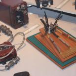
The ovoscope device can be either purchased and quite expensive, or homemade. Private farmers often make it on their own and use it effectively on the farm.
The inspection technique is quite simple. The egg is taken in the right hand and brought to the ovoscope, turning along the longitudinal axis. Proper ovoscoping of chicken eggs will allow you to carefully examine all the shortcomings.
In poultry farms, this procedure is carried out in a special room. Eggs are brought on egg carriers to the hatchery, from where containers with contents are sent to a room for further sorting.
After ovoscoping, eggs suitable for incubation are placed in trays and sent for disinfection, from where they will go directly to the incubator for growing.

Before laying eggs in the incubator, you should be alert to the following defects:
- spotted marble structure of the shell, which indicates a lack or excess of calcium,
- light streaks appearing as a result of damage,
- large air chamber, as well as a chamber at the sharp end and side,
- blood clots,
- dark spots (a sign of mold colonies),
- foreign objects (feathers, grains of sand),
- the contents are orange-red in color without a visually noticeable yolk (most likely, the yolk has broken and mixed with the white),
- two yolks,
- the yolk moves freely around the egg and does not return to its place,
- the yolk is fixed in one place (it is possible that it has dried up).
VIDEO INSTRUCTION
Throughout the incubation period, ovoscopy is carried out several times. This allows you to track the development of the embryo and discard eggs that are unsuitable for further incubation. It is not recommended to remove eggs from the incubator for more than 25 minutes.
Stages of ovoscopy
3rd day of incubation
On the third day of incubation, the egg is clearly translucent and you can see:
- yolk,
- air chamber at the blunt end of the egg.
It is not yet possible to determine whether it is fertilized or not.
4th day of incubation
When ovoscoping you can see:
- air chamber at the blunt end,
- the beginning of the development of blood vessels,
- slight fetal heartbeat.
5th day of incubation
When illuminated you will see:
- air chamber at the blunt end,
- the blood vessels have increased by more than half the egg, they are clearly visible - this means that the embryo is actively developing.
6th day of incubation
Well visible:
- air chamber,
- blood vessels filled almost the entire egg,
- the movements of the embryo itself are visible.


7th day of incubation
When illuminated you will see:
- fetal movements,
- well-developed blood vessels (filled almost the entire egg),
- air chamber.
11th day of incubation
When ovoscoping you can see:
- air chamber,
- blood vessels are clearly visible, completely filling the entire egg,
- the egg is no longer as translucent as on the seventh day and has a darker shade.
15th day of incubation
The following changes are noticeable:
- the egg no longer has the same lumen as on the eleventh day,
- the translucent part has blood vessels,
- The air chamber is clearly visible.
19th day of incubation
During ovoscopy you will see that:
- the egg has practically no lumen,
- the embryo is almost fully developed, but is not yet ready for hatching,
- the air chamber is clearly visible.


The fact that the development of the embryo is disrupted and the egg will have to be discarded is indicated by:
- Detachment of the subshell membrane. The air chamber moves to the side and blood spots instead of blood vessels can be seen throughout the egg.
- Blood rings. The embryo died during the period from the first to the sixth day of incubation, as a result of which blood veins appear in the form of rings.
- Frozen fruit. It can be determined from the seventh to the fourteenth day of incubation. The embryo looks like a spot, the blood vessels are not visible.
- “Zadohlik” is the popular name for eggs from which no chicks have hatched after incubation. The reasons may be disturbances in temperature, humidity levels, or hypothermia.
- Orange color. The yolk broke and mixed with the white.
- Infertile. After the sixth day of incubation, no blood vessels have appeared, only the yolk and the air cushion are visible.
- Lack of calcium in the shell. Can be detected in the first days of incubation by small spots throughout the shell.
- Mold colonies. On the ovoscope they appear as dark spots. They are not even recommended for consumption, as they were obtained from a sick bird.
Development of an embryo in a chicken egg from 1 to 21 days Development of an embryo in a chicken egg from 1 to 21 days Development of an embryo in a chicken egg from 1 to 21 days. Day 1: 6 to 10 hours – The first kidney-shaped cells (prebud) begin to form 8 hours – Appearance of the primitive streak. 10 o'clock - The yolk sac (embryonic membrane) begins to form. Functions: a) blood formation; b) digestion of the yolk; c) absorption of yolk; d) the role of food after hatching. Mesoderm appears; the embryo is oriented at an angle of 90° to the long axis of the egg; The formation of the primary bud (mesonephros) begins. 18 hours – The formation of the primary intestine begins; Primary germ cells appear in the germinal crescent. 20 hours – The vertebral ridge begins to form. 21:00 – The neural groove, the nervous system, begins to form. 22 hours – The first pairs of somites and the head begin to form. 23 to 24 hours – Blood islands, circulatory system, yolk sac, blood, heart, blood vessels begin to form (2 to 4 somites). Day 2: 25 hours – Eyes appear; the spinal column is visible; the embryo begins to turn on its left side (6 somites). 28 hours – Auricles (7 somites). 30 hours - The amnion (embryonic membrane around the embryo) begins to form. The primary function is to protect the embryo from shock and adhesion, and is also responsible, to some extent, for protein absorption. The choion (embryonic membrane that fuses with the allantois) begins to form; Heartbeat begins (10 somites). 38 hours – Midcerebral flexure and embryonic flexure; Heartbeat and blood begins (16 to 17 somites). 42 hours – The thyroid gland begins to form. 48 hours – The anterior pituitary gland and pineal gland begin to develop. Day 3: 50 hours – The embryo turns on its right side; The allantois (embryonic membrane that merges with the chorion) begins to form. Functions of the chorioallantois: a) respiration; b) protein absorption; c) absorption of calcium from the shell; d) storage of kidney secretions. 60 hours - Nasal recesses, pharynx, lungs, the kidneys of the forelimbs begin to form. 62 hours - The hind buds begin to form. 72 hours – Middle and outer ear, trachea begins; The growth of the amnion around the embryo is completed. Day 4: The tongue and esophagus begin to form; the embryo separates from the yolk sac; The allantois grows through the amnion; the amnion wall begins to contract; the adrenal glands begin to develop; the pronephros (non-functioning kidney) disappears; The secondary kidney (metanephros, definitive or final kidney) begins to form; The glandular stomach (proventriculus), the second stomach (gizzard), the blind outgrowth of the intestine (ceca), and the large intestine (large intestine) begin to form. Dark pigment is visible in the eyes. Day 5: The reproductive system and sex differentiation are formed; The thymus, bursa of Fabricius, and duodenal loop begin to form; The chorion and allantois begin to merge; mesonephros begins to function; first cartilage Day 6: The beak appears; voluntary movements begin; The chorioallantois lies opposite the shell of the blunt end of the egg. Day 7: Fingers appear; ridge growth begins; egg tooth appears; Melanin is produced, and the absorption of minerals from the shell begins. The chorioallantois adheres to the inner shell membrane and grows. Day 8: Feather follicles appear; the parathyroid gland begins to form; bone calcification. Day 9: Growth of the chorioallantois is 80% complete; the beak begins to open. Day 10: The beak hardens; the fingers are completely separated from each other. Day 11: The abdominal walls are installed; the intestinal loops begin to protrude into the yolk sac; downy feathers are visible; Scales and feathers appear on the paws; the mesonephros reaches maximum functionality, then begins to degenerate; The metanephros (secondary kidney) begins to function. Day 12: Chorioallantois completes enveloping the containing egg; The water content in the embryo begins to decrease. Day 13: The cartilaginous skeleton is relatively complete, the embryo increases heat production and oxygen consumption. Day 14: The embryo begins to turn its head towards the blunt end of the egg; calcification of long bones accelerates. Turning the eggs doesn't matter any further. Day 15: Loops of intestine are easily visible in the yolk sac; Amnion contractions stop. Day 16: Beak, claws and scales are relatively keratinized; the protein is practically used and the yolk becomes a source of nutrition; downy feathers cover the body; the intestinal loops begin to be retracted into the body. Day 17: The amount of amniotic fluid decreases; location of the embryo: head towards the blunt end, towards the right wing and beak towards the air chamber; definitive feathers begin to form. Day 18: Blood volume decreases, total hemoglobin decreases. The embryo must be in the correct position for hatching: the long axis of the embryo coincides with the long axis of the egg; head at the blunt end of the egg; the head is turned to the right and under the right wing; the beak is directed towards the air chamber; legs pointing towards the head. Day 19: Retraction of the intestinal loop is completed; the yolk sac begins to retract into the body cavity; amniotic fluid (swallowed by the embryo) disappears; the beak can break through the air chamber and the lungs begin to function (pulmonary respiration). Day 20: The yolk sac is completely retracted into the body cavity; the air chamber is pierced by the beak, the embryo emits a squeak; The circulatory system, respiration and absorption of the chorioallantois are reduced; the embryo may hatch. Day 21: The withdrawal process: the circulatory system of the chorioallantois stops; the embryo breaks through the shell at the blunt end of the egg using an egg tooth; the embryo slowly turns from the egg counterclockwise, breaking through the shell; the embryo pushes and tries to straighten its neck, comes out of the egg, is freed from debris and dries. More than 21 days: Some embryos are unable to hatch and remain alive in the egg after 21 days.
During the incubation period, the embryo changes its position several times at a certain time and in a certain sequence. If at any age the embryo takes an incorrect position, this will lead to a developmental disorder or even the death of the embryo.
According to Cuyo, initially the chicken embryo is located along the minor axis of the egg in the upper part of the yolk and faces it with its abdominal cavity, and its back towards the shell; on the second day of incubation, the embryo begins to separate from the yolk and simultaneously turn onto its left side. These processes begin from the head part. Separation from the yolk is associated with the formation of the amniotic membrane and the immersion of the embryo in the liquefied part of the yolk. This process continues until approximately day 5, and the embryo remains in this position until the 11th day of incubation. Until the 9th day, the embryo makes vigorous movements due to contractions of the amnion. But from this day on, it becomes less mobile, since it reaches a significant weight and size, and the liquefied part of the yolk is used by this time. After the 11th day, the embryo begins to change its position and gradually, by the 14th day of incubation, takes a position along the major axis of the egg, the head and neck of the embryo remain in place, and the body descends down to the sharp end, turning at the same time to the left .
As a result of these movements, the embryo lies along the major axis of the egg at the time of hatching. Its head is facing the blunt end of the egg and tucked under the right wing. The legs are bent and pressed to the body (between the thighs of the legs there is a yolk sac that is retracted into the body cavity of the embryo). In this position, the embryo can be released from the shell.
The embryo can make movements before hatching only in the direction of the air chamber. Therefore, he begins to protrude his neck into the air chamber, stretching the embryonic and shell membranes. At the same time, the embryo moves its neck and head, as if freeing it from under the wing. These movements lead first to the rupture of the membranes by the supraclavicular tubercle, and then to the destruction of the shell (pecking). Continuous movements of the neck and pushing away from the shell with the legs lead to rotational movement of the embryo. In this case, with its beak, the embryo breaks off small pieces of the shell until its efforts are sufficient to break the shell into two parts - a smaller one with a blunt end and a larger one with a sharp one. Releasing the head from under the wing is the last movement, and after this the chick is easily freed from the shell.
The embryo can take the correct position if the eggs are incubated in a horizontal as well as a vertical position, but always with the blunt end up.
When large eggs are placed vertically, the growth of the allantois is disrupted, since the inclination of the eggs by 45° is not sufficient to ensure its correct location at the sharp end of the egg, where by this time the white is pushed back. As a result, the edges of the allantois remain open or closed so that the white ends up at the sharp end of the egg, uncovered and not protected from external influences. In this case, the protein sac is not formed, the protein does not penetrate into the amnion cavity, as a result of which the embryo may starve and even die. The protein remains unused until the end of incubation and can mechanically impede the movements of the embryo during hatching. According to the observations of M. F. Soroka, from eggs of ducks with complete and timely closure of the allantois, a high hatching rate of ducklings was obtained with the shortest average duration of the incubation period. The protein in eggs with an untimely closed allantois remained unused even on the 26th day of incubation (in eggs with a timely closed allantois, the protein disappeared already by the 22nd day of incubation). The weight of the embryo in these eggs was approximately 10% less.
Good results can be obtained by incubating duck eggs in a vertical position. But a higher hatching percentage can be obtained if the eggs are moved to a horizontal position during the period of growth of the allantois under the shell and the formation of the albumen sac, that is, from the 7th to the 13th-16th day of incubation. In the case of a horizontal position of duck eggs (M. F. Soroka), the allantois is positioned more correctly, and this leads to an increase in hatching by 5.9-6.6%. However, this increases the number of eggs with the shell pecked at the sharp end. Moving duck eggs from a horizontal position after the closure of the allantois to a vertical position led to a decrease in pecking at the sharp end of the eggs and to an increase in the percentage of ducklings hatching.
According to Yaknyunas, at the Brovary hatchery and poultry station, the hatchability of ducklings reached 82% in the case when the trays were not replenished with eggs after removing waste during the first viewing. This made it possible to incubate duck eggs from the 7th to the 16th day of incubation in a horizontal or highly inclined position, after which the eggs were again placed in a vertical position.
To ensure that the position of the embryo changes correctly and the shells are positioned correctly, periodic rotation of the eggs is used. Rotating the eggs has a beneficial effect on the nutrition of the embryo, on its respiration and thereby improves development conditions.
In a stationary egg, the amnion and embryo may adhere to the shell during the early stages of incubation before being covered by the allantoic membrane. At later stages, the allantois and the yolk sac may become fused, which precludes the possibility of the latter being safely retracted into the body cavity of the embryo.
The disruption of the closure of the allantois in chicken eggs under the influence of insufficient egg rotation was noted by M. P. Dernyatin and G. S. Kotlyarov.
When incubating chicken eggs in a vertical position, it is customary to turn them 45° in one direction and 45° in the other. Turning of eggs begins immediately after laying and continues until hatching begins.
In the experiments of Byerly and Olsen, they stopped turning chicken eggs on the 18th and 1-4th days of incubation and obtained the same hatching results.
In duck eggs, a small rotation angle (less than 45°) leads to impaired growth of the allantois. If the vertically positioned eggs are not tilted sufficiently, the white remains almost motionless and, due to the evaporation of water and an increase in surface tension, becomes so tightly pressed to the shell that the allantois cannot penetrate between them. When the eggs are positioned horizontally, this happens very rarely. Rotating large geese eggs only 45° is completely insufficient to create the necessary conditions for allantois growth.
According to Yu. N. Vladimirova, with an additional rotation of goose eggs by 180° (twice a day), normal growth of the embryo and the correct location of the allantois were observed. Under these conditions, hatchability increased by 16-20%. These results were confirmed by A. U. Bykhovets and M. F. Soroka. Subsequent experiments showed that it is necessary to additionally rotate geese eggs by 180° from the 7-8th to the 16-19th day of incubation (the period of intensive allantois growth). Further rotations of 180° are significant only for those eggs in which the closure of the edges of the allantois is for some reason delayed.
In sectional incubators, the air temperature at the top of the eggs is always higher than the temperature at the bottom of the eggs. Therefore, turning the eggs here is also important for more uniform heating.
At the beginning of incubation, there is a large difference in temperature - at the top of the egg and at the bottom of it. Therefore, frequent turns of eggs 180° can lead to the fact that the embryo will many times fall into the zone of an insufficiently heated part of the egg and this will worsen its development.
In the second half of incubation, the temperature difference between the top and bottom of the eggs decreases and frequent turning can promote heat transfer by moving the hotter upper part of the eggs to a lower temperature zone (G. S. Kotlyarov).
In sectional incubators with one-sided heating, turning the eggs instead of 2 to 4-6 times a day improved incubation results (G. S. Kotlyarov). With 8 egg turnings, embryo mortality decreased, mainly in the last days of incubation. The increase in the number of turnings led to an increase in the number of dead fetuses. When the eggs were turned 24 times, there were many dead embryos in the first days of incubation.
Funk and Forward compared the results of incubating chicken eggs by rotating the eggs in one, two, and three planes. The embryos in eggs that were rotated in two and three planes developed better and the chicks hatched several hours earlier than in eggs that were rotated in one plane as usual. When eggs were incubated in four positions (rotation in two planes), the hatch from eggs with low hatchability increased by 3.1/o, from eggs with average hatchability - by 7-6%, and from eggs with high hatchability - by 4-5%. When turning eggs with good hatchability in three planes, the hatchability increased by 6.4%.
In cabinet incubators, eggs from chickens, turkeys and ducks are incubated in a vertical position. It is advisable to keep large duck eggs in a horizontal or inclined position from the 7th to the 15th day of incubation. Geese eggs are incubated in a horizontal or inclined position. Turning of eggs begins immediately after laying in the incubator and ends when they are transferred to hatching or one day earlier. The eggs are turned every two hours (12 times a day). When positioned vertically, the eggs are rotated 45° to either side of the vertical position. Eggs in a horizontal position are also rotated 180° once or twice a day.


