Internal structure of a cat, organs diagram. Anatomical structure of a cat. Reproductive system of cats
The variety of unique physical capabilities of cats is largely due to the skeleton. Lightning-fast tree climbing, balancing at great heights, crawling, various types of jumps and safe landings - all this is possible thanks to the features of the cat's anatomy and, in particular, the skeleton, so we suggest taking a closer look at its features and structure.
The general structure of the cat's skeleton is similar to that of other mammals, with the exception of some differences in the shape and arrangement of individual bones, which is associated with the horizontal position of the spine and maximum adaptability to the lifestyle of a predator. In addition, differences in the shape and structure of individual bones may be due to breed characteristics. For example, Siamese have narrower and longer bones compared to Persian cats. From the photo below you can understand what a cat’s skeleton looks like without taking into account breeding characteristics.

The skeleton of a cat consists of an average of 244-250 bones. Some sources mention the figure 230-236, since some fused bones are considered as one whole. How many bones a cat has is influenced by the length of the animal's tail, since it contains almost a tenth of all the bones in a cat's body (a "normal" tail has about 26 vertebrae).
Scull
Due to the smaller number of teeth compared to other predators, the cat's skull is characterized by a rounded shape. Its size will depend on belonging to a particular breed or other hereditary characteristics. Representatives of the Persian, exotic and Himalayan breeds are brachycephalic - owners of a shortened skull, which is why they have an abnormal structure of the palate, larynx and trachea. This explains the common problems in these breeds with impaired nasal breathing, snoring and poor tolerance to exercise and heat.
The skull consists of 29 bones, with the brain part being formed from 11, and the facial part from 13 bones. The bones of the skull themselves are larger in size compared to the facial bones. Characteristic features also include large eye sockets and narrowly spaced fangs, adapted for hunting small animals. The main attribute of a predator, which is a cat, is a powerful jaw, which is equipped with different types of teeth. They allow you to grab and hold resisting prey, bite and grind food, and defend yourself if necessary.

Spine
The cat's spine is incredibly flexible because it is formed from small, movable bones. It is represented by many vertebrae, which are divided into several sections:
- Cervical spine - consists of larger 7 vertebrae, which are responsible for support and mobility of the head. Two of them with their own names - epistropheus (axial) and atlas - have the property of rotating 180°. They are connected by a thin process, so they are among the cat’s vulnerable spots: with blows and falls, there is a high risk of rupture of the connection, respectively, fracture of the cervical vertebrae and death.
- The thoracic region consists of 13 vertebrae, to which 12 pairs of rib bones are attached on both sides. Of these, the first 5 pairs are called true, since they are attached to the sternum, and the remaining ones are called false, since they look like arches.
- Lumbar - formed from the 7 largest vertebrae, the size of which increases as they approach the tail. They have special protrusions on the sides on which the muscles and internal organs of the abdominal cavity are attached.
- Sacral region - in contrast to the superflexible lumbar region, it is characterized by a rigid intervertebral connection of three fused vertebrae. This need is due to the fact that the hind limbs are attached to this area, which bear the main load during the movement of the animal (especially jumping).
- The caudal region plays one of the key roles in maintaining body balance during jumps or falls from a height. Strong muscle ligaments provide these animals with ideal “jumping ability,” and cartilaginous pads between the vertebrae provide the possibility of various movements (bending and rotation). The number of tail vertebrae varies depending on the breed, and some breeds may have none at all.

Limb structure
There are two sections in the skeleton of a cat’s limbs:
- A belt of the forelimbs (shoulder), the peculiarity of which is the elastic fastening of the limbs, which is necessary for cats for safe jumping and comfortable landing. It is represented by the scapula, humerus, radius and ulna (forming the forearm), and the hand. The latter consists of the wrist, metacarpus and phalanges of the fingers, of which there are only 5 on the forelimbs.
Another unique feature of feline anatomy is the absence of a full collarbone. It is represented by two non-functional bones that are not attached to the shoulder joint, but are located freely inside the muscles. The shoulder blades are attached to the spine by muscles, ligaments and tendons, due to which the shoulders have virtually no restrictions in movement.
Interesting! Due to the unique structure of the collarbone, a cat is able to crawl into even the narrowest holes if the animal’s head fits in, since it is the latter that is the most voluminous, but not subject to deformation, part of the body.
- The girdle of the hind limbs, which, unlike the shoulder girdle, is rigidly and motionlessly attached to the sacrum. It includes: the pelvic and femur bones, the kneecap, the tibia and fibula, the tarsal and metatarsal bones, to which the phalanges of the fingers are attached. The pelvic bones of the hind legs are longer and better developed compared to the front legs, and the metatarsal bones are more massive, which is associated with the characteristics of the animal’s movement (in particular, jumping). Due to this structure of their limbs, cats can move quickly in horizontal and vertical planes, which is why they are excellent tree climbers. The hind legs rest on the phalanges of the 4 toes. Like other mammals, cats' elbows bend backward and knees bend forward. That part of the paw that can be mistaken for a knee bent back is actually the heel, and the true knee is located in the lower abdomen of the animal.

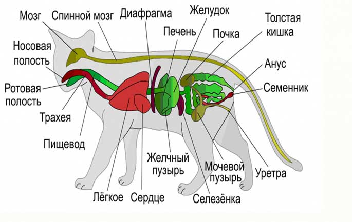
The gracefulness of cats is admired by many people. Pets have flexibility, unique hearing and sense of smell. Animals received such abilities due to their body structure. Knowing the structure of a cat’s internal organs is useful for every owner who cares about the health of their pet.
Respiratory system
The task of the respiratory organs is to ensure gas exchange and deliver oxygen to the tissues. The process of releasing excess moisture also goes through them. The respiratory system participates in heat exchange and removes excess heat and harmful gases.
Respiratory organs of a cat:
- nasopharynx;
- bronchi;
- trachea;
- lungs.
Bengals and other breeds weigh up to 6 kg, the Maine Coon can weigh up to 13 kg
The nasal cavity is surrounded by mucous membrane, which performs the function of smell. Thanks to the villi on the epithelium, the nose serves as a filter that cleans the incoming air from dust and dirt. The larynx has vocal cords that allow the animal to meow.
A cat's lungs are made up of many alveoli. The left lung is slightly larger in volume than the right (8 and 11 cm3, respectively).
Digestive system
The cat's insides form the digestive system, which is responsible for taking in food, processing and eliminating undigested residues. A cat’s body processes food within a day.
Participating in this process are:
- oral cavity;
- pharynx;
- esophagus;
- stomach;
- small and large intestines;
- kidneys and liver.
A cat's stomach is more acidic than a human's, so it can process rough food. The structure of a cat's intestines does not allow it to digest carbohydrates well. This must be taken into account when planning your diet.

Digestive organs
Digestion begins as soon as the food enters the oral cavity. Saliva softens food and promotes quick chewing. Then the food passes through the pharynx and esophagus into the stomach. There, the active breakdown of foods into liquid, carbohydrates, proteins and fats begins. The contents then pass into the duodenum and small intestine. Unprocessed residues form feces and pass through the rectum.
Interesting! The pet's stomach is in constant activity. This is why animals often eat, but little by little.
Reproductive system of cats
The internal structure of a cat and a tom differ in the reproductive system. The cat's genitals provide the formation and transfer of seminal fluid, which includes sperm.
Diagram of the male reproductive system:
- prostate;
- vas deferens;
- scrotum;
- penis;
- testes.
The testes produce sperm and produce testosterone. Sperm is produced throughout the cat's life, or until castration.
Internal and external genitalia of a cat
The reproductive system of a female cat consists of the following organs:
- ovaries;
- the fallopian tubes;
- uterus;
- vagina;
- vestibule of the vagina;
- external genitalia.
The ovaries are a paired organ located in the lumbar region. This organ is responsible for the formation of hormones and the maturation of germ cells. Pets regularly ovulate, which allows them to become pregnant.
Important! If the owner does not plan to breed cats, then it is advisable to sterilize the cat. Otherwise, hormonal imbalances, tumor development, cystitis and other diseases are possible.
The fertilization process takes place in the fallopian tubes, after which the egg passes into the uterus. The uterus is a hollow organ that consists of a cervix, body and horns. The vagina is an organ that connects the cervix and the external genitalia. At the onset of puberty, the cat's ovaries increase in size.

The structure of the reproductive system of cats
The vulva serves as the external organ of the reproductive system in cats. It is located below the anus.
Cardiovascular system and heart
The cardiovascular system is one of the main ones in the animal’s body. It is responsible for vital processes, transports blood throughout the body and saturates tissues with oxygen.
Circulatory system
The anatomical structure of the blood of cats differs from other animals. It cannot be supplemented or replaced. Red blood cells, which are part of the blood, provide the body's organs with oxygen. As blood passes through the body, it travels through the right ventricle and into the pulmonary artery. Oxygenated blood returns to the left side of the atrium and then to the ventricle. In total, cats have 2 circles of blood circulation.
Interesting! The weight of the heart depends on the weight of the pet and usually accounts for 0.6% of the total figure.
urinary system
When studying the structure of a cat, internal organs and their functions, it is worth noting the urinary system. It consists of the following organs:
- kidneys;
- ureters;
- bladder;
- urethra.
The kidneys are a paired organ located on the right and left in the lumbar region. They perform the following functions:
- regulate blood volume and extracellular fluid;
- control ionic balance in the body;
- stabilize the acid level in the blood;
- participate in blood clotting and blood pressure regulation;
- remove excess metabolic products from the body;
- regulate metabolism.
First, primary urine is formed, which contains glucose, vitamins and amino acids. It then passes through the convoluted canals and accumulates in the renal pelvis. From this point on, urine is considered secondary. It enters the ureters and bladder. As a result, urine comes out through the urethra. Spontaneous leakage is facilitated by a muscular organ - the sphincter.
Important! In healthy animals, urine has virtually no color. If it turns orange or brown, this may indicate urolithiasis.
In cats, the urethra is wider and shorter than in cats. Because of this feature, females are less likely to experience urinary problems.
Endocrine system
The endocrine system is responsible for the production of hormones that are carried throughout the body through the blood. Thus, the activity of the body is monitored. Hormones can make organs work faster or vice versa - slower.
The amount of hormones in the blood is constantly monitored and adjusted to always meet the body's needs.
In textbooks and atlases you can find the following division of the endocrine system:
- glandular;
- diffuse.
The glandular system includes:
- hypothalamus;
- pituitary;
- pineal gland;
- thyroid;
- thymus;
- adrenal glands;
- gonads.
Interesting! The diffuse system is scattered throughout the body, but accumulates most in the gastrointestinal tract.
Nervous system
The nervous system of cats consists of the following organs:
- brain;
- spinal cord;
- nerve trunks and endings.
Nervous activity is carried out through the senses. Under the skin there are many nerve endings that can change your pet's behavior. This system is closely related to hormones, so it quickly responds to internal and external events.
To understand how a cat and its nervous system works, it is necessary to study the classification. This part of the body is usually divided into 2 classes: central and peripheral. The first consists of the brain and spinal cord. With their help, nerve impulses are carried out in the body. The peripheral nervous system receives information about pain, pressure, touch, and also transmits commands to the muscles.
Musculoskeletal system
The location of a cat's organs in the musculoskeletal system is throughout the body. The movement apparatus consists of bones, muscles, ligaments and cartilage that form the skeleton.
The axial skeleton includes:
- scull;
- spine;
- thoracic region
The skeleton of a cat consists of 230 bones.

The dexterity and mobility of cats is ensured by the special structure of the skeleton.
Sense organs
The sense organs are represented by a group of important components, which include: vision, smell, taste, hearing. The correct functioning of this system ensures the health of the cat, as well as survival in different conditions.
Visual
Cat eyes are the largest of all pets. Developed vision allows the animal to see not only small details and prey, but also navigate in the dark. The cornea protrudes forward, so the viewing angle is 250°.
Interesting! Experts have found that cats are able to distinguish up to 6 colors.
Auditory
Cats' hearing allows them to detect sound with a frequency of up to 65 kHz. The ear canal consists of 3 parts:
- Outer ear. This part is located on the pet’s head. With its help, sounds are captured and collected, and then transmitted to the eardrum.
- The middle ear transmits the signal from the membrane to the inner ear.
- The inner ear converts sound vibrations into nerve impulses.
Taste buds
Felines are able to distinguish almost all tastes, except sweet. Special papillae located on the tongue help recognize food. Each papilla contains about 30,000 taste buds.
Smell and touch
The sense of touch is accomplished through the whiskers. They are located in front of the pet's face. Animals are able to perceive 2 times more odors than humans. The main organ of smell is the nose. However, there is an additional organ on the upper palate - Jacobson organ.
Interesting! A cat's nose has a unique imprint comparable to a human finger.
The structure of a cat's muscles
Muscles are made up of fibers that contract under the influence of nerve impulses. The ends of the muscles are attached to the bones by tendons. The contraction and relaxation of muscles causes the bones in the joints to move, causing them to flex and extend.

There are about 500 muscles in a pet's body
The anatomical features of a cat and its internal organs make this animal a real predator. Smooth movements, clear and high jumps are ensured by the work of muscles and ligaments. Sharp fangs help chew the coarsest food, and hearing and smell allow them to capture information.
1. Oral cavity (Cavum oris)
Food, entering the initial section of the digestive apparatus through the oral opening, enters the oral cavity, the skeleton of which is the upper and lower jaws, palatine and incisive bones. The hyoid bone, located inside the oral cavity, serves as a place of fixation for the muscles of the tongue, pharynx and larynx. The oral cavity extends orally from the lips, and aborally ends with the pharynx and passes into the pharynx. The dental edge of the closed jaws and lips form the vestibule of the oral cavity. Behind the vestibule is the oral cavity itself. The vestibule communicates with the external environment through the oral fissure. The oral fissure begins at the junction of the upper and lower lips, called the angle of the mouth.
Appearance of the oral cavity
Lips- upper and lower muscle-skin folds, covered on the outside with hair and on the inside with mucous membrane. Outside, the upper lip is sagittally divided by a deep groove - a filter, passing towards the nasal septum. On the upper lip there are hard whiskers, collected in 2 side tufts - mustaches.
Cheeks are a continuation of the lips behind their commissure and form the lateral walls of the oral cavity. The cheeks of cats are relatively small, thin, and covered with hair on the outside. Their inner surface is smooth, and the ducts of the salivary glands open on it.
Teeth- durable organs of the oral cavity that serve to capture and hold food, bite it, crush and grind it, as well as defend and attack.


Adult cats have 30 teeth, 16 of which are in the upper jaw and 14 in the lower jaw. Cats are carnivores by nature, which largely reflects the arrangement of their teeth. Cats have six front teeth and two canines in each jaw. These teeth are involved in the process of biting into meat and then tearing it. Cats have only 6 premolars and 2 molars in the upper jaw and 4 premolars and 2 molars in the lower jaw. Cats are also characterized by an increased size of the upper 4th molar (also called the “carnivore tooth”) and the 1st lower incisor. Due to the arrangement of these “carnivorous teeth”, eating food occurs according to the “scissor principle”, which is extremely effective when cutting raw meat.
STRUCTURE OF TEETH
A tooth consists of dentin, enamels And cement.
Schematic representation of the cutter:

Dentine- tissue that forms the basis of the tooth. Dentin consists of a calcified matrix penetrated by dentinal tubules containing processes of odontoblast cells lining the tooth cavity. The intercellular substance contains organic (collagen fibers) and mineral components (hydroxyapatite crystals). Dentin has different zones that differ in microstructure and color.
Enamel- a substance covering dentin in the crown area. It consists of crystals of mineral salts, oriented in a special way to form enamel prisms. Enamel does not contain cellular elements and is not tissue. The normal color of enamel is from white to cream with a yellowish tint (distinguishable from plaque).
Cement- tissue covering dentin in the root area. The structure of cement is close to bone tissue. Consists of cementocyte and cementoblast cells and a calcified matrix. Nutrition of cement occurs diffusely from the periodontium.
Inside there is tooth cavity, which is divided into coronalcavity And root canal, opening with the above hole in the apex of the tooth. Fills the dental cavity dental pulp, consisting of nerves and blood vessels immersed in loose connective tissue and providing metabolism in the tooth. Distinguish coronal And root pulp.
Gum- mucous membrane that covers the dental edges of the corresponding bones, tightly fused with their periosteum.
The gum covers the tooth in the neck area. It is abundantly supplied with blood (tendency to bleeding), but relatively poorly innervated. The grooved depression located between the tooth and the free edge of the gum is called the gingival sulcus.
The periodontium, alveolar wall and gums form supporting apparatus of the tooth - periodontium.
Periodontium- provides attachment of the tooth to the dental alveolus. It consists of the periodontium, the wall of the dental alveoli and the gums. The periodontium performs the following functions: supporting and shock-absorbing, barrier, trophic and reflex.
The teeth are distributed as follows: 12 incisors (I), 4 canines (C), 10 premolars (P) and 4 molars (M). Thus, the dental formula is as follows:
![]()
All teeth are of pronounced short-crown type.
There are 4 types of teeth: incisors, fangs And permanent teeth: preradical(false, small molars), or premolars And truly indigenous, or molars that do not have milk precursors.
Teeth arranged in order in a row form top
and lower dental arches (arcades)
.
Incisors- small, with uneven edges and 3 protruding points. Each root is single. The lateral incisors are larger than the medial ones, and the incisors of the upper jaw are larger than those of the lower jaw.
Schematic representation of the incisors:

Behind the incisors are located fangs. These are long, strong, deep-set teeth with a simple root and a rounded crown. When the jaws are closed, the lower canines lie laterocaudal to the upper ones. Behind the fangs on each jaw there is an edge free from teeth.
Schematic representation of fangs:

Molars of the upper dental arch.
Premolars are located behind the diastema; there are 3 pairs of them on the upper jaw
and 2 pairs on the bottom. The first premolar of the upper jaw is small,
with a simple crown and a simple root. The second premolar is larger, it has 4 projections - a large central one, a small cranial one
and 2 small caudal ones. The most massive tooth is the third premolar: it has 3 large protrusions located along the length
and small projections lying on the medial side of the first; the root of the tooth has 3 processes.
Schematic representation of premolars:

Upper dental arcade of a seven-month-old cat:

Molars located caudal to the last premolar in the upper jaw. These are small teeth with 2 projections and 2 roots.
Schematic arrangement of molars:

Molars of the lower dental arch.
In the lower arcade 2 premolar; they are identical in size and shape. The crown of each premolar bears 4 projections - one large, one small in front and two more behind. Each premolar has
2 roots.
Molar the lower jaw is the most massive in the arcade and has
2 protrusions and 2 roots. The molars sit obliquely in the sockets, so that when the jaws are closed, the teeth of the upper jaw adjoin the lower teeth from the inside.
The lower dental arcade of a seven-month-old cat:

Baby teeth appear in kittens soon after birth.
They are smaller in size than the permanent ones and less developed. Their color
milky white. There are fewer primary teeth than permanent teeth because molars have no predecessors.
The dental formula of primary teeth is as follows: ![]()
MECHANICAL DIGESTION
Digestion in the oral cavity occurs mainly mechanically; when chewing, large fragments of food are broken into pieces and mixed with saliva.
Mechanical digestion also increases the area exposed to digestive enzymes. The arrangement of teeth is closely related to the natural diet of various animal species and indicates their natural feeding behavior and preferred feeding patterns.
ORAL CAVITY
The oral cavity itself is separated from above, from the side of the nasal cavity, by the hard palate, from the pharynx by the soft palate, and is limited in front and on the sides by dental arcades.
Solid sky arched like a vault. Its mucous membrane forms 7 - 8 caudally concave transverse ridges - palatine ridges, between which the papillae are located. In the anterior part behind the incisors there is a small incisive papilla;
to the right and left of it lie the slit-like nasopalatine canals, which are the excretory ducts of the nasopharynx organ.
In the aboral direction, in the area of the choanae, the hard palate passes into the soft palate without a visible boundary.
Soft palate or velum- is a continuation of the hard palate and is a fold of the mucous membrane that covers the entrance to the choanae and pharynx. The soft palate is based on special muscles: the levator velum palatine, the tensor velum palatine, and the palatine muscle that shortens it after the act of swallowing. The velum palatine hangs from the end of the bony palate and, in a calm state, its free edge touches the root of the tongue, covering the pharynx, the exit from the oral cavity into the pharynx.
The free edge of the velum is called the arch of the palate. The palatine arch, together with the pharynx, forms the velopharyngeal arches, and with the root of the tongue - the palatoglossus arches. Aborally on the sides of the root of the tongue, in the tonsil sinuses there is one palatine tonsil.
SALIVARY GLANDS
Cats have 5 pairs of salivary glands: parotid, submandibular, sublingual, molar and infraorbital.
Layout of the salivary glands of a cat:

1 - parotid
2 - submandibular
3 - sublingual
4 - radical
5 – infraorbital
Parotid salivary gland located ventral to the external auditory canal under the cutaneous muscles. It is flat, has a lobular structure, and borders orally with the large masseter muscle. The excretory ducts of the individual lobules of the gland merge to form the common parotid (stenon) duct. It passes cranially as part of the fascia covering the large masticatory muscle, at the cranial edge of the muscle it turns inward, goes under the mucous membrane and opens into the buccal vestibule of the mouth opposite the last premolar with the salivary papilla. Along the duct there are one or more small accessory parotid salivary glands.
Submandibular gland rounded, lies ventral to the previous one near the large masseter muscle and consists of individual glandular lobules connected by connective tissue. The excretory duct of the submandibular gland is located on its inner surface, it stretches forward under the base of the tongue and opens at the bottom of the oral cavity with a sublingual wart, next to which the duct of the sublingual gland opens.
Sublingual gland elongated, conical, its base is adjacent to the submandibular gland, stretching 1-1.5 cm along its duct. The excretory duct of the sublingual gland is located on the ventral side; in its course it accompanies the duct of the submandibular gland, following first dorsally and then ventrally from it.
Indigenous salivary gland, absent in other domestic animals, in the cat it is located at the cranial edge of the large masseter muscle, between the mucous membrane of the lower lip and the orbicularis oris muscle. It is a flat formation that widens caudally and tapers orally. The anterior edge of the gland is visualized at the level of the canine. It has several ducts that open directly into the oral mucosa.
Orbital or zygomatic gland Of all domestic animals, only dogs and cats have it. It has a round shape and reaches a length of 1.5 cm. It is located medial to the zygomatic arch in the lower part of the orbit. The ventral edge is located behind the molar. Its large excretory duct and additional small ducts open into the oral cavity 3 - 4 mm caudal to the upper molar.
ENZYMATIVE DIGESTION
Saliva is secreted into the oral cavity by five pairs of salivary glands. Typically, a small amount of saliva is present in the mouth, but its flow may increase if the animal sees or smells food.
Salivation continues as food enters the oral cavity, and its effect is enhanced by the chewing process.
Saliva is 99% water, while the remaining 1% is mucus, inorganic salts and enzymes. Mucus acts as an effective lubricant and promotes swallowing, especially dry food. Unlike humans, cats lack the starch-digesting enzyme amylase in their saliva, which prevents the rapid absorption of starch in the mouth. The absence of this enzyme is consistent with the observed carnivorous behavior of cats that tend to consume foods low in starch.
Language- a muscular, movable organ lying at the bottom of the oral cavity.
Tongue and dorsally open pharynx:

Language in cats it is elongated, flat, widened in the middle and slightly narrowed at the end. When the oral cavity is closed, the tongue completely fills it. In terms of external shape, the tongue of cats is long, wide and thin.
The root of the tongue extends from the molars to the epiglottis and is closely connected to the hyoid bone.
The body of the tongue is almost twice as long as the root; it is located between the molars and has a dorsal back and 2 lateral surfaces. At the border with the apex below, the body forms a median fold containing parts of both geniohyoid muscles, this is the frenulum of the tongue. Folds are directed from the caudal end of the body to the epiglottis. The tip of the tongue rests with its free end against the incisor teeth.
On the back of the tongue and in the region of its apex, the mucous membrane is dotted with many rough, keratinized filiform papillae; their apices are directed caudally. Fungiform papillae are located on the surface of the dorsum, the largest of them lie along the edges of the tongue. Large ridge-shaped, or grooved, papillae in two caudally converging rows of 2-3 in each are located at the root of the tongue. The ventral surface and lateral edges of the tongue are smooth, soft, and free of papillae.
The muscles of the tongue consist of longitudinal, transverse and perpendicular bundles. The first ones go from the root of the tongue to its apex, the second ones - from the middle connective tissue septum of the tongue to the sides, the third ones run vertically from the back of the tongue to the bottom surface. These are the actual muscles of the tongue, located in its thickness;
with their help, the tongue can be shortened, thickened and flattened. In addition, there are muscles that connect the tongue to the bones of the oral cavity.
Genioglossus muscle passes from the symphysis of the mandible, where it originates on the medial surface; its fibers pass dorsally, located above the geniohyoid muscle, diverge; of these, the cranial ones reach the tip of the tongue, the caudal ones end at the root of the tongue. Dorsally, the muscle is mixed with the muscle of the same name on the opposite side.
Function: pulls the root of the tongue forward and the top of it to the side.
Lingual lateral muscle arises from the mastoid process of the temporal bone, from the ligament connecting the edge of the external auditory canal and the angular process of the mandible, and from the proximal part of the cranial horns of the hyoid bone. It passes into the lateral part of the tongue between the digastric and lingual main muscles, then, diverging, goes forward to the tip of the tongue, where it ends.
Function: pulls the tongue back with bilateral action, shortening it when swallowing; with unilateral action, turns the tongue to the side.
2. Pharynx (Pharynx)
Pharynx a mobile muscular-cavitary organ in which the digestive tract crosses, going through the pharynx from the oral cavity to the pharynx and further to the esophagus and the respiratory tract - through the choanae to the pharynx and further to the larynx.
Appearance of the pharynx:

Due to the fact that the cross-section of the digestive and respiratory tracts occurs in the pharynx, its mucous membrane, with the help of folds - the velopharyngeal arches, is divided into the upper, respiratory, and lower, digestive parts. The respiratory part is a continuation of the choanae, and is therefore called the nasal part of the pharynx, or nasopharynx. Near the choanae, a paired opening of the auditory tubes opens into the lateral wall of the pharynx. The digestive, or laryngeal, part in front borders the pharynx, being separated from it by the velum palatine, and is a caudal continuation of the oral cavity, rests against the epiglottis at the back and then, located on top of the larynx, follows towards the esophagus, which lies in this area above the trachea.
The muscles of the pharynx are striated, represented constrictors And dilators.
Cranial constrictor The pharynx consists of 2 paired muscles - the pterygopharyngeal and glossopharyngeal.
Pterygopharyngealmuscle flat, triangular, begins at the apex of the uncinate process of the pterygoid bone. Heading caudally, the muscle diverges under the medial constrictor. Some of the fibers are attached to the median suture of the pharynx, the dorsal fibers are attached to the base of the pterygoid bone, the ventral ones run along the length of the pharynx and end on the larynx.
Glossopharyngeal muscle begins on the geniohyoid muscle, passes as a thin ribbon outside the cranial horns of the hyoid bone, turns dorsally and attaches to the middorsal suture of the pharynx.
Middle, or sublingual, constrictor pharynx - a thin muscle covering the middle part of the lateral surface of the pharynx. It begins with two heads - on the cranial horns and the free caudal horn of the hyoid bone; attaches to the dorsal suture of the pharynx and the base of the sphenoid bone.
Caudal or laryngeal constrictor The pharynx begins on the lateral side of the thyroid and cricoid cartilages. The fibers run dorsally and cranially and attach to the pharyngeal suture.
Stylopharyngeal muscle begins at the apex of the mastoid process of the temporal bone. The ribbon-shaped abdomen extends ventrocaudally and is attached to the dorsal wall of the pharynx and larynx. Laterally, the muscle is covered by the middle and caudal constrictors. Contraction of the pharyngeal muscles underlies the complex act of swallowing, which also involves the soft palate, tongue, esophagus and larynx. At the same time, the pharyngeal levators pull it upward, and the compressors successively backwards narrow its cavity, pushing the food bolus into the esophagus. At the same time, the larynx rises and tightly covers the epiglottis, due to pressure on it with the root of the tongue. In this case, the muscles of the soft palate pull it upward and caudally so that the velum palatine rests on the palatopharyngeal arches, separating the nasopharynx. During breathing, the shortened velum palatine hangs obliquely downward, covering the pharynx, while the epiglottis, built of elastic cartilage and directed upward and forward, provides access to a stream of air into the larynx.
3. Esophagus (Oesophagus)
Esophagus It is a cylindrical tube following the pharynx, flattened at the top and bottom.
Esophageal endoscopy:


It is the initial section of the foregut and in structure is a typical tube-shaped organ. The esophagus is a direct continuation of the laryngeal part of the pharynx.
Usually the esophagus is in a collapsed state. The mucous membrane of the esophagus along its entire length is collected in longitudinal folds that straighten out as the food coma passes.
The submucosal layer contains many mucous glands that improve the sliding of food. The muscular layer of the esophagus is a complex multi-level striated layer. The outer membrane of the cervical and thoracic parts of the esophagus is connective tissue adventitia, and the abdominal part is covered with visceral peritoneum. The attachment points of the muscle layers are: laterally - the arytenoid cartilages of the larynx, ventrally - the annular cartilage, and dorsally - the tendon suture of the larynx.
The diameter of the esophagus is relatively constant throughout its entire length and reaches 1 cm during the passage of the food bolus. The esophagus is divided into cervical, thoracic and abdominal sections. Upon exiting the pharynx, the esophagus is located dorsal to the larynx and trachea, covering the bodies of the cervical vertebrae from below, then descends to the left side of the trachea and in the area of its bifurcation returns again to the midline. In the chest cavity, it lies in the mediastinum, passing over the base of the heart and under the aorta. It enters the abdominal cavity through the esophageal opening of the diaphragm, which lies approximately 2 cm ventral to the vertebral column. The abdominal region is very short.

1 - language
2 - pharynx and larynx
3 - esophagus in a collapsed state
4 – stomach
During the process of swallowing, a lump of unchewed food formed by the tongue enters the esophagus. The esophagus does not secrete digestive enzymes, but the cells of the esophagus secrete mucus, which serves to lubricate peristalsis, the automatic wave-like muscle contractions that are stimulated by the presence of food in the esophagus and allow it to move through the gastrointestinal tract. The process of moving food from the mouth to the stomach takes only a few seconds.
4. Stomach (Ventriculus)
Stomach is the organ of the digestive tract where food is retained and subjected to chemical processing. The cat's stomach is single-chamber, intestinal type. It is an extension of the digestive tube behind the diaphragm.

1 - pyloric part of the stomach
2 - cardiac part of the stomach
3 - fundic part of the stomach
4 - exit of the duodenum
5 - cardial opening (entrance of the esophagus)
Appearance of the opened stomach:

TOPOGRAPHY OF THE CAT'S STOMACH
The stomach is located in the anterior part of the abdominal cavity to the left of the midline, in the plane of the IX-XI intercostal space and in the region of the xiphoid process. The anterior, or diaphragmatic, wall is adjacent to the diaphragm only dorsally; the cardiac part of the stomach does not touch the diaphragm, so a small segment of the esophagus passes into the abdominal cavity. The posterior, visceral wall is adjacent to the intestinal loops.
Contrast radiograph of a cat's stomach:

STRUCTURE OF A CAT'S STOMACH
Diagram of the cross section of the stomach indicating the anatomical and functional elements:

In the enlarged initial part of the stomach, which lies on the left, there is the inlet of the esophagus. In the narrowed-elongated part lying to the right and below there is a second opening leading to the duodenum, the pyloric opening, and the pylorus.
In accordance with this, the cardiac and pyloric parts of the stomach are distinguished. The concave and convex sections located between them are called the lesser and greater curvature. The concave lesser curvature faces cranially and to the right. The convex greater curvature is directed caudally and to the left. The middle part of the stomach on the side of the greater curvature is called the fundus of the stomach.


In an empty stomach mucous membrane collected in longitudinal folds running parallel to each other. The surface of the gastric mucosa is about 1/5 - 1/6 of the total surface of the intestinal mucosa.
Muscularis The stomach is well developed and is represented by three layers.
Ultrasound image of the wall of a healthy stomach:

The superficial thin longitudinal layer is directed from the esophagus to the pylorus. In the area where the bottom and pyloric glands are located, the circular, or circular, layer of fibers reaches its greatest expression. In the left part of the stomach, the internal oblique layer predominates. As they approach the pylorus, the muscle walls thicken and, at the border with the duodenum, break off in the form of a thickened annular ridge. This strong muscular sphincter is called the sphincter muscle, or constrictor pylorus. In the constrictor area, the mucous membrane is also collected in longitudinal folds.
The outside of the stomach is covered serosa, which at the lesser curvature passes into the lesser omentum, in the area of greater curvature into the greater omentum. The first connects the stomach to the liver through the hepatogastric ligament. This ligament on the left merges with the ligament of the liver and esophagus and on the right - with the ligament of the liver and duodenum. The greater omentum, from the stomach to the lower back, forms the omental sac.
On the right, near the kidney, at the caudal vena cava and portal vein, there is an entrance to the omental sac. The spleen, located between the layers of the greater omentum, connects to the stomach through the gastrosplenic ligament.
During embryonic development, the stomach, as part of a straight digestive tube, undergoes two 180° rotations. One in the frontal plane counterclockwise and the other in the segmental plane.
FUNCTIONS OF THE STOMACH
The stomach has several functions: it serves to temporarily store food and controls the rate at which food enters the small intestine.
The stomach also secretes enzymes necessary for the digestion of macromolecules.
The stomach muscles regulate motility, allowing food to move aborally (away from the mouth), and aid digestion by mixing and grinding food.
PHASES OF STOMACH SECRETION
Gastric secretion is regulated by complex processes of nervous and hormonal interaction, due to which secretion is produced at the right time and in the required volume. The secretion process is divided into three phases: cerebral, gastric and intestinal.
Brain phase
The medullary phase of secretion is initiated by the anticipation of food intake and the sight, smell and taste of food, which stimulates the secretion of pepsinogen, although gastrin and hydrochloric acid are also released in small quantities.
Gastric phase
The gastric phase is initiated by mechanical stretching of the gastric mucosa, a decrease in acidity, as well as products of protein digestion. In the gastric phase, the main secretion product is gastrin, which also stimulates the secretion of hydrochloric acid, pepsinogen and mucus. Gastrin secretion slows down sharply if the pH drops below 3.0 and may also be controlled by peptic hormones such as secretin.
or enteroglucagon.
Intestinal phase
The intestinal phase is initiated by both mechanical distension of the intestinal tract and chemical stimulation with amino acids and peptides.
5. Small intestine (Intestinum tenue)
Small intestine is a narrowed section of the intestinal tube and consists of many loops that occupy most of the space of the abdominal cavity. The total length of the intestine is almost 4 times the length of the body and is about 1.98 m, with the small intestine accounting for 1.68 m and the large intestine 0.30 m. The mucous membrane of the small intestine is velvety due to the presence of villi. The muscular coat is represented by a longitudinal and circular layer of smooth muscle fibers. The serous membrane passes to the intestine from the mesentery.
According to its position, the small intestine is divided into duodenum, jejunum and ileum. Their length is, respectively, 0.16; 1.45; 0.07 m.

Ultrasound of the small intestine:

The wall of the thin section is richly vascularized. Arterial blood flows through the branches of the cranial mesenteric artery, and to the duodenum also through the hepatic artery. Venous drainage occurs in the cranial mesenteric vein, which is one of the roots of the portal vein of the liver.
Lymphotok from the intestinal wall comes from the lymphatic sinuses of the villi and intraorgan vessels through the mesenteric (intestinal) lymph nodes into the intestinal trunk, which flows into the lumbar cistern, then into the thoracic lymphatic duct and the cranial vena cava.
Nervous support The thin section is represented by branches of the vagus nerve and postganglionic fibers of the solar plexus from the semilunar ganglion, which form two plexuses in the intestinal wall: intermuscular (Auerbach) between the layers of the muscular layer and submucosal (Meissner) in the submucosal layer.
Control of intestinal activity by the nervous system is carried out both through local reflexes and through vagal reflexes involving the submucosal nerve plexus and intermuscular nerve plexus.
Bowel function is regulated by the parasympathetic nervous system. Control is directed from the medullary portion of the vagus nerve to the small intestine. The sympathetic nervous system (control directed from the ganglia in the paravertebral sympathetic trunk) plays a less important role. The processes of local control and coordination of motility and secretion of the intestine and associated glands are of a more complex nature; nerves, paracrine and endocrine chemicals take part in them.
TOPOGRAPHY
The thin section begins from the pylorus of the stomach at the level of the 12th rib, is covered ventrally by the leaves of the greater omentum, and is limited dorsolaterally by the thick section. There are no clear boundaries between the sections of the small intestine, and the identification of individual sections is mainly topographical in nature. Only the duodenum is most clearly distinguished, which is distinguished by its large diameter and topographic proximity to the pancreas.
MININGS OF THE INTESTINAL
The functional features of the small intestine leave an imprint on its anatomical structure.
There are mucous membrane and submucosal layer, muscular (external longitudinal and internal transverse muscles) and serous membranes of the intestine.

Mucous membrane forms numerous devices that significantly increase the suction surface.
These devices include circular folds, or Kirkring folds, in the formation of which not only the mucous membrane is involved, but also the submucosal layer and villi, which give the mucous membrane a velvety appearance.
The folds cover 1/3 or 1/2 of the circumference of the intestine. The villi are covered with a special bordered epithelium, which carries out parietal digestion and absorption. The villi, contracting and relaxing, perform rhythmic movements with a frequency of 6 times per minute, due to which they act as a kind of pumps during suction.
In the center of the villus there is a lymphatic sinus, which receives fat processing products.
Each villus from the submucosal plexus contains 1-2 arterioles, which break up into capillaries. Arterioles anastomose with each other and during absorption all capillaries function, while during a pause there are short anastomoses. Villi are thread-like outgrowths of the mucous membrane, formed by loose connective tissue rich in smooth myocytes, reticulin fibers and immunocompetent cellular elements, and covered with epithelium. The length of the villi is 0.95-1.0 mm, their length and density decreases in the caudal direction, that is, in the ileum the size and number of villi are much smaller than in the duodenum and jejunum.
The mucous membrane of the thin section and villi is covered with a single-layer columnar epithelium, which contains three types of cells: columnar epithelial cells with a striated border, goblet exocrinocytes (secrete mucus) and gastrointestinal endocrinocytes.
Mucous membrane of the thin section is replete with numerous parietal glands - common intestinal, or Lieberkühn glands (Lieberkühn's crypts), which open into the lumen between the villi. The number of glands averages about 150 million (in the duodenum and jejunum there are 10 thousand glands per 1 cm 2 of surface, and 8 thousand in the ileum). The crypts are lined with five types of cells: epithelial cells with a striated border, goblet glandulocytes, gastrointestinal endocrinocytes, small borderless cells of the crypt bottom (stem cells of the intestinal epithelium) and enterocytes with acidophilic granules (Paneth cells). The latter secrete an enzyme involved in the breakdown of peptides and lysozyme.
The duodenum is characterized by tubular-alveolar duodenal, or Bruner's glands, which open into crypts. These glands are a continuation of the pyloric glands of the stomach and are located only on the first 1.5-2 cm of the duodenum.
The final segment of the thin section (ileum) is rich in lymphoid elements, which lie in the mucous membrane at different depths on the side opposite to the attachment of the mesentery, and are represented by both single (solitary) follicles and their clusters in the form Peyer'splaques. Plaques begin in the final part of the duodenum.
The total number of plaques is from 11 to 25, they are round or oval in shape, length from 7 to 85 mm, and width from 4 to 15 mm. The lymphoid apparatus takes part in the digestive processes. As a result of the constant emigration of lymphocytes into the intestinal lumen and their destruction, interleukins are released, which have a selective effect on the intestinal microflora, regulating its composition and distribution between the thin and thick sections. In young organisms, the lymphoid apparatus is well developed, and the plaques are large. With age, a gradual reduction of lymphoid elements occurs, which is expressed in a decrease in the number and size of lymphatic structures.
Muscularis represented by two layers of smooth muscle tissue: longitudinal And circular, and the circular layer is better developed than the longitudinal one. The muscular layer provides peristaltic movements, pendulum-like movements
and rhythmic segmentation, which moves and mixes the intestinal contents.
Serosa forms the mesentery on which the entire thin section is suspended. At the same time, the mesentery of the jejunum and ileum is better expressed, and therefore they are combined under the name mesenteric colon.
INTESTINAL FUNCTIONS
Digestion of food is completed in the small intestine under the action of enzymes produced by the wall ( liver and pancreas) and wall ( Lieberkühnand Brunner's) glands, absorption of digested products into the blood and lymph, and biological disinfection of incoming substances.
The latter occurs due to the presence of numerous lymphoid elements enclosed in the wall of the intestinal tube.
The endocrine function of the thin section is also great, which consists in the production of some biologically active substances by intestinal endocrinocytes (secretin, serotonin, motilin, gastrin, pancreozymin-cholecystokinin, etc.).
PARTS OF THE SMALL INTESTINE
It is customary to distinguish three sections of the thin section: the initial segment or duodenum, middle segment or jejunum and the end segment or ileum.
DUODENUM
Structure
Duodenum- the initial section of the thin section, which is connected to the pancreas and the common bile duct and has the form of a loop facing caudally and located under the lumbar spine.
The duodenum accounts for 10% of the total length of the small intestine. This section of the thin section is characterized by the presence of duodenal (Bruner's) glands and a short mesentery, as a result of which the intestine does not form loops, but forms 4 pronounced convolutions.

Topography
The duodenum, leaving the stomach, rotates so that it forms an acute angle (cranial bend). Initially, it is directed caudally and slightly to the right, but soon acquires a caudal direction, located in the right hypochondrium. Approximately 10 cm caudal to the pylorus, the intestine makes a U-shaped bend, passing 4 - 5 cm forward and to the left, then passes into the jejunum without pronounced boundaries. Between the branches of the U-shaped bend is the duodenum of the pancreas. Approximately 3 cm from the pylorus, the intestine receives the common bile and pancreatic duct. At the confluence of the duct on the mucous membrane there is a small papilla, the apex of which bears an oval opening. The confluence of the accessory duct is located 2 cm caudal to the main pancreatic duct.
JEJUNUM
Structure
Jejunum- the longest part of the thin section. Makes up to 70% of the length of the thin section.
The intestine got its name due to the fact that it has a half-dormant appearance, that is, it does not contain voluminous contents. It is larger in diameter than the ileum located behind it and is distinguished by a large number of vessels passing through a well-developed mesentery.
Due to its considerable length, developed folds, numerous villi and crypts, the jejunum has the largest absorption surface, which is 4-5 times greater than the surface of the intestinal canal itself.
Endoscopy of the jejunum:

Topography
Its loops hang on the elongated mesentery and form numerous curls, occupying a vaguely defined area of the abdominal cavity. Caudally it passes into the ileum.
ILEUM
Structure
Ileum- the final part of the thin section, reaching a length of up to 20% of the length of the thin section. Its structure is no different from the jejunum. Its diameter is relatively constant, in the caudal part the walls are thinner. The ileum is characterized by the accumulation of numerous lymphoid elements that lie in its wall (Peyer's patches). In the right iliac region it flows into the colon, forming a valve (valve). The valve with its protruding part of the mucous membrane is directed into the lumen of the colon. In the area of the valve, the muscle layer is significantly thickened, the mucous membrane is free of villi. During normal peristalsis, the valve periodically expands and allows contents to pass into the large intestine.
Endoscopy of the ileum: 
Topography
The ileum is suspended on the folded mesentery. It is separated from the lower abdominal wall only by the omentum.
WALL GLANDS. LIVER
Liver- the largest gland in the body, it is a parenchymal organ of red-brown color. Its absolute weight in adult cats averages 95.5 g, i.e. 3.11% relative to the total weight of the animal.
Five tubular systems are formed in the liver: 1) bile ducts; 2) arteries; 3) branches of the portal vein (portal system); 4) hepatic veins (caval system); 5) lymphatic vessels.
Appearance of an isolated liver:

The shape of the liver is irregularly rounded with a thickened dorsal margin and sharp ventral and lateral margins. The pointed edges are dissected ventrally by deep grooves into lobes. The surface of the liver is smooth and shiny due to the peritoneum covering it, only the dorsal edge of the liver is not covered with peritoneum, which in this place passes onto the diaphragm, and thus is formed extraperitonealfield liver.
Located under the peritoneum fibrous membrane. It penetrates the organ and divides it into lobes.
The main sagittal notch divides the liver into right and left lobes; in the same notch there is a round ligament, the continuation of which is the falciform ligament connecting the liver with the diaphragm and the transverse coronary ligament.
Each lobe of the liver is further divided into medial and lateral parts. The left medial lobe is small. The left lateral lobe, which with its sharp end covers most of the ventral surface of the stomach, is significantly larger in size. The right medial (cystic) lobe is extensive; on its posterior surface there is a gallbladder with the cystic duct. Right lateral lobe - located dorsal and caudal to the vesical lobe and is deeply split into caudal and cranial parts. The first is elongated and reaches the caudal end of the right kidney, adjacent to its ventral surface; the dorsal surface of the second is in contact with the adrenal gland. In addition to those listed, at the base of the right lateral lobe there is an elongated triangular caudate lobe; it lies at the omental sac and partially covers its entrance.
Schematic representation of the liver and gallbladder:

The liver is a polymer organ in which several structural and functional elements can be distinguished: hepatic lobule, sector, (a section of the liver supplied by a branch of the portal vein of the 2nd order), segment (a section of the liver supplied by a branch of the portal vein of the 3rd order), hepatic acini(adjacent areas of 2 adjacent lobules) and portal hepatic lobule(areas of 3 adjacent lobules).
The classic morphofunctional unit is hepatic lobule hexagonal in shape, located around the central vein of the hepatic lobule.
The hepatic artery and portal vein, having entered the liver, are repeatedly divided into lobar, segmental, etc. branches up to interlobulararteries and veins, which are located along the lateral surfaces of the lobules along with interlobularbile duct, forming hepatic triads. From these arteries and veins branches arise that give rise to sinusoidal capillaries, which flow into the central veins of the lobule.
The lobules consist of hepatocytes, which form trabeculae in the form of two cellular cords. One of the most important anatomical features of the liver is that, unlike other organs, the liver receives blood from two sources: arterial- along the hepatic artery, and venous- along the portal vein.
One of the most important functions of the liver is bile formation process, which led to the formation of the bile ducts. Between the hepatocytes forming the lobules there are bile ducts that flow into the interlobular ducts.
The interlobular bile ducts merge to form the hepatic excretory duct; there may be several of them. The excretory cystic duct also departs from the gallbladder; it connects with the hepatic duct, forming the common bile duct, which opens together with the pancreatic duct
into the duodenum. At the end of the bile duct lies the sphincter of Oddi, which also covers the pancreatic duct.
Gallbladder It is an elongated pear-shaped sac that lies in the cleft of the right medial lobe of the liver so that the apex is visible from the front. Its extended end is free and directed caudoventrally. When moving to its free end, the peritoneum forms 1 - 2 ligament-like folds. The length of the cystic duct is about 3 cm.
At the point where it enters the intestine, the duct has bile duct sphincter(sphincter of Oddi). Thanks to the presence of the sphincter, bile can flow directly into the intestines (if the sphincter is open) or into the gallbladder (if the sphincter is closed).
The anterior, or diaphragmatic, surface is slightly convex and adjacent to the diaphragm, the posterior, or visceral, surface is concave. The lateral and ventral edges are called the sharp edges of the liver, the dorsal edge is called the blunt edge of the liver. Most of the organ is located in the right hypochondrium. Approximately in the center of the visceral surface of the liver, vessels and nerves penetrate into it, and the bile duct emerges - this is the gate of the liver. The caudal vena cava passes along the blunt edge, fused with the liver. To the left of it is the notch for the esophagus.
Blood supply the liver receives through the hepatic arteries, portal vein, and venous outflow occurs through the hepatic veins
into the caudal vena cava.
Innervation The liver is supplied by the vagus nerve through the extra- and intramural ganglia and the sympathetic hepatic plexus, represented by postganglionic fibers from the semilunar ganglion. The phrenic nerve takes part in the innervation of the peritoneum covering the liver, its ligaments and the gallbladder.
LIVER FUNCTIONS
The liver is a multifunctional organ that takes part in almost all types of metabolism. The digestive function of the liver is reduced to the process of bile formation, which promotes the emulsification of fats and the dissolution of fatty acids and their salts. The liver plays a barrier and disinfecting role, is a depot of glycogen and blood (up to 20% of the blood is deposited in the liver), and in the embryonic period it performs a hematopoietic function.
In the animal body, the liver performs many functions, takes part in almost all types of metabolism, plays a barrier and disinfecting role, is a depot of glycogen and blood, and performs a hematopoietic function in the embryonic period. The digestive function of the liver is reduced to the process of bile formation, which promotes the emulsification of fats and the dissolution of fatty acids and their salts. In addition, bile increases the activity of enzymes in intestinal and pancreatic juices and stimulates peristalsis.
WALL GLANDS. PANCREAS
Pancreas flat, variable in outline, about 12 cm long, 1 - 2 cm wide, consists of individual small lobules connected into one whole by loose connective tissue, has a pale pink color.
Appearance of the pancreas:

According to the structure of the iron, it belongs to the complex tubular-alveolar glands of mixed secretion. The gland does not have clear contours, since it does not have a capsule, is stretched along the initial section of the duodenum and the lesser curvature of the stomach, covered with peritoneum ventro-caudally, the dorsal part is not covered with peritoneum.
The pancreas consists of exocrine lobules And endocrine parts.
Schematic representation of the pancreas:


Located in the initial loop of the duodenum. The gland is curved in the middle almost at a right angle: one half lies at the greater curvature of the stomach, its free end touches the spleen, the other half lies in the omentum of the duodenum.
There are usually 2 ducts in the gland. The main duct is short, formed as a result of the fusion of ducts that collect pancreatic juice from both halves of the gland; together with the common bile duct, it flows into the duodenum approximately 3 cm from its beginning. The accessory duct is formed as a result of the connection of branches anastomosing with the main duct; opens approximately 2 cm caudal to the main one, sometimes absent.
Blood supply The glands provide branches of the splenic, hepatic, left gastric and cranial mesenteric arteries, and venous drainage occurs in the portal vein of the liver.
Innervation carried out by branches of the vagus nerve and the sympathetic plexus of the pancreas (postganglionic fibers from the semilunar ganglion).
FUNCTIONS OF THE PANCREAS
The pancreas is responsible for both exocrine,
and for endocrine functions, but in the context of this section only exocrine digestive functions are considered.
The exocrine pancreas is responsible for secreting digestive hormones and large volumes of sodium bicarbonate ions, which neutralize the acidity of the chyme that comes from the stomach.
Secretion products:
Trypsin: breaks down whole and partially digested proteins
on peptides of various sizes, but does not cause the release of individual amino acids.
- chymotrypsin: breaks down whole and partially digested proteins into peptides of various sizes, but does not cause the release of individual amino acids.
- carboxypeptidases: breaks down individual amino acids
from the amino terminus of large peptides.
- aminopeptidase: breaks down individual amino acids
from the carboxyl end of large peptides.
- pancreatic lipase: hydrolyzes neutral fat
into monoglycerides and fatty acids.
- pancreatic amylase: hydrolyzes carbohydrates, converting them
into smaller di- and trisaccharides.
6. Large intestine (Intestinum crassum)
Schematic representation of the large intestine:

Colon is the terminal portion of the intestinal tube and consists of blind, colon And straight intestines and ends in the anus. It has a number of characteristic features, which include relative shortening, volume, and low mobility (short mesentery). The large intestine is distinguished by its width and the presence, on the border with the small intestine, of a peculiar outgrowth - the cecum. The cat has no muscle cords. The mucous membrane, due to the absence of villi, does not have a characteristic
for a slimy velvety feel.
Cross section of the colon wall

Large stenosing malignant tumor in the colon of an old cat with tenism and vomiting:

Blood supply The colon is supplied by the branches of the cranial and caudal mesenteric arteries, and the rectum is supplied with blood by three rectal arteries: cranial(branch of the caudal mesenteric artery), middle and caudal(branches of the internal iliac artery).
Venous drainage from the cecum, colon and cranial portion of the rectum occurs into the portal vein of the liver. From the middle and caudal portions of the rectus cat into the caudal vena cava, bypassing the liver.
Innervation the thick section is provided by branches vagus(transverse position of the colon) and pelvic nerves(blind, most of the colon and rectum). The caudal part of the rectum is also innervated by the somatic nervous system through the pudendal and caudal rectal nerves of the sacral spinal plexus. Sympathetic innervation is carried out through the mesenteric and rectal plexuses, which are formed by postganglionic fibers of the semilunar and caudal mesenteric ganglia.
Muscle control from the nervous system is carried out both through local reflexes and through vagal reflexes involving the submucosal nerve plexus and the intermuscular nerve plexus, which is located between the circular and longitudinal muscle layers. Normal bowel function is regulated by the parasympathetic nervous system. Control is directed from the medullary part of the vagus nerve to the anterior part and from the nuclei of the sacral spine
through the pelvic nerve to the peripheral part of the large intestine.
The sympathetic nervous system (control directed from the ganglia in the paravertebral sympathetic trunk) plays a less important role. The processes of local control and coordination of motility and secretion of the intestine and associated glands are of a complex nature, involving nerves, paracrine and endocrine chemicals.
The loops of the large intestine are located in the abdominal and pelvic cavities.
Contrast radiography of the large intestine:

MININGS OF THE INTESTINAL
The structure of the large intestine consists of several layers: mucous membrane, submucosallayer, muscle layer(2 layers - outer longitudinal layer and inner circular layer) and serosa.
The epithelium of the cecum does not contain villi, but has numerous goblet cells on the surface that secrete mucus.
Mucous membrane It does not have villi or circular folds, which is why it is smooth.
The following types of cells are distinguished in the mucous membrane: intestinal epithelial cells with a striated border, goblet enterocytes, borderless enterocytes - the source of restoration of the mucous membrane, and single intestinal endocrinocytes. Paneth cells, present in the small intestine, are absent in the large intestine.
General intestinal(Lieberkühn's) glands are well developed, lie deep and close to each other and there are up to 1000 glands per 1 cm2.
The openings of the liberkühn glands give the mucous membrane an uneven appearance. In the initial part of the thick section, there is an accumulation of lymphoid elements that form plaques and lymphatic fields. An extensive field is located in the cecum at the confluence of the ileum, and plaques are located on the body of the cecum and at its blind end.
Muscularis in the thick section it is well developed, which gives the entire thick section a thick appearance.
FUNCTIONS OF THE LARGE INTESTINE
Undigested food debris enters the large intestine and is exposed to the microflora inhabiting the large intestine. The digestive capacity of the large intestine of cats is negligible.
Some excreta are released through the mucous membrane of the large intestine ( urea, uric acid) And heavy metal salts, mainly in the initial part of the colon, water is intensively absorbed. The thick section is functionally more of an organ of absorption and excretion than of digestion, which leaves an imprint on its structure
PARTS OF THE LARGE INTESTINE
The large intestine consists of three main parts: cecum, colon And rectum.
CECUM
StructureThe cecum is a blind outgrowth at the border of the thin and thick sections. The iliac foramen is well defined and represents an obturator mechanism.
The cecum has no locking mechanism
and is not clearly expressed. The average length of the intestine is 2-2.5 cm. Its structure resembles a short but wide pocket, ending with a pointed lymphoid end.
Topography
The cecum is suspended on the mesentery on the right in the lumbar region under the 2nd-4th lumbar vertebrae. The cecum forms a pouch, closed at one end, located below the junction of the large and small intestine. In cats, the cecum is a vestigial organ.
COLON
StructureColon - with a length (about 23 cm) and volume, it represents the main part of the large intestine. Its diameter is 3 times greater than the ileum, which flows into it at a distance of 2 cm
from the cranial end. The colon, unlike the small intestine, does not twist in loops. It distinguishes between the ascending, or right, knee, the transverse (diaphragmatic) knee and the descending, or left, knee, which goes into the pelvic cavity, forming a weak gyrus, after which it passes into the rectum.
Topography
The intestine is suspended on a long mesentery and runs in a simple rim from right to left.
RECTUM
StructureThe rectum is small (about 5 cm in length). The intestine has smooth, elastic and thick walls with a uniformly developed muscle layer. The mucous membrane is collected in longitudinal folds and contains modified Lieberkühn glands and numerous mucous glands that secrete large amounts of mucus. In the initial part it is suspended on a short mesentery; in the pelvic cavity it expands somewhat, forming an ampoule. Under the root of the tail, the rectum opens outward into the anus.
Topography
It lies under the sacral and partly under the first caudal vertebrae, ending with the anus.
Anus
The anus is surrounded by a double muscular sphincter. It is formed by striated muscles, the second is a continuation of the smooth muscle layer of the rectum. Besides,
A number of other muscles are attached to the rectum and anus:
1) rectocaudalis muscle represented by a longitudinal layer of rectal musculature, which passes from the walls of the rectum to the first caudal vertebrae;
2) lifteranus originates from the ischial spine and goes from the side of the rectum to the muscles of the anus;
3) suspensory ligament of the anus originates from the 2nd caudal vertebra and in the form of a loop covers the rectum from below.
Constructed of smooth muscle tissue. In males it passes into the retractor of the penis, and in females it ends in the labia.
The perineal part of the rectum is called anal canal. The mucous membrane ends near the anus with a ring anorectal line. The anus is delimited from the outer integument by a circular cutaneous-anal line. Between them in the form of a belt
with longitudinal folds there is a columnar zone.
On the sides of the anus in the sinuses, the anal glands open outward, secreting an odorous fluid.
Surely every owner of a mustachioed four-legged pet will be interested and useful to know “what his miracle consists of” and how much the cat’s anatomy differs from that of a human. Cats, as you know, belong to the class of mammals, just like us, and therefore we should have a lot in common. But to learn more about what a cat’s skeleton is and how all vital processes take place in the body of our beloved purrs, our educational article will help you!
[Hide]
Cat skeleton
Since ancient times, the cat has been considered the standard of grace and elegance. It is unlikely that anyone can compare with her in the ability to climb trees, in agility and the ability to land on her soft cat paws. Nature created our beloved purrs as ideal dexterous predators, but we turned them into pampered pets. However, in necessary situations, the cat quickly “remembers” its purpose, and the cat’s skeleton and its muscles help her in this.
Scull
The cat's skull has almost equally pronounced facial and brain sections. This tells us that the intelligence of domestic predators is very well developed. The bite of the purr is straight and pincer-like, and the size of the jaw is impressive compared to the small dimensions of the animal, which makes the predator dangerous and unpredictable. Cats' teeth tend to change, but by seven months the animal should acquire 30 permanent teeth.

The main role is played by the fangs, which are quite long and sharp, and the auxiliary role is played by the incisors. The cat's skull has very large eye sockets, because keen-sighted cat eyes are impressive in size.
Torso bones
Cats have an unusually flexible spine. This flexibility is created due to the fact that it consists of small movable bones that have a high density. The larger bones make up the cervical region; it has 7 vertebrae, two of which have poetic names - the atlas and the epistropheus. These vertebrae have the property of rotating 180 0 .
The thoracic region consists of 13 vertebrae, to which 12 pairs of ribs are attached on both sides. 8 pairs of them end up attached to the sternum, and 5 pairs are not attached to anything. This ensures the flexibility of the cat's body and its ability to turn around even in very limited space.
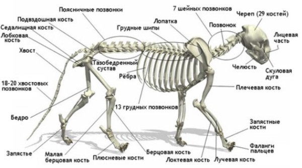
Next comes the lumbar region, which consists of 7 vertebrae, becoming larger as they approach the tail. The vertebrae of the lumbar region have many strong protrusions, because the muscles and tendons that hold all the organs of the abdominal cavity are attached to them. In the sacral region there are 3 powerful fused vertebrae. And the longest is the caudal section, 21-23 decreasing towards the end of the vertebrae; some breeds, which are characterized by a shortened tail, have fewer caudal vertebrae.
One of the leading features of the cat's skeleton is the structure of its collarbones. The fact is that they are in a rudimentary state and do not limit the animal’s movements, as, for example, happens in dogs. Thanks to the “underdeveloped” collarbones, a cat can fit into any gap, as long as its head fits through.
Limb bones
Our fellow cats walk on their toes, and the lower back of their paw was once a foot. The cat's front paws have 5 toes, the outer phalanx of which forms the basis for the claw. The first finger is a rudiment and the claw cannot be removed from it.
The hind limbs of cats are longer, and the joints there are stronger, this allows the cat to withstand sudden strong loads. In addition, this structure of the limbs allows the cat to develop tremendous speed both horizontally and vertically. This is why cats are such good poison dart climbers.

The cat's hind legs have fewer toes - 4, and the fifth is also a rudiment. Based on how many fingers a cat has, she may have polydactyly (more fingers than normal) or oligodactyly (not enough fingers).
Internal organs
The internal structure of a cat is a set of all the same vital systems that are inherent in other mammals. Let's look at them in order.
Circulatory and respiratory systems
The circulatory system of a cat is not particularly different; the animal's pulse at rest ranges from 100 to 150 beats per minute and can be measured by pressing the femoral artery. Normally, blood in an animal’s body should be approximately 7% of its mass; cat blood is specific and clots faster than human blood.
With each beat, a cat's heart pumps about 3 ml of blood. The blood circulation of a cat occurs similarly to that of a person: in the lungs the blood is saturated with oxygen, and in the digestive organs with useful substances. After which the heart carries fresh blood through the arteries to all organs. And through the veins, blood flows back to the heart so that it again sends it to the lungs to be enriched with the necessary oxygen.

The respiratory system, in addition to supplying the blood with oxygen, is also involved in thermoregulation. The breathing rate in cats is 20-30 breaths per minute, in kittens about 40 breaths, and inhalation is through the nose. The air inhaled by a cat through the nose is first warmed and filtered, then passes through the pharynx into the larynx, trachea and lungs of the animal. There is an assumption that a cat makes purring sounds using pocket-like folds that are located in the larynx.
Excretory and digestive systems
The cat's digestive system also has much in common with humans. It begins with the mouth and ends with the rectum and sphincter. Between them are the pharynx, esophagus, stomach, small and large intestines. The pancreas and liver are also considered components of the digestive system.
It is noteworthy that the cat's stomach can digest fairly large pieces of food, which the cat bites off thanks to its strong and sharp incisors and fangs. The cat's intestine is approximately 3 times longer than the animal's body and is 1-1.8 m. The cat also has a cecum, but the murka does not have an appendix.
Fluid is removed from the cat's body through the urinary system - the kidneys, bladder and ureters. Urine formation begins in the kidneys, which also regulate blood chemistry. Urine then moves into the bladder through the ureters, from where it is removed from the body. The process of urination occurs under the control of the closing muscle, which prevents spontaneous urination. The following video will help you understand the structure of a cat by literally looking inside the cat's body!
Reproductive system
The purpose of the reproductive system is obvious - it is the continuation of the cat family. A cat's reproductive organs are the gonads, testicles, vas deferens and penis. In a cat, these are the ovaries, uterus, oviducts and external genitalia. The time for male cats to reach puberty is 6-8 months, however, the safe age for mating, when full-fledged offspring can be expected, is at least 10 months. During puberty, the behavior of cats changes greatly and they show in every possible way their readiness to reproduce.

Sense organs
Nature has endowed our smaller brothers with unusually developed sense organs. Our pets see, hear and smell much more keenly than we do.
Eye
A cat's eyes are much larger in relation to the size of its body than those of a human. The cornea of the cat's eye is more convex, which means that the quality of the image perceived by the cat's eye is higher. Cats can distinguish colors, it is believed that they see at least 3 colors - red, green and blue. The pupil of a cat's eye, just like a human's, is able to expand and contract thanks to a special constrictor muscle. Our mustachioed friends have unusually acute vision, but they don’t see what’s going on under their noses; the optimal distance for the perception of information by a cat’s eye is 2-6 m.
The structure of a cat's eye is distinguished by the presence of a special vascular layer called tapetum, thanks to which the cat's eyes can see in the dark and glow mystically. In addition, our pets may have differently pigmented irises, which is why they are so “different-eyed.”

Ear
The structure of a cat's ear gives it the ability to perceive sounds in the range from 30 hertz to 45 kilohertz, and purrs can also detect ultrasound. Almost all cats have erect ears, with the exception of some breeds. Cats, unlike humans, can actively move their ears; 27 muscles help them do this. All purrs have a fold of skin on the inside of the ear, which some call the “third ear.” It is noteworthy that sometimes completely white cats are born deaf due to gene mutations.
Nose
A cat's nose is considered one of the most vulnerable organs of a purr's body, especially its tip. By the way, the tip of the nose is completely devoid of vegetation and can be of different colors depending on the breed of the cat. Cats' sense of smell is quite well developed because they have a larger number of olfactory receptors compared to us.

In terms of their ability to recognize odors, cats are macrosomatic animals, while humans are microsomatic creatures and the number of odors they perceive is very limited. However, compared to dogs, cats still have weaker olfactory abilities.
Photo gallery
Video “Cats from a scientific point of view”
A very interesting and educational video with a selection of little-known facts about our furry pets to complete our tour of cat anatomy!
Sorry, there are no surveys available at this time.The internal structure of a cat, in terms of the functioning and arrangement of internal organs, is in many ways similar to the internal structure of other species of mammals. But cats have differences that are unique to this type of animal.
Blood circulation and breathing
Circulatory system
Cats do not have any special differences from the circulatory system of many mammals. You can measure your cat's pulse by pressing on the femoral artery, which is located on the inside of the cat's thigh. A cat's normal pulse ranges from 100 to 150 beats per minute. The pulse, breathing rate and temperature of kittens are much higher than those of an adult animal.
The elastic walls of the veins actively relax and contract as the heart pushes blood through the arteries. This is called a pulse. The walls of veins are thinner than the walls of arteries, so they are more susceptible to damage. There is no pulse in the veins, but due to the valves located in the veins, blood moves through them in one direction - towards the heart.
Different parts of the body need different amounts of blood. For example, the brain requires 15 to 20% of the blood contained in a cat's body. About 40% of the blood is consumed by the muscles at rest, but during the flight from an enemy or rival, or the pursuit of prey, blood can circulate in them up to 90% of the total blood, i.e. blood can even flow into the muscles from the brain.
From the heart, arteries carry bright red blood throughout the body, enriched in the lungs with oxygen, and in the digestive system with nutrients. The veins carry carbon dioxide-rich dark blood to the lungs, kidneys and liver.
The pulmonary vein and pulmonary artery are exceptions. Capillaries and pulmonary arteries carry oxygenated blood to the pulmonary alveoli, where oxygen is absorbed from the air the cat inhales. The pulmonary veins return fresh blood to the heart, which pumps it through the arteries throughout the cat's body. Oxygen, in exchange for carbon dioxide, enters the cells, and the veins carry return blood to the heart so that it pumps it back into the lungs for new oxygenation.
Respiratory system of a cat
The respiratory system of a cat plays the main vital function - it is the effective supply of oxygen to the blood. It also provides thermoregulation, removing excess water. A cat's normal body temperature is between 38 and 39°C, higher than that of humans, and in small kittens the temperature can reach up to 40°C. Under the influence of the arching of the diaphragm and pectoral muscles, the expansion of the chest creates negative pressure in the chest, due to this the lungs inflate and draw in air through the nose, and during physical exertion they draw in through the mouth. In cats, the respiratory rate is approximately 20–30 breaths per minute; in kittens it can be higher, up to 40 breaths. The respiratory organs of a cat are the nasopharynx, nose, trachea, bronchi and lungs.
 The air inhaled by a cat first passes through the frontal sinuses of the olfactory apparatus of the cat's nose, where it is moistened, warmed and filtered. The air passes through the respiratory-digestive tract (pharynx) into the larynx, and through the trachea reaches the cat's lungs. The reason for the appearance of such a pleasant cat purring has not yet been fully studied. We can presumably say that these sounds arise with the help of pocket-like folds that are located in the cat’s larynx.
The cat’s larynx consists of a cartilaginous tube, which, thanks to the vibration of the vocal cords located in it, participates in sound production and protects the trachea from food entering it.
A straight cartilaginous tube, the trachea, is constantly kept open by the C-shaped cartilage. Attached to the esophagus is an “open” piece of cartilage through which bolus food passes. During eating, the nasal cavity is closed by the soft palate, and the trachea is closed by the epiglottis. The trachea is divided within the lungs into the main bronchus and the lobar bronchus, which in turn are divided into many bronchioles, which end in the alveoli and air sacs. Oxygenated blood circulates around the alveoli.
The shape of a cat's lungs is a truncated cone, the apex of which is located in the area of the first ribs, and the base is concave, corresponding to the dome of the diaphragm, which is divided into the left and right lungs. Each of the ribs is divided into three lobes: 1 - upper cranial, 2 - middle, 3 - lower caudal (largest). The left lung of a cat is slightly larger than the right lung, due to the additional lobe on it. The volume of the left lung of a cat is on average 11 cm, and the volume of the right lung is 8 cm. The lungs of cats are similar in structure to a bunch of grapes, and the alveoli are berries.
Cat's heart
In fact, the cat's heart, like the human heart, is a paired pump that is designed to pump blood. For example, the body of an average cat, whose weight is about 3.2 kg, contains about 200 ml of blood. With each beat, 3 ml of blood passes through the heart. In structure, the hearts of other mammals are similar to the heart of a cat, but in a cat it is slightly smaller in relation to its body size.
 Blood enters the right side of the heart through the circulatory system, which pushes it to the lungs through the pulmonary artery to saturate it with oxygen. Blood saturated with oxygen enters the left side of the heart from the lungs. Then the heart pumps blood into the aorta, from where it spreads throughout the animal’s body.
The right side of the heart and the left side have an atrium - the upper chamber, and a ventricle - the lower chamber, which is the main blood pump. The atrioventricular (or tricuspid) valve, at the time of contraction of the right atrium, prevents the return of blood from the right ventricle into it. The mitral valve also performs a similar function in the left side of the heart. The muscles of the ventricles are connected to the valves by means of tendons, which prevent the ventricles from being pushed towards the atria when the ventricles contract.
Blood of cats
Cats have specific blood that cannot be replaced or supplemented with blood from other animals. Cats' blood clots faster than human blood.
Yellowish plasma makes up the bulk of the total blood volume, with red blood cells accounting for 30 to 45%, and platelets and white blood cells making up the rest. Plasma is like a “transport” part of the blood, which carries nutrients from the digestive system, including cell waste. The composition and volume of plasma is maintained by fluid that is absorbed into the colon.
The endocrine system and the cat's brain
Information is transmitted to the cat's brain by glands and all sensory organs that produce hormones. The brain processes all chemical signals and sends commands to the entire body through the nervous system. Although the weight of the brain does not exceed 1% of the weight of the entire body, its work requires a lot of energy, so it receives up to 20% of the blood that the heart pumps.
Cat brain
In a cat, the brain consists of a billion neuronal cells, and each cell has up to 10 thousand connections with other cells. In a seven-week-old kitten, messages are transmitted in the brain at a speed of 386 km/h, but as the animal ages, the speed of message transmission decreases.
 The cat's brain is anatomically similar to that of other mammals. The cerebellum is responsible for coordinating motor activity and also controls all muscles. Responsible for the cat’s consciousness (emotions, learning and behavior) is the cerebral hemisphere, the trunk of which connects them with the peripheral nervous system. From the brain, information is delivered to all parts of the cat’s body along the main highway – the spinal cord. The parietal lobe of a cat's brain processes information it receives from its senses. The occipital lobe of the brain controls tactile and visual signals, and the olfactory bulb processes odors.
The temporal lobe of the brain is responsible for a cat's memory and behavior. The pineal gland produces the hormone melatonin, which regulates wakefulness and sleep, and also maintains the rhythm of the animal’s life. Controls the autonomic nervous system and secretes various hormones (for example, a hormone such as oxytocin, which stimulates the process of childbirth in a cat and the secretion of breast milk) - the hypothalamus. Growth hormones are produced and regulated by the pituitary gland. The frontal lobe of the brain controls the cat's voluntary movements, and connects the right and left hemispheres of the cat's brain - the corpus callosum.
Endocrine system of the cat
One of the main systems of endocrine glands in the regulation of the body is the endocrine system, which is localized in various tissues, organs and the central nervous system of the cat. The endocrine system exerts a regulatory influence through hormones of high biological activity, which ensure the life processes of the cat’s entire body - development, growth, reproduction and behavior. The pituitary gland and hypothalamus are the central link in the endocrine system. The adrenal glands, the thyroid gland, as well as the ovaries of cats and the ovaries of cats are a peripheral link in the endocrine system.
Most body functions are regulated by hormones produced by the cat's brain - the hypothalamus produces the hormone ADH (antidiuretic hormone), which regulates the concentration of urine. The hypothalamus also produces corticoliberin and oxytocin, which secrete the following hormones:
ACTH (adrenocorticotropic) hormone, which causes the cat's adrenal glands to release cortisol in response to danger or stress
TSH (thyroid stimulating) hormone, which primarily stimulates the activity of the thyroid gland, which controls the metabolic rate of all substances
MSH (melanocyte stimulating) hormone, which in the pineal gland of the brain accelerates the synthesis of melatonin
FSH (follicle stimulating) hormone, which controls the production of sex hormones, sperm and eggs in cats
LH (luteinizing) hormone, which controls the formation of sex hormones, sperm and eggs in cats
 Next to the kidneys are the adrenal glands, which consist of an inner medulla and cortex. The adrenal cortex produces various hormones, including cortisol, which play an important role in shaping the body's response to injury and regulating metabolism. The adrenal medulla produces the hormones norepinephrine and epinephrine (norepinephrine and epinephrine), which control the dilation of blood vessels and heart rate.
The hypothalamus stimulates the production of corticoliberin by an unfamiliar smell;
Corticotropic hormone, in turn, stimulates the pituitary gland to produce adrenocorticotropic hormone (ACTH), which is transmitted to the adrenal glands through the blood;
ACTH, having entered the adrenal glands, stimulates the production of cortisol in the adrenal cortex, and at this time adrenaline is produced in the adrenal medulla;
Suppresses the production of corticoliberin - cortisol, which is produced by the adrenal cortex, to control the protective reaction.
In the biofeedback system, a vital element is the cat's adrenal glands, which influence its behavior and control its reactions. A cat's mood, tamability, and sociability determine feedback mechanisms.
Reproductive system of cats
 Excess water and decay products are removed from the animal's body by the kidneys and urinary tract in the form of urine; also, part of the genitourinary system is the urethra, which flows into the cat's penis, and into the cat's vagina, two ureters, and the bladder.
The genital system is designed for reproduction. In a cat, it includes the sex glands, testicles in the scrotum, vas deferens, which flow into the urethra and the cat’s penis. In a cat, these are the ovaries, uterus, tubes and external organs near the anus - the vulva and vagina. Ovulation occurring in a cat provokes the cat to mate.
By 6 – 8 months of age, a male cat reaches puberty. This does not mean that the body’s development and growth have ended by this age; it indicates that the animal has already reached physiological maturity, which can be used for reproduction. Depending on the breed of the cat, its physiological maturity manifests itself already at the age of 10 months to 1.5 years. Mating can be carried out only from this age of the cat, in this case you can count on the appearance of full-fledged and healthy offspring, and without harm to its health.
Nervous system of a cat
The nervous system functions in close connection with the endocrine system and directs all the vital functions of the animal. The cat's nervous system reacts quickly to both external and internal events. A cat can control some nervous processes consciously, and others at a subconscious, deeper level.
 The nervous system is divided into two parts: the central part and the peripheral part. But the nervous system actually works as one whole; many elements of the nervous system can be attributed to both the central system and the peripheral system.
The nervous system consists of the brain and the spinal cord - a command center, like a “highway”, for conducting nerve impulses in both directions. Information about touch, temperature, pain and pressure is received by the peripheral nervous system, which transmits all instructions to the muscles. The peripheral nervous system consists of peripheral, spinal and cranial nerves.
The cranial nerves are responsible for transmitting information from the senses and contracting the facial muscles. Spinal nerves extend along the entire length of the spinal cord, connecting individual parts of the body with the central nervous system.
Nerve cells of a cat's body
The nervous system consists of nerve cells, neurons, and the cells that support them, which produce myelin.
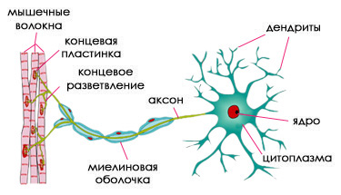 Dendrites are branches extending from the neuron body that receive information from other cells. Each neuron cell has one axon (a long extension) that sends messages directly to organs or other nerve cells. All these messages are carried by chemical substances - transmitters, or neurotransmitters, which are produced in axons. Each cell in a neuron sends messages to other cells.
The fatty protective membrane is myelin, which covers large axons and increases the speed of transmission of all messages between nerves. A nerve fiber consists of a myelin sheath, an axon, and a cell that produces myelin.
In the central nervous system, myelin is produced by oligodendrocyte cells, and in the peripheral nervous system by neurolemmocyte cells. At birth, few nerves have a myelin sheath, but nerves in kittens myelinate very efficiently and quickly.
Reflexes and conscious control
Many functions of the animal’s nervous system are under volitional (voluntary) control. When the animal sees the prey, it controls its muscles so that it can jump on it more accurately. Messages to the brain are transmitted by sensory nerves, and instructions from the brain are transmitted by motor nerves, which cause them to work as needed for the cat to jump accurately. However, such forms of activity as the regulation of breathing and heart rate, internal organs, and digestion processes can occur involuntarily.
Such involuntary activity is regulated by the autonomic nervous system, consisting of two parts - parasympathetic and sympathetic. The first part inhibits activity, the second part stimulates.
When the animal is resting, involuntary activity is controlled by the parasympathetic nervous system - the animal's pupils are constricted, breathing and heartbeat are regular and slow. The sympathetic nervous system comes into play when the animal is nervous - the sympathetic part activates the pituitary gland and hypothalamus of the brain, thereby stimulating the work of the adrenal glands, preparing a defense reaction. Blood comes from the internal organs of the muscles; the fur stands on end, the heartbeat accelerates, the pupils dilate so that the animal can see better - the subcutaneous erector muscles work.
Digestive and excretory system of cats
The cat's digestive system has a number of unique properties that have a significant impact on the process of digesting food. A cat, like all mammals, uses two mechanisms to digest food:
Chemical - food is broken down into nutrients, which are absorbed into the blood through the walls of the small intestine;
Mechanical - food is crushed by teeth.
The digestive system has a barrier function, which, being one of the important functions, prevents various viruses and harmful bacteria from entering the cat’s body.
The complete digestion cycle (digestion of food, absorption of necessary nutrients and excretion of undigested food residues) lasts 24 hours.
The structure of the cat's digestive system and its functioning
The digestive organs include the oral cavity, pharynx, stomach, esophagus, large and small intestines, and rectum.
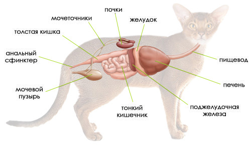 In the process of digestion, the endocrine glands, namely the pancreas, liver and gall bladder, also play an important role.
The oral cavity performs the functions of biting and chewing food. The teeth in the oral cavity are durable organs that serve to capture, hold, bite and grind food, as well as for attack and defense. Saliva consists of 1% mucous substances and 99% water.
The cat, being a predator by nature, tears, gnaws and cuts meat food with its teeth, after which, practically without chewing, it swallows it. The salivary glands in the mouth moisten food so that it passes more easily into the stomach through the esophagus. In the oral cavity, under the influence of saliva, food begins to break down. This process of digestion is called mechanical.
Esophagus:
The cells of the esophagus secrete the mucus necessary for lubrication and ensure easy passage of food through the gastrointestinal tract.
The esophagus, which is relatively elastic and expands, carries food into the stomach.
Stomach:
Food is retained and processed;
Gastric juices are released: (pepsin promotes the breakdown of proteins), mucous substances (perform the function of protecting the walls of the stomach), gastric acid (creates an acidic environment in the stomach favorable for the digestion of proteins);
Muscle activity (promotes mixing of food with gastric juice).
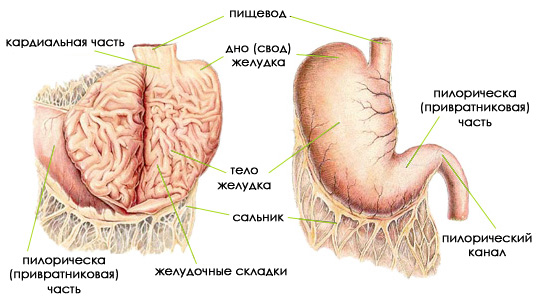 Cats have a single-chamber stomach, consisting of:
cardinal part, in which the inlet of the esophagus is located;
pyloric part, in which there is an opening leading to the duodenum.
Next to the cardinal part there is a convex upper section of the stomach, which is called the fundus of the stomach. The body of the stomach is the largest section.
The pyloric part is the gastric section, which is adjacent to the pyloric canal and connects the lumen of the duodenum and the lumen of the stomach.
In an empty stomach, the mucous membrane is collected in longitudinal gastric folds.
The cat's stomach is covered on the outside with a serous membrane that passes into the omentum. The serosa connects the stomach with the ligament of the esophagus, liver and duodenum.
The mechanics of digestion are controlled by hormones secreted by the thyroid, pancreas and parathyroid glands.
The main function of the thyroid gland is to regulate metabolic rate. An overactive thyroid gland may be accompanied by weight loss, increased heart rate, or uncontrolled appetite. On either side of the thyroid gland are the parathyroid glands, which produce a hormone for the absorption of calcium, which is so necessary for muscle contraction. The pancreas produces insulin, a hormone that circulates in the blood and regulates the amount of glucose.
A cat's digestive process is adapted to frequent food consumption in small portions. Food stays in the cat's stomach, where it undergoes chemical processing.
The cardinal part of the cat's stomach promotes the secretion of gastric juices:
acid, which breaks down dietary fiber;
enzymes, which break down proteins and ensure the digestion of practically unchewed food. In addition, the stomach secretes mucus, which protects the intestines and stomach walls from caustic enzymes.
The gastric muscles regulate motility and allow food to pass into the small intestine, thus aiding digestion.
Small intestine:
In the small intestine, enzymes break down fats, proteins and carbohydrates. Due to reduced amylase activity in cats, carbohydrates are absorbed less efficiently than in dogs.
The small intestine occupies most of the abdominal cavity and consists of many loops. Conventionally, according to position, the small intestine can be divided into three sections: ileum, duodenum and jejunum.
The final stage of digestion occurs in the cat's small intestine, which is 1.6 meters long. Food is mixed as a result of contraction of the stomach muscles and is pushed out in small portions into the duodenum, which in turn receives enzymes from the pancreas and bile from the gallbladder, which helps break down fats.
Digestion of food occurs throughout the small intestine. Nutrients are absorbed into the lymph and blood through the walls of the small intestine.
The largest gland in a cat's body is liver where the blood delivers nutrients. The liver converts these nutrients into essential amino acids and fatty acids. A cat, unlike a human or a dog, requires animal protein to produce a full range of liver acids. Therefore, to maintain vital functions, the cat needs to eat meat, otherwise it may die.
 The liver performs a barrier function, in other words, it promotes the breakdown of toxic substances and prevents the spread of viruses and bacteria.
The liver is divided by a fibrinous membrane into left and right lobes, which are in turn divided into lateral and medial parts. In size, the left lateral lobe is significantly larger than the relatively small left medial lobe and covers at one end most of the ventral gastric surface.
The right medial lobe, in contrast to the left, is large; the gallbladder is located on its back side. At its base there is an elongated caudate lobe, on the right side of the anterior section of which there is the caudate process, and on the left - the papillary process
The liver performs one of the most important functions - the production of bile. The gallbladder is located in the cleft of the right medial lobe and is pear-shaped. The liver is supplied with blood through the hepatic arteries and portal vein, and venous outflow is carried out into the caudal vena cava through the hepatic veins.
Colon
What happens in the colon:
Absorption of electrolytes and water;
Fiber fermentation.
Rectum:
Intake of bacteria, water, undigested food residues and minerals;
Emptying the rectum. This process is completely controlled by the cat, but can be disrupted by clinical and nutritional changes.
After the nutrients are absorbed, undigested food remains enter the large intestine. The large intestine consists of the colon, rectum and cecum, and ends in the anus. In a cat, the length of the large intestine is 30 cm.
The cecum has a length of 2-2.5 cm and is a blind outgrowth at the border of the large and small intestines and is a vestigial organ. The iliac foramen serves as an obturator mechanism.
The colon is the longest section of the large intestine, its length is 20-23 cm. It does not wind in loops like the small intestine, but bends slightly before moving into the rectum, which is approximately 5 cm long. The mucous membrane has many mucous glands that secrete necessary for lubricating dry waste, a large amount of mucus. The rectum opens outward under the root of the tail with the anus, on the sides of which there are anal glands that secrete an odorous fluid.
Excess fluid is removed from the cat’s body using the organs of the urinary system: kidneys, bladder and ureters. Urine is formed in the kidneys, and here nephrons filter out unnecessary substances brought from the liver.
The kidneys maintain the chemical balance of the blood, regulate blood pressure, promote the release of the hormone erythropoietin, and activate vitamin D.
See also on our website: | | | | |