Purulent lymphadenitis, ICD code 10. Axillary lymphadenitis: causes, symptoms, treatment. How to treat with folk remedies at home
Submandibular lymphadenitis can occur after hypothermia or for a number of other reasons. Its treatment can be conservative or surgical and is prescribed only by a doctor.
Lymphadenitis under the jaw - causes
The submandibular lymph nodes are responsible for processing lymph that leaves the head, including from the oral cavity. Lymphadenitis refers to the inflammatory process in the lymph node. Under the jaw, this pathology extremely rarely occurs primarily; more often it is secondary in nature, that is, it becomes a consequence of the underlying disease. First, an inflammatory process occurs in a neighboring organ, and then the infection spreads to the regional lymph node. Disease code by ICD-10 – L.04.0. Lymphadenitis of the face, head, neck.

Depending on the type of course, lymphadenitis occurs:
- sharp– develops with vivid symptoms, lasts no more than 1-3 weeks;
- chronic– accompanied by periodic remissions and exacerbations.
The disease can be serous, not accompanied by purulent processes, or purulent, in which the patient requires surgical intervention. It appears with equal frequency in children and adults. The reasons may be as follows:

In adults, lymphadenitis can be caused by specific infections - tuberculosis, syphilis. In childhood, trauma to the tonsil and throat also contributes to the development of pathology.
Clinical picture of lymphadenitis
In children under 3 years of age, symptoms of the disease cannot appear, because the lymph nodes finally develop only at this age. In other patients, at the initial stage the pathology does not show any signs, but after a couple of days the lymph nodes enlarge, become hard, tight to the touch, and their palpation is very unpleasant. If the disease is not treated at this stage, it becomes acute. The lymph node becomes sharply painful, becomes inflamed, and gives “lumbago” - periodic severe pain radiating to the ear.

In the affected area of the neck, there is redness and swelling (edema) of the skin. Sometimes the skin becomes burgundy, and the swelling spreads to the entire side of the neck. There is pain when swallowing, weakness, and body temperature rises. It is difficult for a person to sleep due to severe pain in the neck, and he loses his appetite. If treatment has not yet begun, lymphadenitis becomes purulent:
- bluish skin;
- trembling of the skin due to the accumulation of pus;
- visible transfusion of pus in the node;
- temperature up to 40 degrees;
- hyperthermia;
- severe pain when moving the jaw.
Chronic lymphadenitis is a consequence of an untreated acute form of pathology, with which the lymph node is constantly hard, enlarged, and slightly painful.
Diagnosis of the disease
Despite the clear clinical signs, it is not always possible to make a diagnosis without a detailed examination. Lymphadenitis must be differentiated from oncological diseases, as well as its serous form from the purulent one - the treatment procedure depends on this. You need to seek help from a therapist, ENT specialist, dentist, or maxillofacial surgeon. The main diagnostic methods and their results are presented below.
For chronic lymphadenitis, the main diagnostic method is ultrasound, based on the results of which the doctor will draw conclusions about the presence of a sluggish inflammatory process.

Treatment of lymphadenitis
Treatment at home is possible if the disease has not reached the purulent stage. Physiotherapeutic techniques are used - electrophoresis of painkillers, absorbable, anti-inflammatory drugs, UHF. The main method of therapy is taking antibiotics. Most often, the causative agents of the disease are staphylococci and streptococci, so broad-spectrum antibiotics from the group of macrolides and penicillins are recommended for treatment.

Will cope with the disease in 7-10 days Amoxiclav, Flemoclav, 5-7 days before – Clarithromycin, Azithromycin. In some cases, doctors recommend combinations of antibiotics from different groups. At the same time, antiseptic rinses of the oral cavity are used (if the cause lies in inflammatory diseases of the oropharynx), lotions are placed on the area of the lymph node with Burov's fluid. In the chronic form of the pathology, immunomodulators (Amiksin, Polyoxidonium) are additionally prescribed. If there is an accumulation of pus in the lymph node, surgery is performed. Under general or local anesthesia, the node is dissected, the pus is removed through drainage, and the cavity is washed with antibiotics.
When several nearby nodes become inflamed, an operation is performed under general anesthesia with opening the area, introducing drainage into the subcutaneous tissue and removing the melted tissue.
Folk remedies for submandibular lymphadenitis
At the very first stage of the disease, when there is no pus in the lymph node yet, along with conservative remedies, you can try alternative treatment according to the following recipes:

Prevention of lymphadenitis under the jaw
Since in most cases the cause of the pathology is infections of the ENT organs and chronic pathologies, they should be treated in a timely manner. For chronic tonsillitis, it is important to undergo therapy with the device 2 times a year. Tonsilor", removing purulent plugs.
As a rule, the diagnosis of “axillary lymphadenitis” frightens patients. This reaction is caused by ignorance of the peculiarities of the course of the disease, which responds well to treatment and does not have an impact on human health in the future, subject to timely diagnosis.
When the disease occurs, there is a strong tugging and swelling in the area of the armpit.
Axillary lymphadenitis (code according to the international classification of diseases ICD-10 - L04.2) is an infectious disease accompanied by inflammation of the axillary lymph nodes and their enlargement to a significant size. The causative agents of the disease are representatives of pathogenic and conditionally pathogenic microflora - diplococci, staphylococci, streptococci, Escherichia coli and Pseudomonas aeruginosa, fungi, etc.
Causes
Lymphadenitis of the axillary region is a direct consequence of infection with viruses, fungi or bacteria that penetrate the lymph nodes in the following ways:
- lymphogenous - through infected lymph;
- hematogenous - through the blood;
- contact – when pathogenic microflora gets into the wound.
The disease can develop against the background of:
- furunculosis;
- tularemia;
- phlegmon;
- brucellosis;
- syphilis;
- gonorrhea;
- eczema;
- AIDS;
- tuberculosis;
- cancer;
- trophic ulcers;
- purulent wounds;
- inflammation of the ovaries in women;
- fungal diseases - microsporia, trichophytosis, sporotrichosis;
- osteomyelitis of the hand bones.
Provoking factors in this case may be:
- reduced immunity - in this case, the body is powerless not only against pathogenic, but also against opportunistic microflora, harmless to a healthy person;
- bad habits – abuse of smoking and alcoholic beverages leads to a decrease in immunity and the accumulation of harmful substances in the body.
Axillary lymphadenitis can develop as a result of cat scratches or bites. In this case, the causative agents will be rickettsia - microorganisms that live in the cat's body.
Symptoms

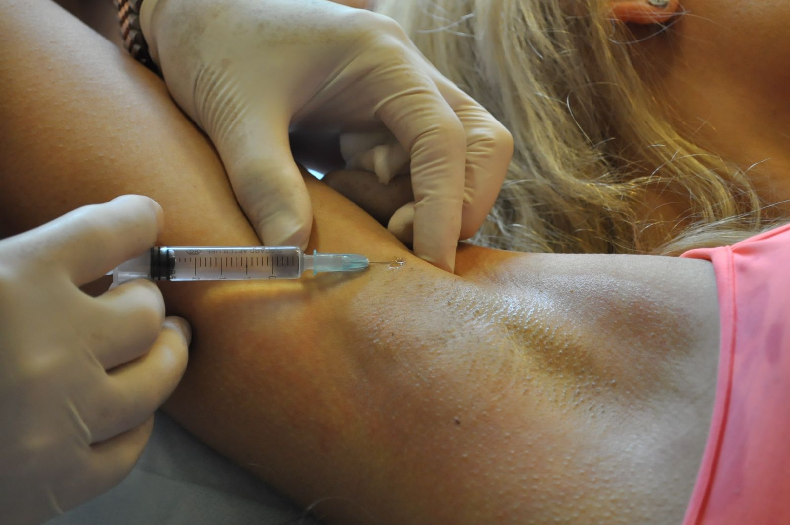
Pain and discomfort under the arms is the first reason to see a doctor
One of the first manifestations of axillary lymphadenitis is pain under the armpit, in the area of the lymph nodes, which appears when you touch the affected area, as well as signs of general intoxication and increased body temperature.
In addition, you may experience:
- swelling and redness of the skin (appears during the acute course of the disease);
- deterioration of appetite, incessant headache, loss of strength due to intoxication of the body;
- abscesses due to suppuration of nodes (can lead to necrotic changes in the structure of tissues and lymph nodes);
- tachycardia with damage to the cardiovascular system;
- gas crepitus, accompanied by a crunching sound when pressed;
- limitation of hand mobility due to damage to nerve tissue.
Diagnostics
Diagnosis of axillary disease is made comprehensively and includes:
- interview and examination of the patient;
- blood and lymph tests;
- puncture of the lymph node to exclude Hodgkin's disease or leukemia;
- computed tomography of the lymphatic system;
- X-ray contrast lymphography – study of problem areas using contrast agents and special equipment;
- lymphoscintigraphy - examination of problem areas using radionuclide substances and special equipment;
- ultrasound examination.
Classification

Simple axillary lymphadenitis occurs unnoticed, without deterioration of well-being or anxiety
The disease is classified according to the nature of its course, clinical picture and type of microorganisms that led to the development of pathology.
Depending on the nature of the course, lymphadenitis is divided into:
- acute, accompanied by pronounced symptoms - swelling, pain, the appearance of compactions in the armpits, a significant increase in body temperature and general intoxication of the body;
- chronic, characterized by a slight increase in lymph nodes (the well-being of patients remains normal, there is no pain on palpation).
Depending on the clinical picture, lymphadenitis is divided into:
- Simple. It proceeds unnoticed, without deterioration of well-being or anxiety. There is no pain or redness of the skin. Body temperature does not rise. There is slight discomfort in the armpit area and a slight increase in size of the lymph nodes.
- Serous. Accompanied by increased discomfort in the armpit, significant enlargement of the lymph node, and pain that appears when touched. The inflamed area turns red and becomes hot to the touch. Nodes and tissues join together into a hot, painful “package”. General health does not deteriorate.
- Purulent. To the above symptoms are added weakness and increased body temperature. Suppuration of the lymph nodes occurs. Fistulas form. Inflammation spreads to nearby tissues.
Depending on the type of microorganisms that led to the development of the disease, lymphadenitis is divided into:
- specific, developed against the background of diseases affecting the lymph nodes - cancer, tuberculosis, brucellosis, syphilis, tularemia;
- nonspecific, developing against the background of weakened immunity under the influence of streptococci, staphylococci, etc.
Depending on the location, axillary lymphadenitis is divided into:
- left-handed;
- right-sided;
- bilateral.
How to cure lymphadenitis of the axillary lymph nodes?

If necessary, treatment of axillary lymphadenitis is carried out surgically
The main directions in the treatment of lymphadenitis under the armpit in women, men and children are:
- drug therapy;
- physiotherapy;
- traditional methods of treatment;
- surgical treatment.
Children are treated in the same way as adults. The dosage of medications is selected taking into account the age and weight of the child.
Drug therapy
Drug treatment for axillary lymphadenitis allows:
- eliminate the root cause of the disease;
- reduce the severity of inflammatory processes in the lymph nodes;
- improve overall well-being.
For this purpose, the following may be prescribed:
- non-steroidal anti-inflammatory drugs;
- antihistamines;
- antibiotics;
- antiviral agents;
- antifungal drugs;
- anti-tuberculosis drugs.
A doctor should prescribe certain medications, including antibiotics, for axillary lymphadenitis. Self-medication in this case is unacceptable due to the fact that it can lead to aggravation of existing health problems.
Physiotherapy
It is necessary to create rest for the affected area, carry out adequate antibiotic therapy and vitamin therapy
Physiotherapy for axillary lymphadenitis can alleviate the general condition of patients, reduce the severity of inflammatory processes in the lymph nodes, and accelerate the restoration of damaged tissues. Patients are usually recommended:
- ultra-high frequency therapy (UHF);
- laser therapy;
- galvanization.
UHF therapy
UHF therapy is a procedure that involves exposing the human body to a high-frequency electromagnetic field and leading to:
- temperature increase in the affected area;
- vasodilation and movement of leukocytes to the affected area;
- proliferation of connective tissue.
The described changes enhance local tissue immunity and contribute to the rapid relief of inflammatory processes.
Indications for UHF therapy are the presence of an acute inflammatory process in the lymph nodes, and contraindications are tumor processes and tuberculosis.
Attention! UHF therapy should not be used for signs indicating infectious processes in the body - increased body temperature, chills, tachycardia, muscle pain, etc.
Laser therapy
Laser therapy is a procedure that involves exposing the human body to waves of a certain frequency in order to:
- improving microcirculation in the inflamed node;
- relieving inflammation;
- pain relief;
- accelerating tissue regeneration.
Indications for using the method are acute and chronic lymphadenitis, and contraindications:
- tumor processes;
- tuberculosis;
- the presence of benign formations in the area of influence.
Galvanization
Galvanization is a procedure that involves exposing the body to an electric current of low voltage and low strength passing through tissues in order to:
- pain relief;
- improving microcirculation in the affected area;
- accelerating the regeneration of tissues and nerve fibers.
Method used:
- in the recovery period after eliminating the cause that led to the development of acute lymphadenitis and reducing the severity of inflammatory processes in the lymph nodes;
- in chronic forms of pathology.
Traditional methods of treatment

Before using any folk remedy, you should consult your doctor for contraindications.
Traditional medicine is used as an adjunct to primary treatment to relieve inflammation, improve overall health and speed up recovery in the early stages of the disease.
The use of traditional medicine is permissible only in combination with taking antibacterial, antiviral or antifungal agents and only after identifying the true cause of lymphadenitis.
The most popular methods of treating axillary lymphadenitis are:
- warming up the lymph nodes;
- the use of herbal preparations and echinacea tincture.
Before using any folk remedy, you should consult your doctor and get his approval.
Warming the lymph nodes is contraindicated in the following cases:
- the presence of tumor processes in the lymph nodes;
- development of adenophlegmon;
- tuberculous lesions of the lymph nodes;
- the presence of signs of intoxication of the body - headaches and muscle pain, elevated body temperature, rapid heartbeat.
Surgical treatment
Surgical treatment is used for the development of purulent complications of lymphadenitis - abscesses and adenophlegmons. Under general or local anesthesia, the purulent focus is opened, the pus and damaged tissue are removed, the wound is washed with antiseptic solutions, sutured and drained (a drainage is inserted into the cavity - a special tube designed for the outflow of fluid and pus and the administration of medications).
Prevention

Proper nutrition is one of the measures to prevent and prevent the development of axillary lymphadenitis
Prevention of axillary lymphadenitis includes:
- protection against infection and timely treatment of viral, fungal and infectious diseases;
- minimizing the likelihood of injury to the armpit area;
- compliance with personal hygiene rules;
- strengthening the immune system;
- maintaining a healthy lifestyle;
- quality food.
Forecast
Timely and adequate treatment of axillary lymphadenitis can completely cure the disease, although it may take a long time. Ignoring the signs of the disease can lead to irreversible changes in the body, even death.
Acute inflammatory swelling of the lymph nodes - spicy always painful. Patients can usually pinpoint the onset of changes.
The lymph nodes- medium density, the skin over them is hyperemic only in severe cases, the swelling is strictly localized. Sometimes a reddened cord - lymphangitis - leads to a skin wound located on the periphery, indicating the cause of the swelling. But even without the presence of lymphangitis, with all local swelling of the lymph nodes, one must always look for the entrance gate of infection, which in most cases is easy to find. There are, however, cases of significant swelling of regional lymph nodes when the inflammatory reaction at the entrance gate has already completely subsided. Experience has shown that if the doctor does not think about the possible cause of the enlarged nodes, significant difficulties arise: for example, in infections of the scalp, swelling of the lymph nodes behind the auricle and occipital nodes is often not correctly recognized as swollen regional lymph nodes simply because the scalp is not examined thoroughly.
In these cases, it is often diagnosed rubella. Swelling of the inguinal lymph nodes in bed patients is often the first symptom of the phlebitis that causes it.
It must therefore be considered serious symptom, if there is no visible reason (balanitis), and one should never assume that we are talking about trifles, even if there seems to be no peripheral infectious focus. Painful swelling of the lymph nodes at the angle of the lower jaw indicates an inflammatory process in the pharynx (tonsillitis, pharyngitis). Associated general symptoms vary depending on the severity of the infection. Most cases proceed without an increase in temperature, while in other cases there is a picture of a general infectious disease with an increase in temperature and leukocytosis. In severe cases, inflamed lymph nodes may undergo purulent melting - lymphadenetic abscess.
Nonspecific chronic inflammatory Lymph node swellings are of clinical interest because they sometimes simulate serious diseases and misdirect the differential diagnosis. In most people, the inguinal lymph nodes are particularly well palpable, sometimes reaching the size of a hazelnut; they are not painful. They should be considered as nodes that have undergone scar changes due to frequent acute inflammation in the genital area (balanitis, vaginitis). Swelling of the lymph nodes at the angle of the lower jaw is also often found, especially in young people, indicating past infections in the nasopharyngeal space.
Tuberculosis of the lymph nodes can manifest itself in various forms.
a) Most often it manifests itself in the form of tuberculosis cervical lymph nodes(cervical lymphomas). In this case, we are usually talking about the oral primary complex. Therefore, mainly children and younger people, up to approximately 25 years of age, become ill. These lymphomas can also be an expression of organ tuberculosis. More than 80% of them are based on tuberculosis infection with the bovi-nus bacillus. At the same time, Wiesmann, among 50 patients infected with the bovinus type bacillus, found lesions in the oral cavity, pharynx and neck organs in 38%, which indicates the preferential localization of the bovinus type bacilli in this area. The primary focus, if you look for it histologically, is very often located in the tonsils, less often in the gums. With tuberculosis of the cervical lymph nodes, the deep cervical nodes located at the angle of the lower jaw are predominantly affected.
The process often involves neighboring nodes, including supraclavicular ones. Usually the process is one-way. But we recently clinically diagnosed lymphogranulomatosis in an 18-year-old girl, Kotopa, who also had many hazelnut-sized lymph nodes palpable on the opposite side, because we too adhered to the rule about the one-sidedness of tuberculous cervical lymphoma, while the biopsy showed tuberculosis. When the primary focus is localized in the gums, the lymph nodes are affected not at the angle of the lower jaw, but somewhat more medially.
For tuberculosis of the cervical lymph nodes they are initially quite dense to the touch, although usually not to the same extent as with lymphogranulomatosis. But it is often impossible to distinguish them from each other. Pressure sensitivity, which is present in most cases, almost always makes it possible to distinguish inflammatory swelling of the lymph node from neoplastic one. Pain and tenderness with pressure are especially pronounced with rapid enlargement of the lymph nodes. This most likely indicates the inflammatory nature of the process. The skin over the lymphoma in the early stages may be completely unchanged. When the knots become larger, that is, they reach approximately the size of a cherry, they almost always soften. Then a bluish color appears above the lymphoma, skin mobility decreases and it appears that the inflammatory process is spreading to the surrounding tissues.
At this stage the diagnosis no doubt. When the node melts, a cold abscess occurs, which leads to the formation of scrofuloderma, which breaks out, leaving behind a fistula. Fistulas of the lymph nodes occur, in addition to tuberculosis, only with actinomycosis of the lymph nodes. Bacteriological examination of pus quickly leads to the correct diagnosis.
General reactions very diverse. In younger people, fever is rarely observed, but in children, even primary tonsillogenous infection often occurs with a high temperature. ROE is slightly accelerated or normal. The Mantoux reaction is almost always positive. There are, however, undoubted cases of tuberculosis of the cervical lymph nodes (bacteria were found) with a negative Mantoux test (up to 1: 100) (Tobler).
b) In addition to classical cases tuberculosis of the cervical lymph nodes, atypical clinical cases are increasingly being observed in which the histologically established diagnosis of tuberculosis is surprising. Unlike cervical tuberculous lymphoma, which, according to its nosological position as a primary complex, affects almost exclusively people under the age of 25 years, the second form can develop at any age. Lymph nodes are very dense, generally do not adhere to the skin, and range in size from a pea to a small hazelnut. In most cases, the cervical lymph nodes are also affected. We are probably talking about hematogenous dissemination. According to my observations, the picture is not the same. With such data, you must always look for the root cause.
In the last cases I observed, it was about tuberculous lesions of the lymph nodes with tuberculous polyserositis, ovarian cancer, lymphogranulomatosis and tuberculosis of the apex of the lungs.
Tuberculosis of the cervical lymph nodes must be differentiated from swelling of gill canal cysts.
In Russia, the International Classification of Diseases, 10th revision (ICD-10) has been adopted as a single normative document for recording morbidity, reasons for the population's visits to medical institutions of all departments, and causes of death.
ICD-10 was introduced into healthcare practice throughout the Russian Federation in 1999 by order of the Russian Ministry of Health dated May 27, 1997. No. 170
The release of a new revision (ICD-11) is planned by WHO in 2017-2018.
With changes and additions from WHO.
Processing and translation of changes © mkb-10.com
Inguinal lymphadenitis
Inguinal lymphadenitis is a type of inflammation of the lymph nodes. The main function of the lymphatic system is to protect the body from various types of external influences. This is accomplished by the formation of special immune cells in it that protect the body from all kinds of infections. Thus, human health directly depends on the state of the lymphatic system. If a person has inflammation of the lymph nodes, this indicates the presence of a dangerous infection in the body.
Inguinal lymphadenitis in men and women is a secondary disease that occurs due to the presence of inflammation in any part of the body. The most common cause of inguinal lymphadenitis is a sexually transmitted disease. Primary inflammation of the lymph nodes occurs very rarely, and its causative agents are pathogenic microflora.
Symptoms of inguinal lymphadenitis
The main symptoms of inguinal lymphadenitis in women and men are:
- thickening and enlargement of lymph nodes in the groin area;
- increased body temperature;
- state of general malaise, weakness;
- the appearance of pain in the groin area and lower abdomen during physical activity and walking;
- redness of the skin around the lymph nodes.
It happens that inguinal lymphadenitis spreads to all lymph nodes. If lymphadenitis is purulent in nature, then it can result in an abscess, in which decomposition of the walls of blood vessels occurs, accompanied by constant bleeding. In this case, immediate treatment of inguinal lymphadenitis is necessary.
Causes of inguinal lymphadenitis
If you suspect lymphadenitis, you must immediately contact a therapist, who, in turn, can refer the patient for consultation with other specialists and for examination.
Sometimes the cause of inguinal lymphadenitis can be a serious disease such as syphilis. Inguinal lymphadenitis in men may be a consequence of metastasis of a malignant tumor of the testicles or penis. In women, inguinal lymphadenitis often occurs as a result of ovarian cysts and various fungal diseases.
In children, inflammation of the inguinal lymph nodes is extremely rare. If it occurs, then this indicates the presence of damage to the skin surfaces of the lower extremities as a result of abrasions, cuts and injuries. If, after healing of all wounds, the lymph nodes continue to become inflamed, it is necessary to urgently show the child to a specialist.
Treatment of inguinal lymphadenitis
Only a doctor can determine the exact diagnosis and the main cause of inguinal lymphadenitis. Therefore, the patient should not self-medicate, but seek advice from a specialist.
Treatment of inguinal lymphadenitis can be conservative or surgical. Conservative treatment is used at an early stage of the disease. The patient is prescribed adequate antibiotic therapy. During treatment, the patient is shown complete rest and warmth, but it is strictly forbidden to warm the inflamed lymph node itself. Heat provokes further progression of the inflammatory process. It should also be taken into account that lymphadenitis can be a consequence of a malignant tumor, and in this case, heating is strictly prohibited, as it promotes the spread of cancer cells. To treat the disease, special local aseptic dressings are widely used.
If inguinal lymphadenitis develops into a purulent form, it can cause necrosis of surrounding tissues. In this case, the only treatment for inguinal lymphadenitis is surgical. The surgeon makes an incision into the inflamed lymph node, extracts pus from there and removes nearby dead tissue. Using antimicrobial and antiseptic drugs, the doctor drains the opened cavity.
To treat the chronic form of inguinal lymphadenitis, it is first necessary to determine the cause of the disease. If the cause is any sexually transmitted disease, then the main treatment should be aimed at eliminating it. As a rule, after eliminating the cause, inflammation of the lymph nodes goes away on its own. If the inflammation does not disappear, the doctor prescribes additional X-ray examination and prescribes treatment aimed at strengthening the patient’s immunity.
Today, doctors try to resort to surgical intervention only in extreme cases, since it has been proven that it can lead to improper drainage of lymph, which, in turn, leads to lymphostasis or elephantiasis.
©g. ICD 10 - International Classification of Diseases, 10th Revision
ICD coding of lymphadenitis
In 2007, the World Health Organization carried out the 10th revision of the classification of diseases to subordinate them to the international coding of diagnoses, resulting in 22 subsections. According to the generally accepted standard for ICD 10, the code for lymphadenitis is L04, with the exception of some diseases that we will consider below.
What is lymphadenitis
Lymphadenitis is a disease of the lymph nodes associated with their inflammation, reaching an infectious-purulent form. Pathology is unpleasant not only due to painful sensations, discomfort, but also because it affects appearance. Most often you can find inflammatory processes in the neck, jaw, and armpits.
The trigger signal is the entry of an infection or pyogenic microorganism into the lymph node. They enter the system from the blood or lymph fluid. The first symptoms most often are pain, accompanied by weakness, malaise, fever, headache, and enlarged lymph nodes.
There are several classifications of this disease, which are also reflected in ICD 10. Depending on the time of occurrence, there are acute and chronic forms. Depending on the location there are:
- submandibular lymphadenitis;
- pathology in the neck area;
- inflammation of the axillary nodes;
- inguinal lymphadenitis.
Patients with such diagnoses are subject to hospitalization. The doctor prescribes medication, physiotherapeutic procedures, and rest.
According to the nature of the infection, a purulent stage can be distinguished, accompanied by constant, throbbing pain, thickening, and redness of the skin in the area of inflammation. This type can lead to serious complications, sepsis, as it quickly spreads to neighboring lymph nodes and penetrates nearby tissues and cells. Purulent pathology requires mandatory surgical intervention and drainage. In the absence of pus, the disease is much easier, does not require surgical intervention, and does not change the condition of the skin.
Classification according to ICD10
Lymphadenitis in ICD 10 can be found in three sections:
- Diseases of the circulatory system include a chronic type of disease numbered I1, nonspecific mesenteric in acute or chronic form - I88.0, nonspecific unspecified - I88.9, as well as other forms of nonspecific pathology - I88.8.
- Diseases of the skin and subcutaneous tissue L04 include an acute form of pathology, numbered according to the location: 0 - face, head and neck area, 1 - torso, 2 - upper extremities (including armpits, shoulder), 3 - lower extremities, pelvic area, 8 – other localizations, 9 – unspecified.
- Enlarged lymph nodes can be considered a symptom rather than a diagnosis, however, it also has a separate classification: R59.0 - well-defined localization, R59.1 - generalized enlargement, lymphadenopathy NOS (except HIV, which is included in B23.1), R59.9 – unspecified form.
Based on the above classification, it is possible to clearly determine where this or that diagnosis belongs. For example, cervical lymphadenitis in ICD 10 refers to L04.0. This approach allows for the standardization of medical documents around the world.
Add a comment Cancel reply
- Scottped on Acute gastroenteritis
Self-medication can be dangerous to your health. At the first sign of disease, consult a doctor.
ICD 10. Class XII (L00-L99)
ICD 10. CLASS XII. DISEASES OF SKIN AND SUBcutaneous Fiber (L00-L99)
Excludes: selected conditions arising in the perinatal period (P00-P96)
complications of pregnancy, childbirth and the puerperium (O00-O99)
congenital anomalies, deformities and chromosomal disorders (Q00-Q99)
diseases of the endocrine system, nutritional disorders and metabolic disorders (E00-E90)
injuries, poisoning and some other consequences of external causes (S00-T98)
lipomelanotic reticulosis (I89.8)
symptoms, signs and abnormalities identified
in clinical and laboratory studies,
not elsewhere classified (R00-R99)
systemic connective tissue disorders (M30-M36)
This class contains the following blocks:
L00-L04 Infections of the skin and subcutaneous tissue
L55-L59 Diseases of the skin and subcutaneous tissue associated with radiation
L80-L99 Other diseases of the skin and subcutaneous tissue
The following categories are marked with an asterisk:
L99* Other disorders of the skin and subcutaneous tissue in diseases classified elsewhere
INFECTIONS OF THE SKIN AND SUBCUTANEOUS FIBER (L00-L08)
If it is necessary to identify the infectious agent, an additional code (B95-B97) is used.
local skin infections classified in class I,
Herpetic viral infection (B00. -)
fissure of the lip commissure [jamming] (due to):
L00 Staphylococcal skin lesion syndrome in the form of burn-like blisters
Excludes: toxic epidermal necrolysis [Lyella] (L51.2)
L01 Impetigo
Excludes: impetigo herpetiformis (L40.1)
Pemphigus neonatorum (L00)
L01.0 Impetigo [caused by any organism] [any location]. Impetigo Bockhart
L01.1 Impetiginization of other dermatoses
L02 Skin abscess, boil and carbuncle
Excludes: areas of the anus and rectum (K61.-)
genital organs (external):
L02.0 Skin abscess, boil and carbuncle of the face
Excludes: external ear (H60.0)
head [any part other than the face] (L02.8)
L02.1 Skin abscess, boil and carbuncle of the neck
L02.2 Skin abscess, boil and carbuncle of the trunk. Abdominal wall. Back [any part other than the gluteal]. Chest wall. Groin area. Crotch. Navel
Excludes: breast (N61)
neonatal omphalitis (P38)
L02.3 Skin abscess, boil and carbuncle of the buttock. Gluteal region
Excludes: pilonidal cyst with abscess (L05.0)
L02.4 Skin abscess, boil and carbuncle of limb
L02.8 Skin abscess, boil and carbuncle of other locations
L02.9 Skin abscess, boil and carbuncle of unspecified localization. Furunculosis NOS
L03 Phlegmon
Included: acute lymphangitis
eosinophilic cellulitis [Velsa] (L98.3)
febrile (acute) neutrophilic dermatosis [Svita] (L98.2)
lymphangitis (chronic) (subacute) (I89.1)
L03.0 Phlegmon of fingers and toes
Nail infection. Onychia. Paronychia. Peronychia
L03.1 Phlegmon of other parts of the extremities
Armpit. Pelvic girdle. Shoulder
L03.3 Phlegmon of the trunk. Abdominal walls. Back [any part]. Chest wall. Groin. Crotch. Navel
Excludes: omphalitis of the newborn (P38)
L03.8 Phlegmon of other localizations
Head [any part other than the face]. Scalp
L03.9 Cellulitis, unspecified
L04 Acute lymphadenitis
Includes: abscess (acute) > any lymph node,
acute lymphadenitis > except mesenteric
Excludes: swollen lymph nodes (R59. -)
disease caused by human immunodeficiency virus
[HIV], manifested as a generalized
Chronic or subacute, except mesenteric (I88.1)
L04.0 Acute lymphadenitis of the face, head and neck
L04.1 Acute lymphadenitis of the trunk
L04.2 Acute lymphadenitis of the upper limb. Armpit. Shoulder
L04.3 Acute lymphadenitis of the lower extremity. Pelvic girdle
L04.8 Acute lymphadenitis of other locations
L04.9 Acute lymphadenitis, unspecified
L05 Pilonidal cyst
Includes: fistula > coccygeal or
L05.0 Pilonidal cyst with abscess
L05.9 Pilonidal cyst without abscesses. Pilonidal cyst NOS
L08 Other local infections of the skin and subcutaneous tissue
Excludes: pyoderma gangrenosum (L88)
L08.8 Other specified local infections of skin and subcutaneous tissue
L08.9 Local infection of skin and subcutaneous tissue, unspecified
BULLOUS DISORDERS (L10-L14)
Excludes: benign (chronic) familial pemphigus
syndrome of staphylococcal skin lesions in the form of burn-like blisters (L00)
toxic epidermal necrolysis [Lyell's syndrome] (L51.2)
L10 Pemphigus [pemphigus]
Excludes: pemphigus neonatorum (L00)
L10.0 Pemphigus vulgare
L10.1 Pemphigus vegetans
L10.2 Pemphigus foliaceus
L10.3 Brazilian bladderwort
L10.4 Pemphigus erythematous. Senir-Usher syndrome
L10.5 Drug-induced pemphigus
L10.8 Other types of pemphigus
L10.9 Pemphigus, unspecified
L11 Other acantholytic disorders
L11.0 Acquired keratosis follicularis
Excludes: keratosis follicularis (congenital) [Darrieu-White] (Q82.8)
L11.1 Transient acantholytic dermatosis [Grover's]
L11.8 Other specified acantholytic changes
L11.9 Acantholytic changes, unspecified
L12 Pemphigoid
Excludes: herpes of pregnancy (O26.4)
impetigo herpetiformis (L40.1)
L12.1 Cicatricial pemphigoid. Benign pemphigoid of the mucous membranes [Levera]
L12.2 Chronic bullous disease in children. Juvenile dermatitis herpetiformis
L12.3 Epidermolysis bullosa acquired
Excludes: epidermolysis bullosa (congenital) (Q81.-)
L12.9 Pemphigoid, unspecified
L13 Other bullous changes
L13.0 Dermatitis herpetiformis. Dühring's disease
L13.1 Subcorneal pustular dermatitis. Sneddon-Wilkinson disease
L13.8 Other specified bullous changes
L13.9 Bullous changes, unspecified
L14* Bullous skin disorders in diseases classified elsewhere
DERMATITIS AND ECZEMA (L20-L30)
Note In this block, the terms “dermatitis” and “eczema” are used interchangeably as synonyms.
Excludes: chronic (childhood) granulomatous disease (D71)
diseases of the skin and subcutaneous tissue associated with exposure to radiation (L55-L59)
L20 Atopic dermatitis
Excludes: limited neurodermatitis (L28.0)
L20.8 Other atopic dermatitis
L20.9 Atopic dermatitis, unspecified
L21 Seborrheic dermatitis
Excludes: infectious dermatitis (L30.3)
L21.1 Seborrheic infantile dermatitis
L21.8 Other seborrheic dermatitis
L21.9 Seborrheic dermatitis, unspecified
L22 Diaper dermatitis
Psoriasis-like diaper rash
L23 Allergic contact dermatitis
Included: allergic contact eczema
diseases of the skin and subcutaneous tissue associated with exposure to radiation (L55-L59)
L23.0 Allergic contact dermatitis caused by metals. Chrome. Nickel
L23.1 Allergic contact dermatitis due to adhesives
L23.2 Allergic contact dermatitis caused by cosmetics
L23.3 Allergic contact dermatitis caused by drugs in contact with skin
If it is necessary to identify the drug, use an additional code for external causes (class XX).
L23.4 Allergic contact dermatitis caused by dyes
L23.5 Allergic contact dermatitis caused by other chemicals
With cement. Insecticides. Plastic. Rubber
L23.6 Allergic contact dermatitis caused by food in contact with skin
L23.7 Allergic contact dermatitis caused by plants other than food
L23.8 Allergic contact dermatitis caused by other substances
L23.9 Allergic contact dermatitis, cause not specified. Allergic contact eczema NOS
L24 Simple irritant contact dermatitis
Included: simple irritant contact eczema
diseases of the skin and subcutaneous tissue associated
L24.0 Simple irritant contact dermatitis caused by detergents
L24.1 Simple irritant contact dermatitis caused by oils and lubricants
L24.2 Simple irritant contact dermatitis due to solvents
L24.3 Simple irritant contact dermatitis caused by cosmetics
L24.4 Irritant contact dermatitis caused by drugs in contact with skin
If it is necessary to identify the drug, use an additional code for external causes (class XX).
Excludes: drug-induced allergy NOS (T88.7)
drug-induced dermatitis (L27.0-L27.1)
L24.5 Simple irritant contact dermatitis caused by other chemicals
L24.6 Simple irritant contact dermatitis caused by food in contact with skin
Excludes: food-related dermatitis (L27.2)
L24.7 Simple irritant contact dermatitis caused by plants other than food
L24.8 Simple irritant contact dermatitis caused by other substances. Dyes
L24.9 Simple irritant contact dermatitis, cause unspecified. Irritant contact eczema NOS
L25 Contact dermatitis, unspecified
Included: contact eczema, unspecified
lesions of the skin and subcutaneous tissue associated
L25.0 Unspecified contact dermatitis caused by cosmetics
L25.1 Unspecified contact dermatitis caused by drugs in contact with skin
If it is necessary to identify the drug, use an additional code for external causes (class XX).
Excludes: drug-induced allergy NOS (T88.7)
drug-induced dermatitis (L27.0-L27.1)
L25.2 Unspecified contact dermatitis due to dyes
L25.3 Unspecified contact dermatitis caused by other chemicals. With cement. Insecticides
L25.4 Unspecified contact dermatitis caused by food in contact with skin
Excludes: food-induced contact dermatitis (L27.2)
L25.5 Unspecified contact dermatitis caused by plants other than food
L25.8 Unspecified contact dermatitis caused by other substances
L25.9 Unspecified contact dermatitis, cause not specified
Dermatitis (occupational) NOS
L26 Exfoliative dermatitis
Excludes: Ritter's disease (L00)
L27 Dermatitis caused by ingested substances
allergic reaction NOS (T78.4)
L27.0 Generalized skin rash caused by drugs and medications
If it is necessary to identify the drug, use an additional code for external causes (class XX).
L27.1 Localized skin rash caused by drugs and medications
If it is necessary to identify the drug, use an additional code for external causes (class XX).
L27.2 Food-related dermatitis
Excludes: dermatitis caused by food in contact with skin (L23.6, L24.6, L25.4)
L27.8 Dermatitis caused by other substances ingested
L27.9 Dermatitis due to unspecified substances ingested
L28 Simple chronic lichen and prurigo
L28.0 Lichen simplex chronic. Limited neurodermatitis. Ringworm NOS
L29 Itching
Excludes: neurotic skin scratching (L98.1)
L29.3 Anogenital itching, unspecified
L29.9 Itching, unspecified. Itching NOS
L30 Other dermatitis
small plaque parapsoriasis (L41.3)
L30.2 Skin autosensitization. Candida. Dermatophytosis. Eczematous
L30.3 Infectious dermatitis
L30.4 Erythematous diaper rash
L30.8 Other specified dermatitis
L30.9 Dermatitis, unspecified
PAPULOSQUAMOUS DISORDERS (L40-L45)
L40 Psoriasis
L40.0 Psoriasis vulgaris. Coin psoriasis. Plaque
L40.1 Generalized pustular psoriasis. Impetigo herpetiformis. Zumbusch's disease
L40.2 Acrodermatitis persistent [Allopo]
L40.3 Palmar and plantar pustulosis
L40.8 Other psoriasis. Flexor inverse psoriasis
L40.9 Psoriasis, unspecified
L41 Parapsoriasis
Excludes: atrophic vascular poikiloderma (L94.5)
L41.0 Lichenoid and smallpox-like acute pityriasis. Mucha-Habermann disease
L41.1 Pityriasis lichenoid chronic
L41.2 Lymphomatoid papulosis
L41.3 Small plaque parapsoriasis
L41.4 Large plaque parapsoriasis
L41.5 Reticular parapsoriasis
L41.9 Parapsoriasis, unspecified
L42 Pityriasis rosea [Gibera]
L43 Lichen ruber flatus
Excluded: lichen planus pilaris (L66.1)
L43.0 Lichen hypertrophic red flat
L43.1 Lichen planus bullous
L43.2 Lichenoid reaction to a drug
If it is necessary to identify the drug, use an additional code for external causes (class XX).
L43.3 Lichen planus subacute (active). Tropical lichen planus
L43.8 Other lichen planus
L43.9 Lichen planus, unspecified
L44 Other papulosquamous changes
L44.0 Pityriasis red hairy pityriasis
L44.3 Lichen ruber moniliformis
L44.4 Infantile papular acrodermatitis [Gianotti-Crosti syndrome]
L44.8 Other specified papulosquamous changes
L44.9 Papulosquamous changes, unspecified
L45* Papulosquamous disorders in diseases classified elsewhere
URTIA AND ERYTHEMA (L50-L54)
Excludes: Lyme disease (A69.2)
L50 Urticaria
Excludes: allergic contact dermatitis (L23.-)
angioedema (T78.3)
hereditary vascular edema (E88.0)
L50.0 Allergic urticaria
L50.1 Idiopathic urticaria
L50.2 Urticaria caused by exposure to low or high temperature
L50.3 Dermatographic urticaria
L50.4 Vibratory urticaria
L50.5 Cholinergic urticaria
L50.6 Contact urticaria
L50.9 Urticaria, unspecified
L51 Erythema multiforme
L51.0 Nonbullous erythema multiforme
L51.1 Bullous erythema multiforme. Stevens-Johnson syndrome
L51.2 Toxic epidermal necrolysis [Lyella]
L51.8 Other erythema multiforme
L51.9 Erythema multiforme, unspecified
L52 Erythema nodosum
L53 Other erythematous conditions
If it is necessary to identify a toxic substance, use an additional external cause code (Class XX).
Excludes: neonatal erythema toxicum (P83.1)
L53.1 Erythema annular centrifugal
L53.2 Erythema marginal
L53.3 Other chronic patterned erythema
L53.8 Other specified erythematous conditions
L53.9 Erythematous condition, unspecified. Erythema NOS. Erythroderma
L54* Erythema in diseases classified elsewhere
L54.0* Erythema marginal in acute articular rheumatism (I00+)
L54.8* Erythema in other diseases classified elsewhere
DISEASES OF THE SKIN AND SUBcutaneous Fiber,
RADIATION EXPOSURE RELATED (L55-L59)
L55 Sunburn
L55.0 First degree sunburn
L55.1 Second degree sunburn
L55.2 Third degree sunburn
L55.8 Other sunburn
L55.9 Sunburn, unspecified
L56 Other acute skin changes caused by ultraviolet radiation
L56.0 Drug phototoxic reaction
If it is necessary to identify the drug, use an additional code for external causes (class XX).
L56.1 Drug photoallergic reaction
If it is necessary to identify the drug, use an additional code for external causes (class XX).
L56.2 Photocontact dermatitis
L56.3 Solar urticaria
L56.4 Polymorphic light rash
L56.8 Other specified acute skin changes caused by ultraviolet radiation
L56.9 Acute skin change caused by ultraviolet radiation, unspecified
L57 Skin changes caused by chronic exposure to non-ionizing radiation
L57.0 Actinic (photochemical) keratosis
L57.1 Actinic reticuloid
L57.2 Diamond-shaped skin on the back of the head (neck)
L57.3 Poikiloderma Siwatt
L57.4 Senile atrophy (flabbiness) of the skin. Senile elastosis
L57.5 Actinic [photochemical] granuloma
L57.8 Other skin changes caused by chronic exposure to non-ionizing radiation
Farmer's leather. Sailor's skin. Solar dermatitis
L57.9 Skin changes caused by chronic exposure to non-ionizing radiation, unspecified
L58 Radiation radiation dermatitis
L58.0 Acute radiation dermatitis
L58.1 Chronic radiation dermatitis
L58.9 Radiation dermatitis, unspecified
L59 Other diseases of the skin and subcutaneous tissue associated with radiation
L59.0 Burn erythema [ab igne dermatitis]
L59.8 Other specified diseases of the skin and subcutaneous tissue associated with radiation
L59.9 Radiation-related disease of skin and subcutaneous tissue, unspecified
DISEASES OF SKIN APPENDIXES (L60-L75)
Excludes: congenital malformations of the external integument (Q84. -)
L60 Nail diseases
Excludes: clubbed nails (R68.3)
L60.5 Yellow nail syndrome
L60.8 Other nail diseases
L60.9 Disease of the nail, unspecified
L62* Changes in nails in diseases classified elsewhere
L62.0* Club nail with pachydermoperiostosis (M89.4+)
L62.8* Changes in nails in other diseases classified elsewhere
L63 Alopecia areata
L63.1 Alopecia universalis
L63.2 Area baldness (band-shaped)
L63.8 Other alopecia areata
L63.9 Alopecia areata, unspecified
L64 Androgenetic alopecia
Included: male type baldness
L64.0 Drug-induced androgenetic alopecia
If it is necessary to identify the drug, use an additional code for external causes (class XX).
L64.8 Other androgenetic alopecia
L64.9 Androgenetic alopecia, unspecified
L65 Other non-scarring hair loss
Excludes: trichotillomania (F63.3)
L65.0 Telogen effluvium hair loss
L65.1 Anagen hair loss. Regenerating miasma
L65.8 Other specified non-scarring hair loss
L65.9 Non-scarring hair loss, unspecified
L66 Scarring alopecia
L66.0 Alopecia macular cicatricial
L66.1 Lichen planus. Follicular lichen planus
L66.2 Folliculitis leading to baldness
L66.3 Abscessive perifolliculitis of the head
L66.4 Folliculitis reticular, cicatricial, erythematous
L66.8 Other cicatricial alopecias
L66.9 Scarring alopecia, unspecified
L67 Abnormalities of hair and hair shaft color
Excludes: knotty hair (Q84.1)
telogen hair loss (L65.0)
L67.0 Trichorrhexis nodosum
L67.1 Changes in hair color. Gray hair. Graying (premature). Hair heterochromia
L67.8 Other abnormalities of hair and hair shaft color. Hair fragility
L67.9 Abnormality of hair and hair shaft color, unspecified
L68 Hypertrichosis
Included: excessive hairiness
Excludes: congenital hypertrichosis (Q84.2)
resistant vellus hair (Q84.2)
L68.1 Hypertrichosis of vellus hair, acquired
If it is necessary to identify the drug causing the disorder, use an additional external cause code (class XX).
L68.2 Localized hypertrichosis
L68.9 Hypertrichosis, unspecified
L70 Acne
Excludes: keloid acne (L73.0)
L70.0 Acne vulgaris
L70.2 Acne pox. Necrotic miliary acne
L71 Rosacea
L71.0 Perioral dermatitis
If it is necessary to identify the drug that caused the lesion, use an additional external cause code (class XX).
L71.9 Rosacea, unspecified
L72 Follicular cysts of the skin and subcutaneous tissue
L72.1 Trichodermal cyst. Hair cyst. Sebaceous cyst
L72.2 Stiatocystoma multiple
L72.8 Other follicular cysts of the skin and subcutaneous tissue
L72.9 Follicular cyst of skin and subcutaneous tissue, unspecified
L73 Other diseases of hair follicles
L73.1 Pseudofolliculitis of beard hair
L73.8 Other specified diseases of follicles. Sycosis of the beard
L73.9 Disease of hair follicles, unspecified
L74 Diseases of merocrine [eccrine] sweat glands
L74.1 Miliaria crystalline
L74.2 Miliaria deep. Tropical anhidrosis
L74.3 Miliaria, unspecified
L74.8 Other diseases of merocrine sweat glands
L74.9 Merocrine sweating disorder, unspecified. Sweat gland damage NOS
L75 Diseases of apocrine sweat glands
Excludes: dyshidrosis [pompholyx] (L30.1)
L75.2 Apocrine miliaria. Fox-Fordyce disease
L75.8 Other diseases of apocrine sweat glands
L75.9 Disorder of apocrine sweat glands, unspecified
OTHER DISEASES OF THE SKIN AND SUBcutaneous Fiber (L80-L99)
L80 Vitiligo
L81 Other pigmentation disorders
Excludes: birthmark NOS (Q82.5)
Peutz-Jigers syndrome (Touraine) (Q85.8)
L81.0 Post-inflammatory hyperpigmentation
L81.4 Other melanin hyperpigmentation. Lentigo
L81.5 Leucoderma, not elsewhere classified
L81.6 Other disorders associated with decreased melanin production
L81.7 Pigmented red dermatosis. Angioma creeping
L81.8 Other specified pigmentation disorders. Iron pigmentation. Tattoo pigmentation
L81.9 Pigmentation disorder, unspecified
L82 Seborrheic keratosis
Black papular dermatosis
L83 Acanthosis nigricans
Confluent and reticulate papillomatosis
L84 Corns and calluses
Wedge-shaped callus (clavus)
L85 Other epidermal thickenings
Excludes: hypertrophic skin conditions (L91. -)
L85.0 Acquired ichthyosis
Excludes: congenital ichthyosis (Q80.-)
L85.1 Acquired keratosis [keratoderma] palmoplantar
Excludes: hereditary keratosis palmoplantaris (Q82.8)
L85.2 Keratosis punctate (palmar-plantar)
L85.3 Xerosis of the skin. Dry skin dermatitis
L85.8 Other specified epidermal thickenings. Cutaneous horn
L85.9 Epidermal thickening, unspecified
L86* Keratoderma in diseases classified elsewhere
Follicular keratosis > due to insufficiency
L87 Transepidermal perforated changes
Excludes: granuloma annulare (perforated) (L92.0)
L87.0 Keratosis follicular and parafollicular, penetrating the skin [Kierle disease]
Hyperkeratosis follicular penetrating
L87.1 Reactive perforating collagenosis
L87.2 Creeping perforating elastosis
L87.8 Other transepidermal perforation disorders
L87.9 Transepidermal perforation disorders, unspecified
L88 Pyoderma gangrenous
L89 Decubital ulcer
Ulcer caused by plaster cast
Ulcer caused by compression
Excludes: decubital (trophic) cervical ulcer (N86)
L90 Atrophic skin lesions
L90.0 Lichen sclerotic and atrophic
L90.1 Schwenninger-Buzzi anetoderma
L90.2 Anetoderma Jadassohn-Pellisari
L90.3 Pasini-Pierini atrophoderma
L90.4 Chronic atrophic acrodermatitis
L90.5 Scar conditions and fibrosis of the skin. Soldered scar (skin). Scar. Disfigurement caused by a scar. Tripe NOS
Excludes: hypertrophic scar (L91.0)
L90.6 Atrophic stripes (striae)
L90.8 Other atrophic skin changes
L90.9 Atrophic skin change, unspecified
L91 Hypertrophic skin changes
L91.0 Keloid scar. Hypertrophic scar. Keloid
Excludes: acne keloids (L73.0)
L91.8 Other hypertrophic skin changes
L91.9 Hypertrophic skin change, unspecified
L92 Granulomatous changes in the skin and subcutaneous tissue
Excludes: actinic [photochemical] granuloma (L57.5)
L92.0 Granuloma annular. Perforated granuloma annulare
L92.1 Necrobiosis lipoidica, not elsewhere classified
Excluded: associated with diabetes mellitus (E10-E14)
L92.2 Facial granuloma [eosinophilic granuloma of the skin]
L92.3 Granuloma of the skin and subcutaneous tissue caused by a foreign body
L92.8 Other granulomatous changes of skin and subcutaneous tissue
L92.9 Granulomatous change of skin and subcutaneous tissue, unspecified
L93 Lupus erythematosus
systemic lupus erythematosus (M32. -)
If it is necessary to identify the drug that caused the lesion, use an additional external cause code (class XX).
L93.0 Discoid lupus erythematosus. Lupus erythematosus NOS
L93.1 Subacute cutaneous lupus erythematosus
L93.2 Other limited lupus erythematosus. Lupus erythematosus deep. Lupus panniculitis
L94 Other localized connective tissue changes
Excludes: systemic connective tissue diseases (M30-M36)
L94.0 Localized scleroderma. Limited scleroderma
L94.1 Linear scleroderma
L94.5 Vascular atrophic poikiloderma
L94.6 Anyum [spontaneous dactylolysis]
L94.8 Other specified localized connective tissue changes
L94.9 Localized connective tissue change, unspecified
L95 Vasculitis limited to the skin, not elsewhere classified
Excludes: creeping angioma (L81.7)
hypersensitivity angiitis (M31.0)
L95.0 Vasculitis with marbled skin. White atrophy (plaque)
L95.1 Erythema sublime persistent
L95.8 Other vasculitis limited to skin
L95.9 Vasculitis limited to skin, unspecified
L97 Ulcer of lower extremity, not elsewhere classified
L98 Other diseases of the skin and subcutaneous tissue, not elsewhere classified
L98.1 Artificial [artificial] dermatitis. Neurotic scratching of the skin
L98.2 Feverish neutrophilic dermatosis Sweet
L98.3 Wells eosinophilic cellulitis
L98.4 Chronic skin ulcer, not elsewhere classified. Chronic skin ulcer NOS
Tropical ulcer NOS. Skin ulcer NOS
Excludes: decubital ulcer (L89)
specific infections classified in headings A00-B99
lower limb ulcer NEC (L97)
L98.5 Mucinosis of the skin. Focal mucinosis. Lichen myxedema
Excludes: focal oral mucinosis (K13.7)
L98.6 Other infiltrative diseases of the skin and subcutaneous tissue
Excludes: hyalinosis of the skin and mucous membranes (E78.8)
L98.8 Other specified diseases of the skin and subcutaneous tissue
L98.9 Lesions of skin and subcutaneous tissue, unspecified
L99* Other lesions of the skin and subcutaneous tissue in diseases classified elsewhere
Nodular amyloidosis. Patchy amyloidosis
L99.8* Other specified changes in the skin and subcutaneous tissue in diseases classified elsewhere
Share the article!
Search
The last notes
Subscription by email
Enter your email address to receive the latest medical news, as well as the etiology and pathogenesis of diseases, their treatment.
Categories
Tags
Website " Medical practice"is dedicated to medical practice, which talks about modern diagnostic methods, describes the etiology and pathogenesis of diseases, and their treatment
All iLive content is reviewed by medical experts to ensure it is as accurate and factual as possible.
We have strict sourcing guidelines and only link to reputable sites, academic research institutions and, where possible, proven medical studies. Please note that the numbers in parentheses (, etc.) are clickable links to such studies.
If you believe that any of our content is inaccurate, out of date, or otherwise questionable, please select it and press Ctrl + Enter.
The inflammatory process in the lymph nodes is often purulent in nature and is called lymphadenitis. A common disease among children and adult patients, it is most often detected in the axillary, submandibular, groin area or neck area.
Based on the severity of the course, lymphadenitis is divided into the following subtypes:
- with the formation of pus and non-purulent;
- acute and chronic type;
- single and multiple lesions (according to the number of affected lymph nodes);
- specific and nonspecific form.
The nonspecific form of the disease is caused by strepto- and staphylococci, as well as other pyogenic microflora. The clinical picture is aggravated by the release of toxins and breakdown products from the primary lesion. The causative agents can be microorganisms from boils, carbuncles, upper respiratory tract infections (sore throat, pharyngitis, bronchitis, etc.), bacteria from erysipelas or trophic ulcers.
A specific pathology is caused by “cat scratch disease,” tuberculosis, syphilis, etc. In this case, the provocateurs of lymphadenitis are specific infectious agents: Candida fungi, Koch’s bacillus, actinomycetes, etc.
Lymphadenitis: code according to ICD-10
The International Classification of Diseases, tenth revision, includes class XII - “Infections of the skin and subcutaneous tissue” with a rubricator in which acute lymphadenitis corresponds to the coding L04. If there is a need to indicate the infectious agent, use additional identification with code B95-B97.
In turn, acute lymphadenitis ICD is divided into:
- L04.0 – pathological lesions are located in the face, neck, and head;
- L04.1 – lymph nodes of the body are inflamed;
- L04.2 – disease detected on the upper extremities (shoulders, armpits);
- L04.3 – identification of affected nodes (the pathology is acute) on the lower extremities (pelvic area);
- L04.8 – localization in other zones;
- L04.9 – acute lymphadenitis of unspecified type.
The nonspecific form of lymphadenitis I88 is included in the heading “Diseases of the veins, lymphatic vessels and nodes”, class IX:
- I88.0 – mesenteric lymphadenitis of nonspecific type (acute/chronic);
- I88.1 – chronic course of the disease, excluding mesenteric;
- I88.8 – other nonspecific lymphadenitis;
- I88.9 is a nonspecific process of an unspecified nature.
ICD-10 code
I88 Nonspecific lymphadenitis
L04 Acute lymphadenitis
I88.1 Chronic lymphadenitis, other than mesenteric
Causes of lymphadenitis
Lymphadenitis is a consequence of infection of the lymph node by pathogenic microorganisms; as a primary and independent disease, it develops extremely rarely. Bacteria that provoke the pathology are: streptococcus, staphylococcus, Pseudomonas aeruginosa, Escherichia coli, pneumococcus. The lymph node enlarges as a result of the accumulation of cells in the inflammatory zone. Microorganisms can also enter the lymph node through lymphatic flow from the original lesion. For example, as a result of caries, purulent rash on the skin, boil, etc.
Often the causes of lymphadenitis lie in diseases of the internal organs. The presence of inflammatory processes in the intestines, infections in the ovaries, and various liver diseases is dangerous due to the hematogenous spread of pathogenic particles (through the bloodstream), settling in the lymph system and causing inflammation of the lymph node.
The contact method of infection is the rarest, when microbes enter the lymph node directly, which is possible when the integrity of the skin is lost (for example, wounded) of the lymph node.
Nonspecific infection is the most common cause of compaction, growth and inflammatory reaction in the lymph nodes. Caused by conditionally pathogenic microorganisms, lymphadenitis is typical for: submandibular, cervical, elbow, inguinal, axillary, femoral, popliteal areas. Favorable conditions for the proliferation of pathogenic microorganisms will be injury, hypothermia, stressful or painful conditions, etc.
Lymph nodes are protective filters that prevent the penetration and reproduction of pathogenic microflora in the human body. When the level of infectious particles (elements of dead cells, microorganisms, tumor components, etc.) is excessively high, the lymphatic system may not be able to cope and an inflammatory process develops. Lymphadenitis indicates a weakened immune system due to various factors - an elderly or, conversely, a young, immature body, mental or physical fatigue, previous illnesses, etc.
Enlarged lymph nodes should not be confused with the inflammatory process in their tissues. The growth of the lymph node is due to the production of a larger number of lymphocytes, in which antibodies are produced to combat a potential threat, which in itself indicates the performance of a protective function by the lymphatic system and does not relate to pathology.
, , , ,
How long does lymphadenitis last?
Remembering the types and characteristics of the course of lymphadenitis, we can answer the question: “How long does lymphadenitis last?” An acute process is characterized by a sudden onset with severe symptoms and a duration of up to two weeks. Inflammation of the lymph nodes of the chronic type is a sluggish, latent pathology without obvious manifestations, which develops for more than a month.
It should be noted that non-purulent and purulent lymphadenitis can occur in both acute and chronic forms. Although the formation of suppuration is often caused by a sharp deterioration in the general condition characteristic of the acute course of the disease. The purulent process requires sanitation and cleaning of the affected tissues. When the lymph node melts after opening the abscess, the cavity is drained. The speed of healing of the wound surface also affects the duration of recovery.
As for specific lymphadenitis, the therapeutic effect is achieved in at least eight months. Depending on the severity of the primary inflammatory process, treatment can last up to one and a half years.
Symptoms of lymphadenitis
The symptoms of the disease largely depend on the type of lymphadenitis and help the specialist make the correct diagnosis, as well as choose the right treatment tactics. Common signs are: swelling, local redness of the skin, temperature, limited mobility of the limb, chills, and an increase in the number of leukocytes in the blood.
The following symptoms of lymphadenitis are distinguished:
- nonspecific chronic inflammation is a sluggish, hidden process that does not manifest itself for a long time. It is characterized by slight swelling of the skin adjacent to the affected lymph node and low-grade fever (37 o C);
- acute lymphadenitis – has pronounced symptoms, namely: severe pain and enlarged nodes, limiting motor ability. Often the condition is aggravated by aching or dull headache, general weakness, fever;
- the state of the purulent process is determined by a twitching, sharp pain syndrome. On palpation the patient feels pain. The skin is red. As the disease progresses, the affected lymph nodes grow together with each other and with adjacent tissues, forming immobile seals;
- serous type pathology - dull pain syndrome is localized in the area of regional lymph nodes, which are enlarged and dense. The initial stage is characterized by the absence of signs of inflammation on the skin; only after destructive processes in the lymph node tissue and accumulation of purulent contents do necrotic areas appear;
- adenophlegmon is the stage into which purulent inflammation passes without proper therapy. The skin has signs of hyperemia, swelling has blurred boundaries with areas of softening. Obvious signs of pathology include high fever, rapid heartbeat, chills, severe weakness, and headache.
It must be remembered that lymphadenitis is a secondary disease that can mask serious problems (plague, tumors, tuberculosis, etc.). Only a competent specialist can differentiate the pathological condition, so it is important to seek advice in a timely manner.
Cervical lymphadenitis
Enlargement of the cervical lymph nodes occurs as a result of infectious and inflammatory processes of the upper respiratory tract (sore throat, pharyngitis, purulent otitis media, etc.). Cervical lymphadenitis occurs mainly in children, as a consequence of influenza, acute respiratory viral infections, and pneumonia. In adulthood, it may indicate serious diseases such as tuberculosis or syphilis.
Submandibular lymphadenitis
In clinical practice, the most common cases of inflammation of the submandibular lymph nodes. This pathology develops due to chronic tonsillitis, gum inflammation or advanced caries. Submandibular lymphadenitis is characterized by a gradual increase in symptoms. If at the first signs of pathology it is possible to determine the source of infection, then recovery occurs quickly.
Inguinal lymphadenitis
Acute lymphadenitis
The presence of an infection in the body, such as a boil, purulent wound or scratch, allows bacteria to enter the lymphatic channel. Lymph brings pathogenic flora to the lymph nodes, which become inflamed. This is how acute lymphadenitis occurs, which manifests itself as sharp, increasing pain, increased temperature and deterioration of the general condition.
, , , , , , ,
Subacute lymphadenitis
A very rare disease, subacute lymphadenitis, in clinical manifestations, is in many ways reminiscent of an acute inflammatory process in the lymph nodes. This pathology is differentiated by the primary immune reaction. The subacute variety is characterized by a more intense red coloration of the skin in the area of the infected lymph node, which has a dense consistency than in acute lymphadenitis. To confirm the diagnosis, a visual examination is not enough, so cytological and histological examination is used.
Cytology detects macrophages with a large number of cellular particles and leukocytes, as well as follicular hyperplasia at the cellular level. The analysis reveals single mastocytes, basophilic cells and a huge number of lymphoblasts. The histological method makes it possible to determine the sharp outline of lymphatic follicles and an increase in blood vessels filled with blood.
In the subacute form, a significant increase in body temperature is possible if pus forms. In other cases, the temperature is close to low-grade fever.
Chronic lymphadenitis
The chronic course of lymphadenitis is a consequence of an acute process or occurs as an independent disease, bypassing the acute stage. This difference is associated with the microorganisms that cause the disease.
Generalized lymphadenitis
Simultaneous inflammation of several lymph nodes or their sequential damage is generalized lymphadenitis. A rather rare disease is a consequence of a primary infectious process, for example, generalized tuberculosis. Often the disease manifests itself and proceeds vividly with severe intoxication, and also progresses quickly. In this case, all groups of lymph nodes are significantly enlarged, inflammation rapidly covers nearby tissues, spreading to internal organs. The generalized form can become chronic, gradually depleting the body's defenses.
Inflammation of the lymph nodes of a generalized type is possible with the following diseases:
- bacterial infections - tuberculosis, syphilis, sepsis, etc.;
- malignant/benign tumors – leukemia, lung cancer, sarcoidosis, etc.;
- autoimmune problems - dermatomyositis, rheumatoid arthritis, lupus, etc.;
- storage diseases – Niemann-Pick and Gaucher diseases;
- reactions to medications and chemical compounds – hay fever, allergic reactions to medications.
Hemorrhagic lymphadenitis
Hemorrhagic lymphadenitis is a special form of inflammation of the lymph nodes, in which dysfunction of capillary permeability leads to saturation of the lymph node with blood. A similar thing is observed with anthrax or plague.
Anthrax inflammation is characterized by lymphangitis and regional lymphadenitis, but enlargement of the lymph nodes occurs painlessly. The inflammatory process has a long course. Initially, nodes located in close proximity to the carbuncle are affected, and then distant ones. However, suppuration of the lymph nodes is extremely rare.
Granulomatous lymphadenitis
Granulomatous lymphadenitis is characterized by the presence of granulomas or the formation of groups of histiocytes. Along the course, the disease is divided into granulomas with epithelioid histiocytes and a purulent process.
The diagnosis is confirmed by bacteriological, immunohistochemical or serological methods, as well as specific skin tests and molecular testing (PCR).
The lesion most often covers regional lymph nodes, into which pathogenic flora collects from the entrance gates of infection, but disseminated infection can develop. The degree of enlargement of the node and the intensity of pain depend on the course of the inflammatory disease, its characteristics and the clinic of the primary focus.
Specific lymphadenitis
Serious diseases such as tuberculosis, syphilis, HIV, plague and others cause infection of the lymphatic system, which is manifested by enlarged lymph nodes. Moreover, the underlying disease may still be in its infancy, and the lymph nodes promptly “signal” about a hidden problem.
Specific lymphadenitis is classified into:
- viral;
- tuberculous;
- actinomycotic;
- fungal;
- syphilitic;
- vaccine, etc.
The specific form of inflammation of the lymph nodes is characterized by a wide range of clinical manifestations. Damage to the cervical nodes often indicates tuberculous lymphadenitis; enlarged nodes in the groin area indicate specific peritonitis. The supraclavicular nodes respond if the primary infection is localized at the pulmonary apex. Pathology of regional lymph nodes is observed after vaccinations. The tumor is detected on one or both sides. “Infantile prurigo” or scrofulosis also causes generalized growth of nodes.
Specific lymphadenitis most often has a chronic form with characteristic periods of exacerbation. Symptoms of the disease vary depending on the type of infection. The pathogen is identified based on a blood test.
Tuberculous lymphadenitis
Penetration of the tuberculosis bacillus into the lymphatic system causes an increase in the nodes in the neck and submandibular zone. Over time, the lymph nodes soften, the pathological process covers nearby cells, and when the capsule of the lymph node is opened, a gray purulent mass with a crumbly consistency is discovered. Often, tuberculous lymphadenitis, developing against the background of primary or secondary tuberculosis, is the cause of symmetrical inflammation of the lymph nodes. The tuberculous form of inflammation rarely spreads to the inguinal lymph nodes.
When differentiating the disease, it is necessary to exclude fistulas in the neck, nonspecific type lymphadenitis, metastasis of malignant tumors, and lymphosarcoma. Microscopic analysis of intracapsular pus helps to establish an accurate diagnosis.
Symptoms of inflammation of the lymph nodes largely depend on the progression of tuberculosis and the degree of tissue damage to the node. Palpation in the initial phase of the lesion does not reveal pain, which is characteristic of the caseous period of decomposition and fistula formation.
Caseous lymphadenitis
Caseous lymphadenitis is a form of tuberculous lymphadenitis, characterized by caseous decay of lymph node tissue. In order to understand the aspects of the formation of this process, one should turn to the concept of primary tuberculosis, which develops when microbacteria enter the lungs. Infection is possible both by aerogenic and nutritional methods. Primary tuberculosis is detected more often in childhood and is divided into stages:
- the appearance of a primary lesion in the lung;
- lymphangitis – spread of infection to the draining lymph vessels;
- lymphadenitis – damage to regional nodes.
In the area of inflammation, tissue necrosis is observed, and serous edema gradually develops, leading to caseous-type pneumonia. The size of the primary tuberculous area depends on the affected area (alveolitis, lobe, acinus or segment). Specific inflammation quite quickly covers the lymphatic vessels adjacent to the primary lesion. Forming towards the root of the lung, lymphostasis and characteristic swelling with tubercles in the peribronchial and perivascular tissue make the hilar nodes accessible to infection. This is how the second stage begins - lymphangitis, which spreads to the regional lymph nodes, where caseous necrosis soon appears. The growth of lymph nodes in size determines the total defeat and the onset of caseous lymphadenitis.
Nonspecific lymphadenitis
Staphylococcal and streptococcal infections are the cause of nonspecific lymphadenitis. The primary source of inflammation is a festering scratch or wound, erysipelas, boils, skin ulcers, etc. Pathogenic microorganisms infect lymph nodes, spreading through the flow of lymph, blood, or directly when a node is injured.
Nonspecific lymphadenitis is classified according to the type of course into:
- acute - more often as a serous form. There may be an enlargement of one or a group of lymph nodes that are painful and elastic in consistency;
- chronic – acts as a primary disease (the result of chronic inflammatory processes: tonsillitis, dental problems, etc.) or is a consequence of acute inflammation of the lymph nodes.
The acute course is characterized by the absence of symptoms or a slight change in the general condition of the patient. The severity of inflammatory signs of lymph nodes largely depends on the primary focus. The progression of the disease from the serous form to the purulent stage is caused by an increase in temperature, weakness, and malaise. The next signs of progression of inflammation will be pain and immobility of the lymph node.
The chronic type of nonspecific process is not characterized by the formation of pus. The lymph nodes remain enlarged for a long time, practically painless and not fused with the surrounding tissues. Sometimes the spread of connective tissue in the lymph node is fraught with problems with lymph circulation, swelling, lymphostasis, and elephantiasis.
Reactive lymphadenitis
Reactive lymphadenitis is a stage of inflammation of the lymph nodes, caused by local disorders in the body. The reactive form is characterized by the development of a pathological focus in the absence of predisposing factors. For example, with tuberculous lymphadenitis, the pathogenic bacillus did not give any symptoms (hidden process), only the diagnosis of enlarged lymph nodes revealed the pathogen.
We can say that the reactive phase often accompanies the acute course of inflammation. However, it also occurs during a chronic disease during an exacerbation period, characterized by an active reaction on the part of the body.
Reactive lymphadenitis manifests itself as a result of the failure of the immune forces of children or due to a prepared organism that already knows the microorganism and has antibodies to suppress it. Let's remember the mantu test, which shows the body's response to the tuberculosis bacillus. The presence of a skin pellet indicates recognition of an infection. A similar immune response mechanism is displayed by the lymph nodes.
The reactive course of inflammation is always a rapid process, meaning a fight against the source of infection, when the remaining parts of the body’s defense system have not yet had time to “engage in confrontation.” The reactive phase changes quite quickly. This is how recovery can occur if the infectious agent was suppressed in time by the body’s immune forces.
Lymphadenitis behind the ear
Quite often, inflammation of the lymph nodes behind the ears is observed. The reason for the increase in size of the node is the complication of purulent and inflammatory processes in the body due to the proliferation of pyogenic agents. Predisposing factors are colds (sore throat, pharyngitis, runny nose, etc.), pathologies of the eyes, ears (fungal infections, herpes, etc.) or allergic reactions.
Lymphadenitis behind the ear can be purulent/non-purulent, occur in acute/chronic form, and involve one or a group of nodes. The clinical picture of the disease manifests itself with the formation of a behind-the-ear lump, which is painful and the pain spreads into the ear, which often confuses the patient. The general condition worsens: a headache appears, a temperature is observed during the purulent course of the disease, the pain syndrome acquires a “shooting” character, and in some cases, redness of the skin of the area of the inflamed lymph node is possible.
The growth of the behind-the-ear nodes in size is sometimes caused by lymphoma and various cancers of the lymph nodes. Active hair loss on the head, annoying itching and peeling of the skin often indicate a fungal infection. In any case, you should not independently diagnose or prescribe treatment. Timely contact with a specialist will save you from mistakes that provoke irreparable complications.
Postauricular lymphadenitis
The growth of lymph nodes behind the ears signals the need to undergo examination. Inflammation of the nodes can indicate both the reaction of the body's defenses and the presence of an infectious agent. Diseases of the throat, ears, eyes, and some allergic manifestations cause the spread of pathogenic flora with the lymph flow. A large volume of pathogenic microorganisms settling in the lymph nodes often provoke postauricular lymphadenitis. The inflammatory process can be a harbinger of serious diseases such as cancer.
Lymphadenitis has a direct connection with the primary lesion of a viral, fungal or viral nature. Thus, peeling of the scalp, severe hair loss and incessant itching are symptoms of a fungal disease. Frequent colds and various diseases of the upper respiratory tract make it possible for microorganisms to enter the lymphatic system. Problems with the oral cavity, untreated or neglected caries, diseases of the organs of vision are also the cause of inflammatory enlargement of the lymph nodes.
The location of the primary and secondary lesions in close proximity to the brain poses a danger to the patient in the form of complications, severe course of the disease and long recovery. Only timely seeking medical help will avoid all negative consequences and restore health in a short time.
Lymphadenitis of the face and neck
The face is the location of the cheek, mandibular, chin, parotid, as well as the smallest lymph nodes located near the nasolabial fold and in the inner corners of the eyes. On the neck there are chains of superficial and deep (retropharyngeal) lymph nodes. The retropharyngeal nodes receive lymph from the posterior parts of the nasal cavity, partly from the palate. Lymph from the oral cavity, paranasal sinuses, teeth, mucous membrane, jaws, salivary glands flows into the lymph nodes of the cheeks, mandibular and submandibular, and chin. Diseases of these organs contribute to the spread of infection through the lymphatic system and cause lymphadenitis of the face and neck.
Inflammation of the nodes of the submandibular, mental and cervical zones can be odontogenic or non-odontogenic. Odontogenic processes are characterized by a pathological relationship with the dental system and often develop against the background of acute periostitis, chronic periodontitis, and acute pericoronitis. Inflammations of the lymph nodes of the non-odontogenic type include otogenic, rhinogenic and stomatogenic (formed as a result of stomatitis, otitis, glossitis, gingivitis, etc.).
Lymphadenitis of the throat
Viruses that provoke various diseases of the upper respiratory tract (sore throat, pharyngitis, laryngitis, etc.) or the oral cavity (stomatitis, diphtheria, etc.) can cause inflammation of the lymph nodes in the neck, as well as in the submandibular area. Being a secondary pathological process, lymphadenitis of the throat often disappears when the underlying cause is treated. For example, the cervical lymph nodes return to normal with appropriate treatment for angina.
Symptoms of inflammation of the lymph nodes in the throat area:
- growth of the node in size - the size varies from a small pea to a chicken egg;
- the presence of pain – discomfort is felt during swallowing or during palpation;
- general malaise – weakness, fever, headache, etc.
Damage to the lymph nodes of the throat area is possible without a concomitant disease, against the background of a significant weakening of the body’s immune forces. In this case, the clinical picture is mild: there is a slight enlargement of the lymph nodes, and slight pain may be present during examination and when swallowing. The patient's body is usually weakened by frequent colds.
Occipital lymphadenitis
Identifying the reasons why occipital lymphadenitis develops helps to quickly eliminate the primary disease and cope with inflammation of the lymph nodes. For example, infections of the scalp, occurring against the background of dermatophytosis or pediculosis, create conditions for damage to the occipital lymph nodes.
Generalized viruses serve as an important diagnostic factor for occipital lymphadenitis that accompanies rubella measles. With this pathology, inflammation of the posterior cervical nodes is also possible, and sometimes a generalized type of lymphadenopathy is formed. Symptoms of a secondary pathological process appear before skin rashes differentiate. In the case of occipital lymphadenitis, the nodes are slightly enlarged, have a soft-elastic consistency, easily move when palpated, without pain.
Diseases that cause the growth of occipital lymph nodes include chicken pox. Characteristic manifestations of the disease will be fever, skin rash, and upper respiratory tract disorders. Specific infections of syphilis and tuberculosis extremely rarely affect the cervical and occipital lymph nodes.
Posterior cervical lymphadenitis
Lymphadenitis of the neck area is dangerous due to its close proximity to the brain, so it is important to diagnose the disease at an early stage and undergo appropriate treatment.
Posterior cervical lymphadenitis is a common finding in rubella. In addition to enlarged lymph nodes, patients complain of limited neck movement due to severe pain. In parallel, inflammation of the nodes near the ears, as well as the occipital region, is observed. An interesting fact is that the pathological growth of lymph nodes occurs before the appearance of the characteristic red rash.
Identifying the causes of the inflammatory process and the increase in the volume of lymph nodes located on the back of the neck is difficult in most cases. This is due to the fact that inflammation can be provoked not only by an infectious disease, but also by more severe illnesses, such as tuberculosis. In the process of differential diagnosis, it is necessary to provide for the possibility of the formation of lymphoma, the development of lymph node metastases due to malignant neoplasms of the neck and head.
Mesenteric lymphadenitis
Disease of the lymph nodes of the intestinal mesentery develops more often in children and occurs with signs of intoxication and characteristic pain in the abdominal area. There can be many reasons for the occurrence of pathology:
- viruses of acute respiratory processes (adeno-/enteroviruses);
- cytomegalovirus;
- causative agent of tuberculosis;
- intestinal infections (campylobacter, salmonella, etc.);
- staphylo- and streptococci;
- Epstein-Barr virus.
It should be noted that there are a large number of lymph nodes in the abdominal cavity, so mesenteric lymphadenitis can occur when any pathogenic microorganisms of a viral or bacterial nature enter them from the primary focus of inflammation.
The disease begins with acute pain to the right of the navel, but the patient's general condition remains stable. As the inflammation progresses, nausea and vomiting are observed, which do not bring relief, and the patient develops a fever. The clinical picture is complemented by stool disturbances. Such symptoms last for several days and require immediate consultation with a specialist, since the lack of proper treatment can lead to serious complications.
Intestinal lymphadenitis
Mesadenitis is called intestinal lymphadenitis, which is classified into the following forms:
nonspecific inflammation:
- simple/purulent;
- pseudotuberculosis/tuberculosis.
by flow type:
- chronic/acute.
Inflammation of the lymph nodes of the peritoneal zone occurs hematogenously (through the bloodstream), directly through the intestines (saliva, sputum). The causative agents of the pathology are infections of the upper respiratory tract, inflammatory processes of the appendix and other parts of the digestive tract. When making a diagnosis using differential diagnostics, the following should be excluded:
- attack of acute appendicitis;
- helminth infection;
- abdominal vasculitis of hemorrhagic type;
- lymphogranulomatosis.
Intestinal lymphadenitis is manifested by pain in the lower abdomen near the navel. The disease is accompanied by fever, nausea, a slight increase in temperature, and impaired excretory function (constipation or diarrhea). If the main focus is in the upper respiratory tract, then the pathological condition is aggravated by the signs of a cold (runny nose, cough, etc.).
Among the complications of the disease are suppuration of the mesenteric nodes, leading to an abscess, sepsis, and intestinal obstruction due to adhesions.
Lymphangitis and lymphadenitis
The secondary inflammatory process in the capillaries and trunks of the lymphatic bed is called lymphangitis. Pathology is observed in purulent-inflammatory diseases as a result of superficial (scratch, wound) or deep damage (carbuncle, boil). Infectious agents in most cases are strepto- and staphylococci, but in clinical practice there are such pathogens as: Proteus, Escherichia coli and tuberculosis coli and other microorganisms.
Quite often, lymphangitis and regional lymphadenitis are simultaneously detected, accompanied by swelling, hyperemia along the lymph vessels, pain, fever, chills and general weakness. Manifestations of superficial lymphangitis outwardly resemble erysipelas; seals in the form of a cord or rosary are felt along the vascular bed. Damage to the deep vessels of the lymphatic system is not accompanied by severe hyperemia, but swelling is clearly visible and pain persists.
For diagnostic purposes, computer thermal scanning, ultrasound angioscanning, determination of the source of infection and isolation of the pathogen are used. In the treatment of lymphangitis, an important place is occupied by the elimination of the primary focus of suppuration and the use of antibiotics. Compresses and ointment dressings are applied locally, mud therapy and X-ray treatment are used.
Odontogenic lymphadenitis
An acute serous inflammatory process in the submandibular lymph nodes without the necessary treatment transforms into a new stage called odontogenic lymphadenitis. The lymph node is enlarged, the pain syndrome is of a shooting nature. Body temperature often rises, appetite and the general condition of the patient worsen. In the case of pus formation, the face can become asymmetrical due to the accumulated infiltrate. The skin becomes swollen with a red tint. Palpation causes discomfort. An acute purulent process covers nearby tissues, and symptoms of intoxication appear.
The cause of the pathology is dental disease. Infection of the oral cavity penetrates not only into the perimaxillary nodes, but also into the parotid, buccal, mental, superficial and deep cervical lymph nodes. The pain in the area of the affected nodes intensifies when moving the head. Difficulty opening the mouth is observed only if the purulent process spreads to the masticatory muscles.
Complications of odontogenic lymphadenitis are an abscess or adenophlegmon.
Lymphadenitis of the cervical lymph nodes
The specific nature of cervical lymphadenitis of tuberculous nature has a number of distinctive features:
- the affected lymph nodes are fused to each other, but do not involve nearby tissues in the pathological process;
- most often occurs without pain, as confirmed by palpation;
- inflammation in most cases is asymmetrical.
Tuberculous lymphadenitis of the cervical lymph nodes most often becomes chronic. Moreover, only in half of the cases the primary focus is found in the lungs. In clinical practice, there are atypical forms of mycobacteria that are difficult to respond to traditional therapy, so lymph nodes often have to be excised.
The presence of primary syphiloma on the head (as a rule, this is the area of the tongue/lips) causes the appearance of cervical lymphadenitis, in which the nodes are densely elastic, not fused with each other and the surrounding tissue, and are mobile. During examination of patients, the latter note the absence of pain, and the size of the inflamed lymph nodes varies in each case.
The cervical nodes of children under 5 years of age become inflamed with Kawasaki disease, accompanied by fever, skin rash, erythema on the soles and palms. The peculiarity of the disease is that it cannot be treated with antibiotics.
Lymphadenitis of the lower extremities
The lower extremities contain two groups of large nodes: popliteal and inguinal, which in turn are divided into superficial and deep. Lymph from the abdominal region, genital area and gluteal area enters the inguinal nodes, so the presence of infection in these organs provokes their inflammation. Viruses and bacteria that cause a pathological lesion in the legs (for example, a purulent wound) can also spread to the popliteal and inguinal nodes. Malignant tumors can metastasize to the nodes of the groin area, thereby causing their growth.
The onset of the disease is determined by enlarged lymph nodes and pain when pressed. Purulent lymphadenitis of the lower extremities causes an increase in temperature and swelling of the affected area. The spread of pus onto nearby tissues is dangerous due to the formation of phlegmon.
Therapy includes the use of antibacterial complexes under the supervision of a specialist. Lack of treatment threatens to spread the infection to neighboring lymph nodes, as well as throughout the body.
Femoral lymphadenitis
The growth of femoral and inguinal lymph nodes is observed in the bubonic form of plague. The disease is accompanied by severe fever, as well as signs of intoxication. A week later, suppuration of the node develops and a lymphatic fistula is formed.
Femoral lymphadenitis is sometimes diagnosed with cat scratch disease, although the axillary and ulnar lymph nodes are more often inflamed. If the pathological process is started, then a fluctuation with purulent softening of the nodes is detected.
The popliteal lymph nodes receive lymph from the area of the feet, so wounds, abrasions on them should be promptly treated, abscesses and other purulent formations should be opened.
The inguinal lymph nodes become inflamed due to chlamydia; the pathology is caused by genital infections (venereal disease, syphilis, AIDS, Nicolas-Favre disease). Moreover, the lesion often involves a group of nodes, welded together and with surrounding tissues.
The causes of femoral lymphadenitis are carbuncles, boils, and thrombophlebitis of the lower extremities. Infections are not only bacterial in nature, but are caused by viruses and protozoa. If it is impossible to determine the root cause of inflammation, then inflammation continues in the lymph node, which can lead to suppuration and melting of the node.
, , , ,
Lymphadenitis of the abdominal cavity
Infectious practice indicates the widespread occurrence of nonspecific mesenteric lymphadenitis, which is most common in childhood, but is also detected in adult patients.
Mesadenitis or lymphadenitis of the abdominal cavity is formed during acute respiratory processes, under the influence of pathogens of intestinal diseases, as well as as a result of the presence of bacteria that provoke mononucleosis and tuberculosis. The peritoneal area is a favorite breeding ground for pathogens. If the immune system cannot cope with the number of viruses, then the main blow falls on the mesenteric lymph nodes.
The first signs of pathology can easily be confused with ordinary indigestion or symptoms of intoxication. The pain is localized near the navel on the right, more towards the lower abdomen. The pain syndrome intensifies as inflammation develops, acquiring an aching, cramping character of varying intensity. The condition is complemented by fever, nausea, vomiting, and impaired excretory function. The formation of pus in the capsule of the affected lymph node is expressed by chills, fever, tachycardia and a sharp deterioration in health.
Lymphadenitis of the abdominal cavity requires qualified treatment, otherwise complications are possible - peritonitis, adhesions of the peritoneal organs, abscess and sepsis.
Lymphadenitis of the lungs
Inflammation of the intrathoracic lymph nodes is a form of primary tuberculosis that develops immediately after infection. This disease has become widespread, especially among children and adolescents, which is associated with vaccination and the insufficiently strengthened immunity of patients in this age group.
Pulmonary lymphadenitis manifests itself in different ways, depending on the location of the inflammation, the degree of spread of the pathological focus and the enlargement of the lymph nodes. Symptoms of the disease:
- cough, as with whooping cough, especially tormenting the patient at night;
- development of a dry, non-productive cough into a wet cough with sputum production;
- noisy breathing;
- irritability;
- night chills;
- feverish condition;
- chest pain;
- signs of intoxication;
- weakness.
Clinical practice knows cases of asymptomatic disease, when pulmonary lymphadenitis is diagnosed only during a routine examination. An increase in superficial nodes helps in differentiating pathology.
Inflammation of the lung tissue is often combined with lymphadenitis and lymphangitis. Damage to the lymph nodes is detected by x-ray methods.
Lymphadenitis of the mammary gland
The mammary gland is conventionally divided into four parts:
- two quadrants outside;
- two quadrants inside.
Lymphatic drainage from the outer quadrants enters the axillary lymph nodes. The inner quadrants of the chest communicate with the parasternal lymph nodes. It should be noted that the mammary gland undergoes changes at various stages of the menstrual cycle, during pregnancy and breastfeeding.
Regional inflammation refers to local damage to the lymph nodes. Symptoms of the disease begin with a pathological process in the nodes of the axilla, mammary glands, sub- and supraclavicular lymph nodes. Lymphadenitis of the mammary gland is most often provoked by streptococci and staphylococci, less often by gono- and pneumococci. The infection comes from primary foci of inflammation located directly on the chest (purulent wounds, skin diseases), or through the lymphogenous/hematogenous route from other affected organs and systems.
The clinical picture includes: enlarged and painful lymph nodes, the skin over the affected node is hyperemic and tense. The patient's condition worsens when purulent discharge forms at the site of inflammation.
Left-sided lymphadenitis
Lymphadenitis is a disease that causes a single inflammation of a lymph node (local lesion) or a group of nodes (regional lesion). Pathology can be detected on one or both sides simultaneously, most often in the armpits and groin. Active growth of lymph nodes in size and severe pain indicate the development of a pathological focus.
A complication after BCG vaccination is considered to be tuberculous inflammation of the lymphatic system, which affects children. The causes of the disease are:
- decreased resistance of the body as a result of frequent colds, severe diathesis, rickets, etc.;
- vaccination of premature babies;
- administering too much vaccine.
The consequence of vaccination will be axillary left-sided lymphadenitis with a characteristic pain syndrome, often accompanied by lymphangitis.
Atypical mycobacteria of the face and oral cavity provoke unilateral inflammation of the lymph nodes of the lower jaw. Damage to the cervical nodes on the left or right is observed due to an infectious and inflammatory process in the body (sore throat, flu, tonsillitis, pneumonia, etc.).
Right-sided lymphadenitis
Right-sided lymphadenitis occurs in various pathologies. For example, the culprits of inflammation of the lymph nodes to the right below the navel are infections of viral etiology such as gastroenteritis, ENT diseases. The attacks occur in an acute form, resembling inflammation of the appendix with severe symptoms of intoxication.
Enlargement of the submandibular lymph nodes on the right side is caused by pathologies of the oral cavity of the same part of the face (untreated caries, gum disease, various chronic diseases).
The only sign of infectious mononucleosis is inflammation of the cervical lymph nodes on one side or symmetrically. With odontogenic lymphadenitis, the patient's face becomes skewed towards the side where the pathological process of the lymph node has formed. If there is an infected tooth on the right side of the jaw, then right-sided lymphadenitis develops. Inflammation begins from nearby nodes (the area of the main collector); later, signs of the disease are detected along the flow of lymph or spread through the bloodstream.
Bilateral lymphadenitis
Severe bilateral lymphadenitis is formed as a result of:
- infectious mononucleosis;
- secondary type syphilis;
- infiltrative lesion;
- detection of toxoplasma;
- phenytoin therapy and other diseases.
When infected with primary syphilis, a symmetrical inflammatory process of the inguinal and submandibular nodes up to the size of a large bean or nut is common. In parallel, pathology is detected in the lymphatic vessel leading to the affected lymph node.
A disease such as rubella provokes an increase in the behind-the-ear, posterior-cervical and occipital nodes, often on both sides. The growth of lymph nodes is observed in the absence of a characteristic red rash. Patients' complaints boil down to pain when turning the neck.
The odontogenic source of infection in acute or chronic leukemia is usually localized in the lymph nodes of the neck, submandibular area and face. In this case, inflammation is often unilateral, but bilateral damage also occurs due to a decrease in the body's defenses.
, , , ,
Supraclavicular lymphadenitis
When the supraclavicular lymph nodes become inflamed, lymphoma, infectious and tumor processes of the breast organs, infections and tumor neoplasms of the peritoneal organs should be excluded. Tumors of the gastrointestinal tract often cause enlargement of the lymph nodes above the collarbone, predominantly on the left. Neoplasms of the genital area, mammary glands, and lungs can also metastasize to the area of the supraclavicular lymph nodes.
An important differential feature is supraclavicular lymphadenitis. For example, the growth of a node in the right supraclavicular fossa is detected with a malignant lower lobe lesion of the lung.
Inspection and diagnosis of inflammation of the supraclavicular nodes is carried out as follows:
- the patient is in a sitting position with his arms down, his gaze directed forward;
- the doctor stands behind the patient;
- the chance of detecting enlarged nodes in the supraclavicular fossa increases in the supine position;
- a Valsalva maneuver is performed to help move the angle closer to the surface of the skin (sometimes even a slight cough helps).
Lymphadenitis in adults
The adult body is able to resist various viruses and bacteria. This becomes possible thanks to the lymphatic system, which provides protective and drainage functions. Enlarged lymph nodes indicate the presence of infection in the body and that the immune system has responded by producing antibodies to pathogens.
Lymphadenitis in adults often occurs asymptomatically or with a slight deterioration in health. First of all, the cervical, submandibular, axillary and inguinal nodes become inflamed. The cause of the pathology is purulent processes in various organs (furuncle, erysipelas, etc.), severe diseases (tuberculosis, plague, etc.). Factors that provoke lymphadenitis in adult patients are: stress, hypothermia, decreased defenses. As a primary disease, inflammation of the lymph nodes is extremely rare with direct infection of the node due to damage (trauma).
The lymph nodes of the groin area become inflamed more often in adulthood and indicate problems with the genital organs and may portend the presence of a sexually transmitted disease. Lymphadenitis of the cervical nodes is considered a rather serious pathology in adults and requires immediate consultation with a dentist or otolaryngologist.
Lymphadenitis in children
Inflammation of the lymph nodes in childhood manifests itself much more clearly and proceeds more intensely than in adult patients. Lymphadenitis in children is provoked by various infections, most often of the upper respiratory tract. Diseases such as: sore throat, ARVI, tonsillitis, caries, etc. contribute to the enlargement of the submandibular lymph nodes. In clinical practice, cases of damage to the cervical, popliteal, and axillary nodes are much less common. Inguinal lymphadenitis in children is an even more rare phenomenon, differentiated as a result of strangulation of the inguinal hernia.
Lymphadenitis in pregnant women
Inflammation of the lymph nodes during pregnancy occurs due to infectious and inflammatory processes and decreased immunity. Lymphadenitis in pregnant women mainly occurs in catarrhal and purulent forms.
Purulent inflammation is dangerous due to its consequences:
- lymphangitis – the disease spreads to the walls of the lymphatic vessels;
- thrombophlebitis - damage to the circulatory system;
- disruption in the functioning of organs or body systems of the expectant mother.
Chronic processes often manifest themselves during pregnancy; for this reason, women are recommended to be examined before conception. A visit to the dentist and otolaryngologist is considered mandatory.
Detection of lymphadenitis after pregnancy requires immediate contact with a specialist. Therapy is carried out with antibiotics, often cephalosporins or macrolides. Purulent formations in the lymph nodes are subject to surgical treatment.
The lack of proper and timely treatment threatens with irreparable consequences for the fetus (developmental delay, infection, etc.). The presence of chronic inflammation can affect subsequent pregnancies.
Why is lymphadenitis dangerous?
The progression of the inflammatory focus in the lymph nodes often entails hemorrhagic, fibrinous, serous purulent changes. Advanced stage lymphadenitis without proper treatment provokes irreversible processes: tissue necrosis, abscess formation, ichorous destruction of lymph nodes, sepsis (blood poisoning).
Simple inflammation does not spread beyond the lymphatic capsule. Pathology with destructive manifestations covers nearby tissues, greatly enlarging the affected area. The nonspecific form of the disease tends to develop into thrombophlebitis with the formation of metastatic foci of infection (septicopyemia, lymphatic fistula).
The chronic inflammatory process is dangerous because in most cases it occurs latently, without pronounced symptoms, indicating a decrease in the body’s defenses and threatening degeneration of the lymph nodes. Characteristic complications of chronic nonspecific lymphadenitis are: swelling, elephantiasis (overgrowth of connective tissue), lymphostasis, dysfunction of lymph circulation.
Consequences of lymphadenitis
Lymphadenitis is dangerous due to the possibility of infection and cancer cells spreading throughout the body through the bloodstream or lymphatic system. The lack of an effective and correct treatment regimen can lead to death.
The consequences of lymphadenitis include impaired lymph circulation, penetration of pus into adjacent tissues, development of elephantiasis, and the formation of torticollis in children.
, , , [
, , ,
Blood test for lymphadenitis
Quantitative and qualitative characteristics of the composition make it possible to identify a blood test for lymphadenitis. In addition to the general analysis, they calculate the leukemia formula and the level of LDH (lactate dehydrogenase), characteristic of leukemia and lymphoma. An excess of ESR indicates inflammatory and tumor processes. A mandatory examination of a peripheral blood smear is required to determine infectious mononucleosis.
Uric acid and transaminases (indicative of hepatitis) in a blood biochemistry test are the basic criteria for systemic pathologies (autoimmune diseases, malignant neoplasms).
Lymph nodes normally have an oval or bean-shaped shape and are located in groups. The characteristics of the nodes (shape, size, structure, number, location) vary depending on the age and individual characteristics of the patient’s body.
Lymphadenitis on ultrasound is noted by a number of features:
- nodes are increased in size;
- the lymph node capsule is tense;
- the vascular pattern is enhanced;
- the cortical and pericortical zones are significantly expanded;
- swelling is determined;
- anechoic areas may be present;
- hyperplastic and fused nodes are observed;
- sometimes metastatic changes are detected.
Thanks to ultrasound, it is possible to identify the relative position of the lymph node and adjacent tissues - the presence of connections, the presence of connective tissue, maintaining the integrity of the capsule of the node, the spread of the tumor process, adherence to nearby organs (for example, a vessel).
Which doctor treats lymphadenitis?
Inflammation of the lymph nodes is a reason to contact a therapist, who will refer you for a consultation with an endocrinologist, surgeon or oncologist. The choice of specialist largely depends on the nature of the course of lymphadenitis and the severity of the initial lesion. An examination by an infectious disease specialist, hematologist or rheumatologist is often required.
The duration of treatment for a specific type of lymphadenitis depends on the severity of the underlying disease and the correctness of the therapeutic regimen.
Prevention of lymphadenitis
Prevention of acute and chronic inflammation of the lymph nodes consists in timely treatment of primary diseases: tonsillitis, tonsillitis, purulent skin lesions, osteomyelitis, rhinitis, gingivitis, boils, carbuncles and various infectious diseases (influenza, ARVI, etc.). Prevention of lymphadenitis also includes anti-caries measures: systematic visits to the dentist for the treatment of caries, stomatitis and other pathological lesions of the oral cavity.
Nursing mothers are advised to carefully monitor the hygiene of the mammary glands and prevent the development of lactostasis. In cases of infectious diseases, it is important to follow all the instructions of the attending physician regarding antibacterial therapy. You should not independently reduce the period of taking antibiotics or replace the prescribed drug with another drug.
Preventive measures to prevent inflammation of the lymph nodes include timely removal of splinters, treatment of abrasions, microcracks and cuts with the application of antiseptic dressings.
Immunocorrective programs help increase the body's defenses and more effectively fight pathogenic microflora.
Prognosis of lymphadenitis
Nonspecific acute lymphadenitis at the beginning of its development, when provided with quality treatment, most often has a favorable prognosis. The destructive course of the inflammatory process ends in the destruction of the lymph node with subsequent scarring. The progression of acute forms of lymphadenitis of the extremities provokes a violation of lymphatic drainage, the formation of lymphostasis, and subsequently – elephantiasis.
The purulent type of inflammation threatens with periadenitis (the pathology spreads to the surrounding tissues); an abscess or phlegmon/adenophlegmon is formed around the melted node, requiring long-term treatment. The outcome of the disease is often thrombophlebitis and lymphatic fistulas.
The prognosis of chronic lymphadenitis is favorable when the root cause of inflammation is established and timely treatment is performed. Negative consequences may include scarring and swelling of the tissue over the affected node. As a result of wrinkling and hardening of the lymph node, a proliferation of connective cells and impaired lymph circulation are observed.
Lymphadenitis can resolve on its own if the underlying purulent-inflammatory disease is treated promptly and effectively. Therefore, at the first symptoms of inflammation of the lymph nodes, you must visit a doctor and strictly follow his instructions.
It is important to know!
Submandibular lymphadenitis can develop in both adults and children. It is important to understand that this disease is rarely primary. What does it mean? This means that the cause of lymphadenitis is an inflammatory process in some other organ, and only then does the infection spread to the lymph nodes.


